
Direct Detection of Lyme Borrelia: Recent Advancement and Use of Aptamer Technology

Acarology Unit, Infectious Disease Research Center, Institute for Medical Research, National Institutes of Health, Ministry of Health Malaysia, Setia Alam 40170, Malaysia
*
Author to whom correspondence should be addressed.
Biomedicines 2023, 11(10), 2818; https://doi.org/10.3390/biomedicines11102818
Submission received: 12 September 2023
/
Revised: 20 September 2023
/
Accepted: 22 September 2023
/
Published: 18 October 2023
(This article belongs to the Section Biomedical Engineering and Materials)
Abstract
:Borrelia burgdorferi sensu lato (B. burgdorferi s.l.), which is predominantly spread by ticks, is the cause of Lyme disease (LD), also known as Lyme borreliosis, one of the zoonotic diseases affecting people. In recent years, LD has become more prevalent worldwide, even in countries with no prior records. Currently, Lyme Borrelia detection is achieved through nucleic acid amplification, antigen detection, microscopy, and in vitro culture. Nevertheless, these methods lack sensitivity in the early phase of the disease and, thus, are unable to confirm active infection. This review briefly discusses the existing direct detection methods of LD. Furthermore, this review also introduces the use of aptamer technology integrated with biosensor platforms to detect the Borrelia antigen. This aptamer technology could be explored using other biosensor platforms targeting whole Borrelia cells or specific molecules to enhance Borrelia detection in the future.
1. Introduction
Lyme disease (LD), or Lyme borreliosis, is caused by a bacterial infection from Borrelia burgdorferi sensu lato (B. burgdorferi s.l.). This vector-borne zoonotic disease is primarily spread by ticks from the genus Ixodes. The typical clinical manifestation of early infection is erythema migrans (EM) rash at the site, which occurs in >80% of patients in both Europe and the United States [1]. LD symptoms may range from asymptomatic to severe and develop into encephalitis and arthritis without prompt treatment. Other non-specific symptoms include fever, malaise, and myalgia; hence they are often misdiagnosed or left untreated. Currently, LD is prevalent in the United States and Europe [1], but diagnosing LD has become challenging as clinical manifestations vary between B. burgdorferi genospecies and disease progression differs between patients [2,3,4]. For example, rheumatological manifestations of LD are common in North America, while neurological manifestations are customary in Europe. In addition, acrodermatitis chronica atrophicans and lymphocytomas, usually caused by Borrelia afzelii or Borrelia garinii, are common in Europe and Asia but extremely rare in the United States [5,6]. In the United States, the new species Borrelia mayonii was recognized in the Upper Midwest region [7].
The global impact of LD has recently expanded to previously undetected regions and countries due to the booming international tourism in endemic nations, where the disease is under-reported by local healthcare practitioners [8]. For instance, LD cases have been documented among tourists in Brazil, Canada, Australia, and Japan [9,10,11,12,13]. The LD reservoir and vector hosts have migrated from their native habitats following the effect of climate change in the north, thus allowing B. burgdorferi to expand its territory northward by 250–500 km in the next 30 years [14]. Several studies have reported increasing LD incidences in various parts of Canada, Europe, and Asia, particularly China [14,15,16,17,18].
Malaysia has a tropical climate and abundant wildlife in local forests, making it an ideal breeding ground for ticks. Ixodes granulatus, a vector for the Borrelia pathogen, has been recorded in numerous areas throughout Peninsular Malaysia, but local LD occurrence is not well-reported [19,20,21,22,23,24]. In addition, Borrelia yangtzensis was isolated from I. granulatus ticks discovered on rodents in Selangor’s recreational forests (18.1%) and Sarawak’s oil palm plantations (72.2%) [21,25]. Clinical case reports from Japan and China have associated these new Borrelia genospecies with LD [26,27]. Moreover, unpublished data from the Acarology Unit, IMR, have reported that 47.4% of Borrelia spp. was isolated from ticks collected from four coastal locations in Selangor, and 73.3% of ticks from recreational forests in Malaysia carried Rickettsia and Borrelia. Therefore, the risk of Borrelia infection in the Malaysian population remains high despite the small number of confirmed LD infections, owing to underdiagnosis or a lack of sensitive detection tools.
Several seroprevalence studies in Malaysia reported that 153 serum samples from patients exhibiting various infectious disease symptoms showed that 16.3% of IgM and 3.3% of IgG were reactive to the complete antigen of B. afzelii. Meanwhile, 8.1% of serum samples among the aborigines in Peninsular Malaysia were reactive to B. burgdorferi s.l. genospecies [28]. These findings indicate the occurrence of co-infections and mixed infections in LD patients, such as leptospirosis, tick typhus, and melioidosis.
The LD diagnostic test comprises a two-tier serology test recommended by the Food and Drug Administration (FDA). Alternative detection assays with various sensitivities are employed in private laboratories as a confirmation tool for this disease. Currently, the direct detection test for LD is not easily accessible to the public. Therefore, this review discusses the existing direct detection methods of LD and the potential of aptamer technology integrated with a biosensor for Borrelia detection in various samples to enhance the sensitivity of detection tools.
2. Current Guidelines for LD Diagnostic Test
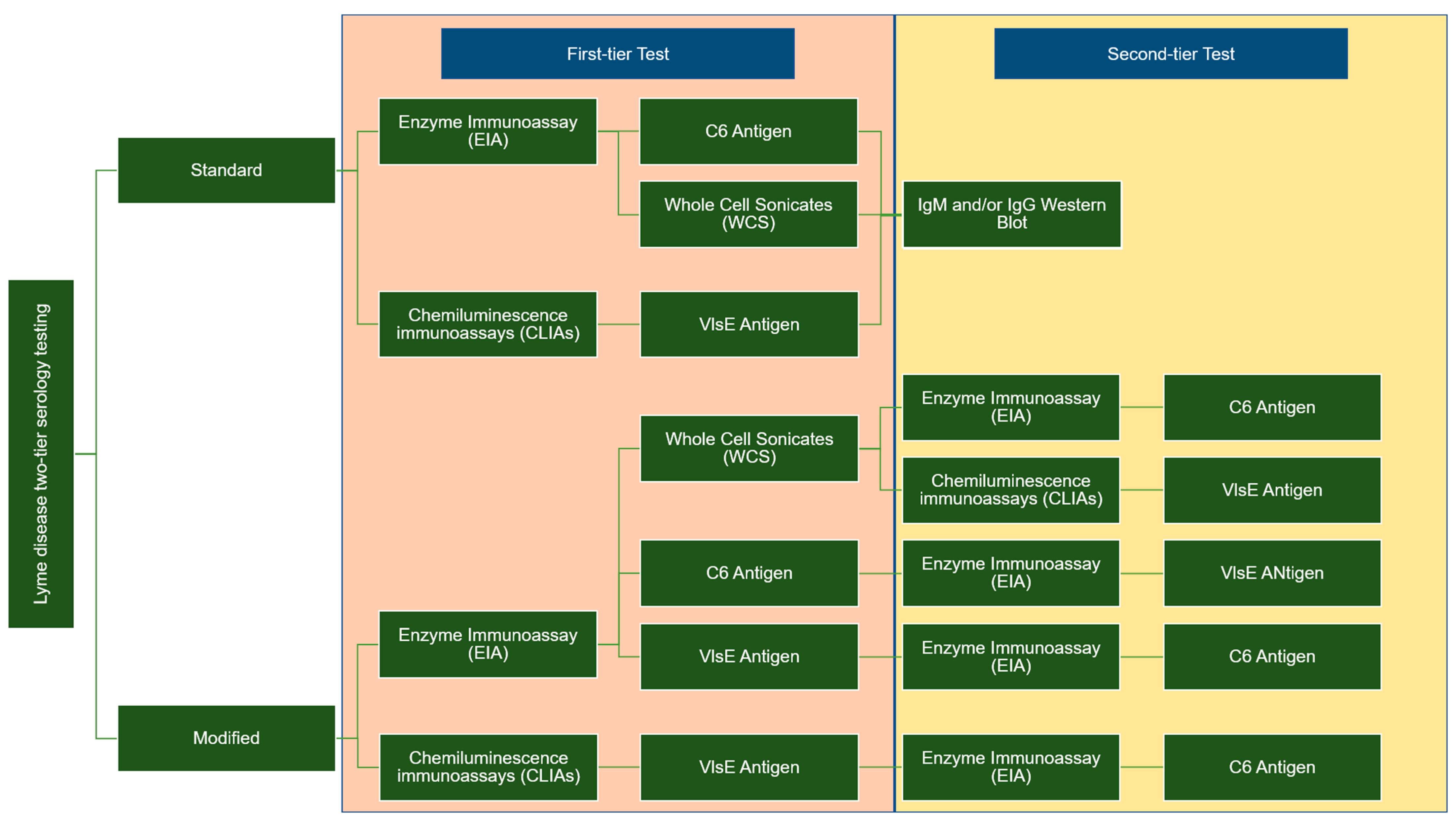
Two types of enzyme immunoassay, known as standard or modified, two-tiered serology testing (STTT/MTTT), are used to detect immunoglobulin (IgM or IgG). These methods differ in terms of the second confirmation assay; the STT uses western blotting, whereas the second enzyme immunoassay (EIA) is utilized for MTT when the sample is positive or ambiguous. Figure 1 summarises the difference between the two-tier STTT and MTTT testing [29,30,31,32].
The STTT sensitivity for LD is less than 50% in early localized infections, whereas the sensitivity can reach up to 100% in the late stages of the disease. The MTTT is consistent and more sensitive in early localized LD than STTT and demonstrated similar sensitivity in detecting late infections and specificities to STTT [33]. The two-tier testing is sufficient to rule in LD among patients who have tested positive in the early stages, but the tool has low predictive value to rule out LD, which necessitates retesting after 30 days [34]. Nevertheless, LD sensitivity in recovering patients treated at stage 1 remained low after 30 days. Resultantly, the diagnosis and treatment of early localized LD solely depend on the clinical symptoms of individuals with a history of exposure to black-legged ticks.
Early LD can be difficult to diagnose, as some individuals with localized B. burgdorferi infections do not have an EM rash and may exhibit symptoms similar to those of other diseases. As both STTT and MTTT cannot distinguish between active and past infection, B. burgdorferi antibodies can persist for months to years after the initial infection [35]. Despite the higher sensitivity than STTT, MTTT sensitivity is still <90%. Thus, patients with early localized LD should be treated based on their clinical presentation rather than serologic data [33].
The main pitfall of this assay is that it requires a complex laboratory infrastructure, producing inter- and intra-laboratory result variability, a long turnaround time, and a high cost for the immunoblot assay [36]. Eventually, this assay will only be based on yes-or-no results for routine cases of suspected Lyme disease. Other limitations include a high background rate of seropositivity in the endemic area, antibody cross-reactivity with other related bacterial infections, and false-positive results due to other medical conditions [37].
3. Direct Detection Method of Borrelia
3.1. Nucleic Acid Amplification
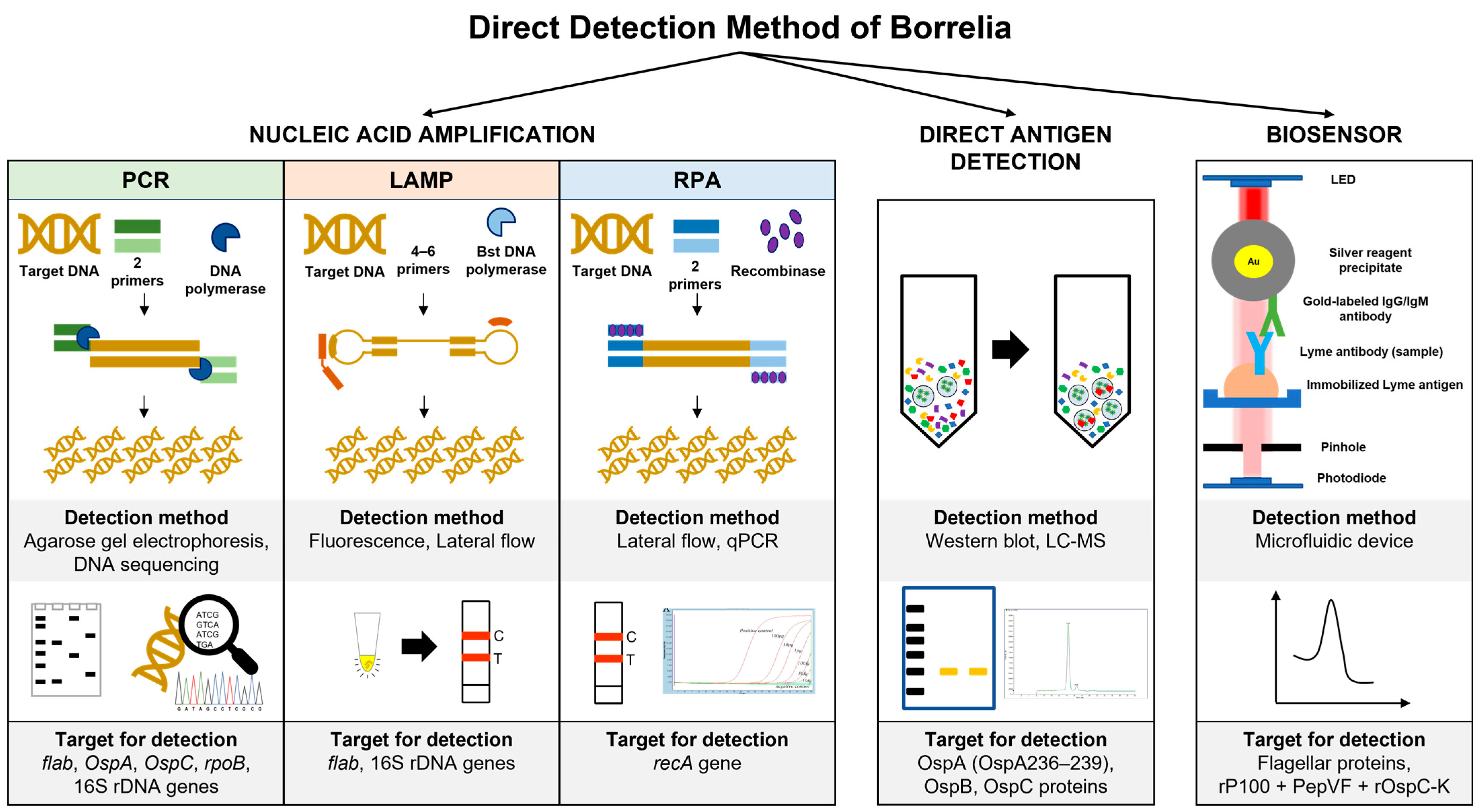
Most commercial kits are developed to target and conserve the genomic sequences of Borrelia species using two-step PCR (nested PCR). Borrelia species can be directly detected in patients’ samples, such as synovial fluids, cerebrospinal fluid (CSF), and blood, using polymerase chain reaction (PCR). The PCR assay targets several genes located on the genome sequence or linear plasmid of Borrelia species, such as flagellin B (flab), outer surface protein A (OspA), OspC rpoB, and 16S rDNA [38,39,40]. These target genes are also useful for phylogenetic analysis of Borrelia from ticks. Nevertheless, this method demonstrates various sensitivity ranges (4–100%) with 93–100% specificity [41]. Furthermore, the sensitivity ranges between 5 and 50% for EM skin biopsies compared to the CSF samples [42,43]. The low DNA recovery in patients’ samples, particularly for younger patients with limited samples, reflects the unsatisfactory sensitivity of the assay.
Several PCR protocols have been established to enhance the sensitivity of the detection method in combination with the enrichment step. For instance, the loop-mediated isothermal amplification (LAMP) assay targets the 16S rRNA and has higher sensitivity (0.2 to 0.02 pg of DNA) in detecting B. burgdorferi s.l. isolated from field-collected ticks compared to conventional and nested PCR [44]. In another study, the LAMP assay targeted the flagellin (fla) gene to detect as few as 20 copies of DNA per reaction and cross-react with 11 related bacteria [45]. Thus, the success rate of the LAMP assay is equivalent to nested PCR. Furthermore, the high amplification efficiency of the LAMP assay is due to the continuous amplification under isothermal conditions, yielding magnesium pyrophosphate as a by-product. The white-coloured precipitation is easily observed by the naked eye or by real-time turbidity monitoring using the conventional photometer [46].
Recombinase polymerase amplification (RPA) is another example of an isothermal amplification method that utilizes the recombinase protein to unwind the double-stranded DNA molecules and the strand-displacing activity to amplify DNA targets. This process takes 20–30 min from 37 °C to 42 °C [47]. The detection limit of the RPA assay targeting the recA gene is five copies, while the sensitivity is 50 femtograms of B. burgdorferi genomic DNA [48].
Even though the PCR sensitivity is high, this assay cannot differentiate between live and dead bacteria within the samples. The positive PCR result is unable to distinguish live bacteria in patients with persistent arthritis after antibiotic therapy [49]. In addition, PCR sensitivity is also highly variable depending on the type of starting materials, DNA extraction methods, target gene location, and PCR amplification method [50,51,52,53,54]. Thus, further evaluation of the PCR assay is still required to improve the assay sensitivity, and the method must be standardized to provide unambiguous diagnostic results.
3.2. Direct Antigen Detection
Borrelia antigen detection is a dependable, quick, accurate, and sensitive method for early LD diagnosis and improved patient care before acquiring disseminated LD. These assays are performed using Borrelia polyclonal antibodies, with the shed surface antigens detectable in the urine, blood, and various organs of infected hosts [55]. Nonetheless, the assay sensitivity, specificity, and accuracy in detecting several antigenic moieties (31 kDa, 34 kDa, 39 kDa, and 93 kDa) are reportedly low in invalidated clinical samples [56]. The assay can be enhanced by performing high-speed centrifugation to separate Borrelia membrane proteins from the rest of the serum proteins. B. burgdorferi cells that have been broken will release membrane vesicles containing membrane proteins into the blood. Precisely, high-speed centrifugation contributes to a low protein detection limit at approximately 4.0 fmol of ospA/mg of serum protein [57].
Hydrogel microparticles are widely used in biomedical research, such as being incorporated with chemical bait to mediate small target molecule sequestration and entrapment. This method helps concentrate the target molecules in the solution and eliminate other molecules through sieving. For instance, Douglas et al. reported that Acid Black 48 dye effectively acts as bait to concentrate the B. burgdorferi OspA and OspB proteins at a limit of detection (LOD) of 700 pg/mL in 10 mL of urine [58]. In another study, Remazol Brilliant Blue dye was used as bait for the Nanotrap technology. The Nanotrap particles were coupled with a Borrelia monoclonal antibody that targeted epitope OspA 236–239, with a detection rate as low as 1.7 pg/mL of Borrelia antigen. These studies supported the shedding of urinary OspA protein, which strongly correlates with the clinical diagnosis of the early and active stage of LD [59].
An earlier study found ten highly conserved amino acids in OspC that are significantly immunodominant and potential immunogens for anti-OspC antibody production [60]. In addition, this peptide contains a common sequence (PVVAESPKKP) found in most pathogenic Borrelia [60,61]. A solid-phase immobilized epitope immunoassay (SPIE-IA) of OspC protein was recently developed [62], despite the high variability of this protein within and between the Borrelia species. The developed SPIE-IA recorded a LOD as low as 17 pg/mL for OspC from infected animal blood samples. Moreover, the study detected a mean value of 10 ng/mL OspC in the plasma of infected mice at seven days post-infection [62].
3.3. Biosensor
Several single multiplexed assays have been developed to replace the second tier of serology assays. For example, the mChip-Ld assay targeted three Borrelia antigens (VlsE, PepVF, and OspC) and showed a sensitivity range between 80% and 100% in sample patients with early Lyme disease and Lyme arthritis, respectively [36]. Several biosensor applications have been developed to detect Borrelia antigen, including field-effect transistors (FETs) and microfluidics [63,64]. The attachment of B. Burgdorferi flagellar antibodies (p41) to nanotubes effectively detected antigens in buffer (1 ng/mL) using atomic force microscopy [63,64]. The assay utilized the turnoff voltage measurement following antigen exposure, which involves shifting the threshold voltage towards the more negative region. Furthermore, the device is highly selective for the target antigen on the flagellar protein. In a different study, the PPO triplex test (rP100 + PepVF + rOspC-K)-based microfluidic diagnostic device (mChip-Ld) was developed for antibody detection during early and late LD stages. The assay recorded 84% sensitivity and 92% specificity, comparable to the lab-based C6 peptide ELISA [64]. Figure 2 summarises the method used for direct detection of genetic material and soluble antigens of Borrelia.
4. Aptamer for Borrelia Antigen Detection
The aptamer is a single-stranded nucleic acid (RNA or DNA) that can bind with various target molecules, including surface proteins, viruses, bacteria, nanoparticles, and small analytes. The aptamer can bind to target molecules with low nanomolar and picomolar affinity and distinguish the target molecule from other similar molecular structures or functional groups [65]. Furthermore, the aptamer can form a compatible tertiary structure that fits into the binding pocket of the target molecules through hydrogen bonds and hydrophobic interaction.
The aptamer can be isolated in vitro via the Systematic Evolution of Ligands by Exponential Enrichment (SELEX). This method allows large oligonucleotide libraries to undergo several enrichment cycles of bound aptamer, which involves binding, washing, and eluting, to produce the best aptamer with high affinity and specificity towards the target molecules.
The use of aptamers as diagnostic tools has been widely explored in managing infectious diseases. Xenobiotic nucleic acid (XNA) is a new aptamer variety that has been extensively studied due to its intrinsic resistance to cellular nucleases [66]. The direct evolution of specialized XNA polymerase has promoted the enzymatic production and enrichment of XNA libraries to synthesize XNA sequences from DNA templates before being transcribed into the DNA [67,68,69]. Aptamer detection has been applied to various targets of infectious diseases, such as SARS-CoV, SARS-CoV-2, Mycobacterium tuberculosis (M. tb), and melioidosis [70,71]. Several aptamers targeting tick-borne diseases have been developed, such as surface protein E of the tick-borne encephalitis virus (TBEV) and the B. burgdorferi outer surface protein (ospA, ospC, and BmpA) associated with LD [72].
There are several advantages to the application of aptamers as a diagnostic tool, such as ease of chemical synthesis and modification, low manufacturing costs, thermal stability, and minimum batch-to-batch variation for mass production. Despite the susceptibility to nuclease degradation, the sugar ring, nucleotide bases, and phosphodiester bond modification can improve aptamer structural stability in the nuclease environment and enhance the binding affinity to the target molecules [73]. The aptamer can be modified at the 3′ or 5′ end with functional groups (thiol, amine, carboxyl) or biomolecules (biotin, fluorophore) for capturing and detection purposes [reviewed by [74,75,76].
Regarding the detection of Borrelia antigen, the aptamer can be used to detect the presence of soluble proteins released by the bacteria or whole bacteria in the collected samples. Until now, various samples have been tested to detect the presence of Borrelia directly or indirectly from blood serum, cerebrospinal fluid (CSF), urine, skin biopsy, and synovial fluid [77,78,79]. This bacteria’s antigen is often shed into the body fluids, which can later be detected using the aptamer. Interestingly, aptamers can function as reporters and capture agents for direct detection. Several surface proteins of Borrelia have potentially been used, such as OspA, OspC, flagellin, BBK32, PepVF, VlsE, BmpA, DbpA, and DbpB [64,80,81].
5. Current Aptamer Development for Borrelia Antigen Detection
Only one study has used aptamer technology to directly detect Borrelia antigen using surface-enhanced Raman spectroscopy (SERS) [82]. SERS is an ultrasensitive method for single-molecule detection, which is a powerful tool for biosensing applications in various fields. Furthermore, SERS peaks have narrow bandwidths and a characteristic molecular “fingerprint” compared to other techniques; thus, this method is ideal for multiplex detection. The unique surface plasmon resonance of metallic nanoparticles is utilized in this technology, which operates as signal-amplifying substrates and eliminates the need for pathogen culture [83]. Surface-bound, highly affine capture biomolecules provide a layer of specificity to these particles, allowing whole-cell pathogen fingerprinting.
The design of highly sensitive Raman aptasensors requires rigorous control of the three-dimensional assembled configuration to maximize SERS enhancement, particularly the local amplification of the electromagnetic (EM) field. Moreover, SERS aptasensors were developed by combining SERS probes with highly sensitive and selective aptamers for recognizing target molecules [84]. In a recent study, SERS accurately identified 91% of serum samples from Lyme patients and 96% of serum samples from symptomatic controls with a LOD of 1 × 10⁻⁴ ng/mL, more than four orders of magnitude lower than serum samples from early LD patients [82].
This assay demonstrated a 50% improvement in sensitivity without significantly reducing specificity, unlike the existing Lyme diagnostic assay. In other studies, SERS identified bacteria from complex solutions with a high capture efficiency for S. aureus (88.89%) and E. coli (74.96%) within 15 min [85,86]. The latest applications of the other SERS-based aptasensor include the detection of the SARS-CoV-2 virus, Salmonella Typhimurium, H3N2 virus, Vibrio parahaemolyticus, Staphylococcus aureus, and influenza A [87,88,89,90,91,92]. Table 1 compares the available detection methods for Lyme disease and their sensitivity.
6. Future Direction of the Use of LD Aptasensor
Integrating the aptamer technology with the available sensor platform might improve the sensitivity and specificity for direct detection of Borrelia antigen. As discussed previously, several biosensor platforms have been developed for direct antigen detection for LD using the antibody, such as microfluidics, lateral flow assays (LFAs), vertical flow assays (VFAs), surface plasmon resonance, and biochips [93]. This antibody-based biosensor can be replaced by an aptamer that might give better performance. The aptamer can easily be modified to conjugate with any nanoparticle, such as gold, graphene oxide, and fluorescence dye, which can later be used as capturing or reporter agents [94,95,96]. Nowadays, the label-free aptamer is used with quencher molecules such as graphene oxide that can turn “on” the sensor when the aptamer binds to a specific target with a detection limit of 1.26 pg/mL [97]. The yes-or-no test might suit LD diagnosis when a low amount of antigen is present in the samples and eliminates the need for an enrichment step. Other than that, aptasensor development for direct detection of Borrelia might be able to differentiate between live and dead bacteria. This could be performed using the whole-bacteria aptamer SELEX (Systematic Evolution of Ligands by Exponential Enrichment) using live Borrelia and heat-killed Borrelia. This whole-cell SELEX was successfully developed to detect live Salmonella Typhimurium and Lactobacillus acidophilus down to 600 CFU mL−1 and 106 CFU mL−1, respectively [98,99]. This aptasensor might give a superior result compared to PCR, which is unable to differentiate dead bacteria from viable bacteria. The use of aptamers for direct detection of Borrelia integrated with available or new sensor technology might change the way LD is diagnosed in the near future.
7. Conclusions
The direct detection of Borrelia antigen or whole bacterial cells is a promising tool in the early identification of LD for improved patient management. The aptamer is an advanced technology with the potential for Borrelia antigen detection. Notably, combining the latest technology with the aptamer could enhance test sensitivity and detection limits and reduce the time required to complete the assay. Furthermore, the test can function alone or complement the conventional serological test practiced in most laboratories. In summary, a fast and convenient assay may facilitate the diagnosis of the fever-like symptom possibly caused by Lyme Borrelia infection.
Author Contributions
Conceptualization, N.A.A.N.K., C.I.M. and M.A.; writing—original draft preparation, N.A.A.N.K., C.I.M. and M.A.; writing—review and editing, C.I.M. and M.A.; visualization, N.A.A.N.K. and C.I.M.; project administration, N.A.A.N.K.; funding acquisition, N.A.A.N.K. All authors have read and agreed to the published version of the manuscript.
Funding
This work is funded by the Ministry of Health, Malaysia, Grant No. (NMRR ID: NMRR-21-02063-AVY; 21-087).
Acknowledgments
The authors would like to thank the Director General of Health, Malaysia, for his permission to publish this article.
Conflicts of Interest
The authors declare no conflict of interest.
References
- Steere, A.C.; Strle, F.; Wormser, G.P.; Hu, L.T.; Branda, J.A.; Hovius, J.W.; Li, X.; Mead, P.S. Lyme borreliosis. Nat. Rev. Dis. Prim. 2016, 2, 16090. [Google Scholar] [CrossRef] [PubMed]
- Ivanova, L.; Christova, I.; Neves, V.; Aroso, M.; Meirelles, L.; Brisson, D.; Gomes-Solecki, M. Comprehensive seroprofiling of sixteen B. burgdorferi OspC: Implications for Lyme disease diagnostics design. Clin. Immunol. 2009, 132, 393–400. [Google Scholar] [CrossRef] [PubMed]
- Baranton, G.; Seinost, G.; Theodore, G.; Postic, D.; Dykhuizen, D. Distinct levels of genetic diversity of Borrelia burgdorferi are associated with different aspects of pathogenicity. Res. Microbiol. 2001, 152, 149–156. [Google Scholar] [CrossRef] [PubMed]
- Stanek, G.; Strle, F. Lyme borreliosis–from tick bite to diagnosis and treatment. FEMS Microbiol. Rev. 2018, 42, 233–258. [Google Scholar] [CrossRef]
- Shapiro, E.D.; Gerber, M.A. Lyme Disease. Clin. Infect. Dis. 2000, 31, 533–542. [Google Scholar] [CrossRef]
- Marques, A.R.; Strle, F.; Wormser, G.P. Comparison of Lyme Disease in the United States and Europe. Emerg. Infect. Dis. 2021, 27, 2017–2024. [Google Scholar] [CrossRef]
- Pritt, B.S.; Mead, P.S.; Johnson, D.K.H.; Neitzel, D.F.; Respicio-Kingry, L.B.; Davis, J.P.; Schiffman, E.; Sloan, L.M.; Schriefer, E.M.; Replogle, A.J.; et al. Identification of a novel pathogenic Borrelia species causing Lyme borreliosis with unusually high spirochaetaemia: A descriptive study. Lancet Infect. Dis. 2016, 16, 556–564. [Google Scholar] [CrossRef]
- Parola, P.; Paddock, C.D. Travel and tick-borne diseases: Lyme disease and beyond. Travel. Med. Infect. Dis. 2018, 26, 1–2. [Google Scholar] [CrossRef]
- Jorge, L.M.A.; De Janeiro, B.P.G.D.R.; Lupi, O.; Hozannah, A.R.; Filho, F.B.; Usp, B. Lyme disease in a Brazilian traveler who returned from Germany. An. Bras. Dermatol. 2017, 92, 148–149. [Google Scholar] [CrossRef]
- Soloria, H.; Adams, D. Lyme Arthritis in a Military Dependent Child transferred from Japan. Mil. Med. 2020, 185, e301–e302. [Google Scholar] [CrossRef]
- Seki, M.; Watanabe, Y.; Kawabata, H. A case of Lyme disease in a Japanese woman. Infect. Drug Resist. 2018, 11, 625–628. [Google Scholar] [CrossRef]
- Vien, V.P.; Bassi, R.; Maxim, T.; Bogoch, I.I. Lyme disease vs Baggio–Yoshinari syndrome in a returned traveller from Brazil. J. Travel Med. 2017, 24, tax055. [Google Scholar] [CrossRef] [PubMed]
- Subedi, S.; Dickeson, D.J.; Branley, J.M. First report of Lyme neuroborreliosis in a returned Australian traveller. Med. J. Aust. 2015, 203, 39–40. [Google Scholar] [CrossRef] [PubMed]
- Simon, J.A.; Marrotte, R.R.; Desrosiers, N.; Fiset, J.; Gaitan, J.; Gonzalez, A.; Koffi, J.K.; Lapointe, F.-J.; Leighton, P.A.; Lindsay, L.R.; et al. Climate change and habitat fragmentation drive the occurrence of Borrelia burgdorferi, the agent of Lyme disease, at the northeastern limit of its distribution. Evol. Appl. 2014, 7, 750–764. [Google Scholar] [CrossRef] [PubMed]
- Nelder, M.; Wijayasri, S.; Russell, C.; Johnson, K.; Marchand-Austin, A.; Cronin, K.; Johnson, S.; Badiani, T.; Patel, S.; Sider, D. The continued rise of Lyme disease in Ontario, Canada: 2017. Can. Commun. Dis. Rep. 2018, 44, 231–236. [Google Scholar] [CrossRef]
- Ogden, N.H.; Lindsay, L.R.; Morshed, M.; Sockett, P.N.; Artsob, H. The emergence of Lyme disease in Canada. Can. Med. Assoc. J. 2009, 180, 1221–1224. [Google Scholar] [CrossRef]
- Stone, B.L.; Tourand, Y.; Brissette, C.A.; Hermance, M.E.; Thangamani, S.; Knox, K.K.; Thomm, A.M.; Harrington, Y.A.; Ketter, E.; Patitucci, J.M.; et al. Brave New Worlds: The Expanding Universe of Lyme Disease. Vector Borne Zoonotic Dis. 2017, 17, 619–629. [Google Scholar] [CrossRef]
- Fang, L.-Q.; Liu, K.; Li, X.-L.; Liang, S.; Yang, Y.; Yao, H.-W.; Sun, R.-X.; Sun, Y.; Chen, W.-J.; Zuo, S.-Q.; et al. Emerging tick-borne infections in mainland China: An increasing public health threat. Lancet Infect. Dis. 2015, 15, 1467–1479. [Google Scholar] [CrossRef]
- Mariana, A.; Zuraidawati, Z.; Ho, T.M.; Kulaimi, B.M.; Saleh, I.; Shukor, M.N.; Shahrul-Anuar, M.S. Ticks (Ixodidae) and other ectoparasites in Ulu Muda Forest Reserve, Kedah, Malaysia. Southeast Asian J. Trop. Med. Public Health 2008, 39, 496–506. [Google Scholar]
- Mariana, A.; Zuraidawati, Z.; Ho, T.M.; Mohd Kulaimi, B.; Saleh, I.; Shukor, M.N.; Shahrul-Anuar, M.S. A survey of ectoparasites in Gunung Stong Forest Reserve, Kelantan, Malaysia. Southeast Asian J. Trop. Med. Public Health 2005, 36, 1125–1131. [Google Scholar]
- Khoo, J.J.; Ishak, S.N.; Lim, F.S.; Mohd-Taib, F.S.; Khor, C.S.; Loong, S.K.; AbuBakar, S. Detection of a Borrelia sp. From Ixodes granulatus Ticks Collected From Rodents in Malaysia. J. Med. Entomol. 2018, 55, 1642–1647. [Google Scholar] [CrossRef]
- Paramasvaran, S.; Sani, A.R.; Hassan, L.; Krishnasamy, M.; Jeffery, J.; Oothuman, P.; Salleh, I.; Lim, K.H.; Sumarni, M.G.; Santhana, R.L. Ectoparasite fauna of rodents and shrews from four habitats in Kuala Lumpur and the states of Selangor and Negeri Sembilan, Malaysia and its public health significance. Trop. Biomed. 2009, 26, 303–311. [Google Scholar]
- Madinah, A.; Fatimah, A.; Mariana, A.; Abdullah, M.T. Ectoparasites of small mammals in four localities of wildlife reserves in Peninsular Malaysia. Southeast Asian J. Trop. Med. Public Health 2011, 42, 803–813. [Google Scholar] [PubMed]
- Mariana, A.; Mohd, K.B.; Halimaton, I.; Suhaili, Z.A.; Shahrul-Anuar, M.S.; Nor, Z.M.; Ho, T.M. Acarine ectoparasites of Panti Forest Reserve in Johore, Malaysia. Asian Pac. J. Trop. Biomed. 2011, 1, 1–5. [Google Scholar] [CrossRef] [PubMed]
- Lau, A.C.C.; Qiu, Y.; Moustafa, M.A.M.; Nakao, R.; Shimozuru, M.; Onuma, M.; Mohd-Azlan, J.; Tsubota, T. Detection of Borrelia burgdorferi Sensu Lato and Relapsing Fever Borrelia in Feeding Ixodes Ticks and Rodents in Sarawak, Malaysia: New Geographical Records of Borrelia yangtzensis and Borrelia miyamotoi. Pathogens 2020, 9, 846. [Google Scholar] [CrossRef]
- Saito, K.; Ito, T.; Asashima, N.; Ohno, M.; Nagai, R.; Fujita, H.; Koizumi, N.; Takano, A.; Watanabe, H.; Kawabata, H. Case report: Borrelia valaisiana infection in a Japanese man associated with traveling to foreign countries. Am. J. Trop. Med. Hyg. 2007, 77, 1124–1127. [Google Scholar] [CrossRef] [PubMed]
- Ni, X.B.; Jia, N.; Jiang, B.G.; Sun, T.; Zheng, Y.C.; Huo, Q.B.; Liu, K.; Ma, L.; Zhao, Q.-M.; Yang, H.; et al. Lyme borreliosis caused by diverse genospecies of Borrelia burgdorferi sensu lato in northeastern China. Clin. Microbiol. Infect. Off. Publ. Eur. Soc. Clin. Microbiol. Infect. Dis. 2014, 20, 808–814. [Google Scholar] [CrossRef]
- Chee-Sieng, K.; Habibi, H.; Nurul-Farhana, M.-R.; Josephine Rebecca, C.; Siti-Sarah, N.; Jefree, J.; Loong, S.-K.; Abd-Jamil, J.; Khoo, J.-J.; Lee, H.-Y. Seroprevalence of Borrelia burgdorferi among the indigenous people (Orang Asli) of Peninsular Malaysia. J. Infect. Dev. Ctries. 2019, 13, 449–454. [Google Scholar]
- Branda, J.A.; Linskey, K.; Kim, Y.A.; Steere, A.C.; Ferraro, M.J. Two-tiered antibody testing for Lyme disease with use of 2 enzyme immunoassays, a whole-cell sonicate enzyme immunoassay followed by a VlsE C6 peptide enzyme immunoassay. Clin. Infect. Dis. Off. Publ. Infect. Dis. Soc. Am. 2011, 53, 541–547. [Google Scholar] [CrossRef]
- Molins, C.R.; Delorey, M.J.; Sexton, C.; Schriefer, M.E. Lyme Borreliosis Serology: Performance of Several Commonly Used Laboratory Diagnostic Tests and a Large Resource Panel of Well-Characterized Patient Samples. J. Clin. Microbiol. 2016, 54, 2726–2734. [Google Scholar] [CrossRef]
- Lipsett, S.C.; Branda, J.A.; McAdam, A.J.; Vernacchio, L.; Gordon, C.D.; Gordon, C.R.; Gordon, C.R.; Nigrovic, L.E. Evaluation of the C6 Lyme Enzyme Immunoassay for the Diagnosis of Lyme Disease in Children and Adolescents. Clin. Infect. Dis. 2016, 63, 922–928. [Google Scholar] [CrossRef] [PubMed]
- Pegalajar-Jurado, A.; Schriefer, M.E.; Welch, R.J.; Couturier, M.R.; MacKenzie, T.; Clark, R.J.; Ashton, L.V.; Delorey, M.J.; Molins, C.R. Evaluation of Modified Two-Tiered Testing Algorithms for Lyme Disease Laboratory Diagnosis Using Well-Characterized Serum Samples. J. Clin. Microbiol. 2018, 56, e01943-17. [Google Scholar] [CrossRef] [PubMed]
- Hatchette, L.; Lindsay, R. Modified two-tiered testing algorithm for Lyme disease serology: The Canadian context. Can. Commun. Dis. Rep. 2020, 46, 125–131. [Google Scholar] [CrossRef] [PubMed]
- Waddell, L.A.; Greig, J.; Mascarenhas, M.; Harding, S.; Lindsay, R.; Ogden, N. The Accuracy of Diagnostic Tests for Lyme Disease in Humans, A Systematic Review and Meta-Analysis of North American Research. PLoS ONE 2016, 11, e0168613. [Google Scholar] [CrossRef]
- Kalish, R.A.; McHugh, G.; Granquist, J.; Shea, B.; Ruthazer, R.; Steere, A.C. Persistence of immunoglobulin M or immunoglobulin G antibody responses to Borrelia burgdorferi 10–20 years after active Lyme disease. Clin. Infect. Dis. Off. Publ. Infect. Dis. Soc. Am. 2001, 33, 780–785. [Google Scholar] [CrossRef]
- Arumugam, S.; Nayak, S.; Williams, T.; Maria, F.S.d.S.; Guedes, M.S.; Chaves, R.C.; Linder, V.; Marques, A.R.; Horn, E.J.; Wong, S.J.; et al. A Multiplexed Serologic Test for Diagnosis of Lyme Disease for Point-of-Care Use. J. Clin. Microbiol. 2019, 57, e01142-19. [Google Scholar] [CrossRef]
- Teny, M.J.; Alan, J.T. Appropriate laboratory testing in Lyme disease. Clevel. Clin. J. Med. 2019, 86, 751. [Google Scholar]
- Das, S.; Hammond-McKibben, D.; Guralski, D.; Lobo, S.; Fiedler, P.N. Development of a sensitive molecular diagnostic assay for detecting Borrelia burgdorferi DNA from the blood of Lyme disease patients by digital PCR. PLoS ONE 2020, 15, e0235372. [Google Scholar] [CrossRef]
- Cerar, T.; Korva, M.; Avšič-Županc, T.; Ružić-Sabljić, E. Detection, identification and genotyping of Borrellia spp. in rodents in Slovenia by PCR and culture. BMC Vet. Res. 2015, 11, 188. [Google Scholar] [CrossRef]
- Cerar, T.; Ružić-Sabljić, E.; Glinšek, U.; Zore, A.; Strle, F. Comparison of PCR methods and culture for the detection of Borrelia spp. in patients with erythema migrans. Clin. Microbiol. Infect. 2008, 14, 653–658. [Google Scholar] [CrossRef]
- Aguero-Rosenfeld, M.E.; Wang, G.; Schwartz, I.; Wormser, G.P. Diagnosis of lyme borreliosis. Clin. Microbiol. Rev. 2005, 18, 484–509. [Google Scholar] [CrossRef] [PubMed]
- Mygland, Å.; Ljøstad, U.; Fingerle, V.; Rupprecht, T.; Schmutzhard, E.; Steiner, I. EFNS guidelines on the diagnosis and management of European Lyme neuroborreliosis. Eur. J. Neurol. 2010, 17, 8–16.e1–4. [Google Scholar] [CrossRef] [PubMed]
- Picha, D.; Moravcova, L.; Zdarsky, E.; Maresova, V.; Hulinsky, V. PCR in lyme neuroborreliosis: A prospective study. Acta Neurol. Scand. 2005, 112, 287–292. [Google Scholar] [CrossRef] [PubMed]
- Yang, J.; Guan, G.; Niu, Q.; Liu, Z.; Li, Y.; Liu, J.; Ma, M.; Ren, Q.; Liu, A.; Luo, J.; et al. Development and application of a loop-mediated isothermal amplification assay for rapid detection of Borrelia burgdorferi s. l. in ticks. Transbound. Emerg. Dis. 2013, 60, 238–244. [Google Scholar] [CrossRef] [PubMed]
- Zhang, L.L.; Hou, X.X.; Geng, Z.; Lou, Y.L.; Wan, K.L.; Hao, Q. Combination of Loop-Mediated Isothermal Amplification Assay and Nested PCR for Detection of Borrelia burgdorferi sensu lato in Human Serum Samples. Biomed. Environ. Sci. BES 2015, 28, 312–315. [Google Scholar]
- Mori, Y.; Nagamine, K.; Tomita, N.; Notomi, T. Detection of loop-mediated isothermal amplification reaction by turbidity derived from magnesium pyrophosphate formation. Biochem. Biophys. Res. Commun. 2001, 289, 150–154. [Google Scholar] [CrossRef]
- Lobato, I.M.; O’Sullivan, C.K. Recombinase polymerase amplification: Basics, applications and recent advances. TrAC Trends Anal. Chem. 2018, 98, 19–35. [Google Scholar] [CrossRef]
- Liu, W.; Liu, H.-X.; Zhang, L.; Hou, X.-X.; Wan, K.-L.; Hao, Q. A Novel Isothermal Assay of Borrelia burgdorferi by Recombinase Polymerase Amplification with Lateral Flow Detection. Int. J. Mol. Sci. 2016, 17, 1250. [Google Scholar] [CrossRef]
- Li, X.; McHugh, G.A.; Damle, N.; Sikand, V.K.; Glickstein, L.; Steere, A.C. Burden and viability of Borrelia burgdorferi in skin and joints of patients with erythema migrans or lyme arthritis. Arthritis Rheum. 2011, 63, 2238–2247. [Google Scholar] [CrossRef]
- van Dam, A.P. Molecular diagnosis of Borrelia bacteria for the diagnosis of Lyme disease. Expert Opin. Med. Diagn. 2011, 5, 135–149. [Google Scholar] [CrossRef]
- Bonin, S. Diagnostic Tools for Assessment in Humans. Open Dermatol. J. 2016, 10, 62–69. [Google Scholar] [CrossRef]
- Ružić-Sabljić, E.; Cerar, T. Progress in the molecular diagnosis of Lyme disease. Expert Rev. Mol. Diagn. 2017, 17, 19–30. [Google Scholar] [CrossRef] [PubMed]
- Trevisan, G.; Bonin, S.; Ruscio, M. A Practical Approach to the Diagnosis of Lyme Borreliosis: From Clinical Heterogeneity to Laboratory Methods. Front. Med. 2020, 7, 265. [Google Scholar] [CrossRef] [PubMed]
- Nolte, O. Nucleic Acid Amplification Based Diagnostic of Lyme (Neuro-)borreliosis—Lost in the Jungle of Methods, Targets, and Assays? Open Neurol. J. 2012, 6, 129–139. [Google Scholar] [CrossRef]
- Dorward, D.W.; Schwan, T.G.; Garon, C.F. Immune capture and detection of Borrelia burgdorferi antigens in urine, blood, or tissues from infected ticks, mice, dogs, and humans. J. Clin. Microbiol. 1991, 29, 1162–1170. [Google Scholar] [CrossRef]
- Klempner, M.S.; Schmid, C.H.; Hu, L.; Steere, A.C.; Johnson, G.; McCloud, B.; Noring, R.; Weinstein, A. Intralaboratory reliability of serologic and urine testing for Lyme disease. Am. J. Med. 2001, 110, 217–219. [Google Scholar] [CrossRef]
- Cheung, C.S.F.; Anderson, K.W.; Benitez, K.Y.V.; Soloski, M.J.; Aucott, J.N.; Phinney, K.W.; Turko, I.V. Quantification of Borrelia burgdorferi Membrane Proteins in Human Serum: A New Concept for Detection of Bacterial Infection. Anal. Chem. 2015, 87, 11383–11388. [Google Scholar] [CrossRef]
- Douglas, T.A.; Tamburro, D.; Fredolini, C.; Espina, B.H.; Lepene, B.S.; Ilag, L.; Espina, V.; Petricoin, E.F.; Liotta, L.A.; Luchini, A. The use of hydrogel microparticles to sequester and concentrate bacterial antigens in a urine test for Lyme disease. Biomaterials 2011, 32, 1157–1166. [Google Scholar] [CrossRef]
- Magni, R.; Espina, B.H.; Shah, K.; Lepene, B.; Mayuga, C.; Douglas, T.A.; Espina, V.; Rucker, S.; Dunlap, R.; Petricoin, E.F.I.; et al. Application of Nanotrap technology for high sensitivity measurement of urinary outer surface protein A carboxyl-terminus domain in early stage Lyme borreliosis. J. Transl. Med. 2015, 13, 346. [Google Scholar] [CrossRef]
- Earnhart, C.G.; Rhodes, D.V.L.; Smith, A.A.; Yang, X.; Tegels, B.; Carlyon, J.A.; Pal, U.; Marconi, R.T. Assessment of the potential contribution of the highly conserved C-terminal motif (C10) of Borrelia burgdorferi outer surface protein C in transmission and infectivity. Pathog. Dis. 2014, 70, 176–184. [Google Scholar] [CrossRef]
- Mathiesen, M.J.; Christiansen, M.; Hansen, K.; Holm, A.; Åsbrink, E.; Theisen, M. Peptide-based OspC enzyme-linked immunosorbent assay for serodiagnosis of Lyme borreliosis. J. Clin. Microbiol. 1998, 36, 3474–3479. [Google Scholar] [CrossRef] [PubMed]
- Dolange, V.; Simon, S.; Morel, N. Detection of Borrelia burgdorferi antigens in tissues and plasma during early infection in a mouse model. Sci. Rep. 2021, 11, 17368. [Google Scholar] [CrossRef] [PubMed]
- Lerner, M.B.; Dailey, J.; Goldsmith, B.R.; Brisson, D.; Johnson, A.C. Detecting Lyme disease using antibody-functionalized single-walled carbon nanotube transistors. Biosens. Bioelectron. 2013, 45, 163–167. [Google Scholar] [CrossRef]
- Nayak, S.; Sridhara, A.; Melo, R.; Richer, L.; Chee, N.H.; Kim, J.; Linder, V.; Steinmiller, D.; Sia, S.K.; Gomes-Solecki, M. Microfluidics-based point-of-care test for serodiagnosis of Lyme Disease. Sci. Rep. 2016, 6, 35069. [Google Scholar] [CrossRef] [PubMed]
- Ruscito, A.; DeRosa, M.C. Small-Molecule Binding Aptamers: Selection Strategies, Characterization, and Applications. Front. Chem. 2016, 4, 14. [Google Scholar] [CrossRef] [PubMed]
- Eremeeva, E.; Fikatas, A.; Margamuljana, L.; Abramov, M.; Schols, D.; Groaz, E.; Herdewijn, P. Highly stable hexitol based XNA aptamers targeting the vascular endothelial growth factor. Nucleic Acids Res. 2019, 47, 4927–4939. [Google Scholar] [CrossRef]
- Pinheiro, V.B.; Taylor, A.I.; Cozens, C.; Abramov, M.; Renders, M.; Zhang, S.; Chaput, J.C.; Wengel, J.; Peak-Chew, S.-Y.; McLaughlin, S.H.; et al. Synthetic genetic polymers capable of heredity and evolution. Science 2012, 336, 341–344. [Google Scholar] [CrossRef]
- Dunn, M.R.; Otto, C.; Fenton, K.E.; Chaput, J.C. Improving Polymerase Activity with Unnatural Substrates by Sampling Mutations in Homologous Protein Architectures. ACS Chem. Biol. 2016, 11, 1210–1219. [Google Scholar] [CrossRef]
- Peng, C.G.; Damha, M.J. Polymerase-directed synthesis of 2′-deoxy-2′-fluoro-beta-D-arabinonucleic acids. J. Am. Chem. Soc. 2007, 129, 5310–5311. [Google Scholar] [CrossRef]
- Chen, Z.; Wum, Q.; Chen, J.; Ni, X.; Dai, J. A DNA Aptamer Based Method for Detection of SARS-CoV-2 Nucleocapsid Protein. Virol. Sin. 2020, 35, 351–354. [Google Scholar] [CrossRef]
- Ahn, D.G.; Jeon, I.J.; Kim, J.D.; Song, M.S.; Han, S.R.; Lee, S.W.; Jung, H.; Oh, J.-W. RNA aptamer-based sensitive detection of SARS coronavirus nucleocapsid protein. Analyst 2009, 134, 1896–1901. [Google Scholar] [CrossRef] [PubMed]
- Kondratov, I.G.; Khasnatinov, M.A.; Potapova, U.V.; Potapov, V.V.; Levitskii, S.A.; Leonova, G.N.; Pavlenko, E.V.; Solovarov, I.S.; Denikina, N.N.; Kulakova, N.V.; et al. Obtaining aptamers to a fragment of surface protein E of tick-borne encephalitis virus. Dokl. Biochem. Biophys. 2013, 448, 19–21. [Google Scholar] [CrossRef] [PubMed]
- Ni, S.; Yao, H.; Wang, L.; Lu, J.; Jiang, F.; Lu, A.; Zhang, G. Chemical Modifications of Nucleic Acid Aptamers for Therapeutic Purposes. Int. J. Mol. Sci. 2017, 18, 1683. [Google Scholar] [CrossRef] [PubMed]
- Odeh, F.; Nsairat, H.; Alshaer, W.; Ismail, M.A.; Esawi, E.; Qaqish, B.; Al Bawab, A.; Ismail, S.I. Aptamers Chemistry: Chemical Modifications and Conjugation Strategies. Molecules 2019, 25, 3. [Google Scholar] [CrossRef]
- Ni, S.; Zhuo, Z.; Pan, Y.; Yu, Y.; Li, F.; Liu, J.; Wang, L.; Wu, X.; Li, D.; Wan, Y. Recent Progress in Aptamer Discoveries and Modifications for Therapeutic Applications. ACS Appl. Mater. Interfaces 2021, 13, 9500–9519. [Google Scholar] [CrossRef]
- Elskens, J.P.; Elskens, J.M.; Madder, A. Chemical Modification of Aptamers for Increased Binding Affinity in Diagnostic Applications: Current Status and Future Prospects. Int. J. Mol. Sci. 2020, 21, 4522. [Google Scholar] [CrossRef]
- Priem, S.; Burmester, G.R.; Kamradt, T.; Wolbart, K.; Rittig, M.G.; Krause, A. Detection of Borrelia burgdorferi by polymerase chain reaction in synovial membrane, but not in synovial fluid from patients with persisting Lyme arthritis after antibiotic therapy. Ann. Rheum. Dis. 1998, 57, 118–121. [Google Scholar] [CrossRef]
- Santino, I.; Berlutti, F.; Pantanella, F.; Sessa, R.; Del Piano, M. Detection of Borrelia burgdorferi sensu lato DNA by PCR in serum of patients with clinical symptoms of Lyme borreliosis. FEMS Microbiol. Lett. 2008, 283, 30–35. [Google Scholar] [CrossRef]
- Johnson, B.J. Laboratory Diagnostic Testing for Borrelia burgdorferi Infection. Lyme Disease: An Evidence-Based Approach; CABI: Wallingford, UK, 2011; pp. 73–88. [Google Scholar]
- Flynn, C.D.; Sandomierski, M.; Kim, K.; Lewis, J.; Lloyd, V.; Ignaszak, A. Electrochemical Detection of Borrelia burgdorferi Using a Biomimetic Flow Cell System. ACS Meas. Sci. Au 2023, 3, 208–216. [Google Scholar] [CrossRef]
- Dattwyler, R.J.; Gomes-Solecki, M. Peptide Diagnostic Agent for Lyme Disease. U.S. Patent 7887815B2, 15 February 2011. Google Patents. [Google Scholar]
- Tabb, J.S.; Rapoport, E.; Han, I.; Lombardi, J.; Green, O. An antigen-targeting assay for Lyme disease: Combining aptamers and SERS to detect the OspA protein. Nanomed. Nanotechnol. Biol. Med. 2022, 41, 102528. [Google Scholar] [CrossRef]
- Luo, X.; Zhao, X.; Wallace, G.Q.; Brunet, M.-H.; Wilkinson, K.J.; Wu, P.; Cai, C.; Bazuin, C.G.; Masson, J.-F. Multiplexed SERS Detection of Microcystins with Aptamer-Driven Core–Satellite Assemblies. ACS Appl. Mater. Interfaces 2021, 13, 6545–6556. [Google Scholar] [CrossRef] [PubMed]
- Xu, X.; Ma, X.; Wang, H.; Wang, Z. Aptamer based SERS detection of Salmonella Typhimurium using DNA-assembled gold nanodimers. Microchim. Acta 2018, 185, 325. [Google Scholar] [CrossRef] [PubMed]
- Zhang, C.; Wang, C.; Xiao, R.; Tang, L.; Huang, J.; Wu, D.; Liu, S.; Wang, Y.; Zhang, D.; Wang, S.; et al. Sensitive and specific detection of clinical bacteria via vancomycin-modified Fe3O4@Au nanoparticles and aptamer-functionalized SERS tags. J. Mater. Chem. B 2018, 6, 3751–3761. [Google Scholar] [CrossRef] [PubMed]
- Pang, Y.; Wan, N.; Shi, L.; Wang, C.; Sun, Z.; Xiao, R.; Wang, S. Dual-recognition surface-enhanced Raman scattering(SERS)biosensor for pathogenic bacteria detection by using vancomycin-SERS tags and aptamer-Fe3O4@Au. Anal. Chim. Acta 2019, 1077, 288–296. [Google Scholar] [CrossRef]
- Zavyalova, E.; Ambartsumyan, O.; Zhdanov, G.; Gribanyov, D.; Gushchin, V.; Tkachuk, A.; Rudakova, E.; Nikiforova, M.; Kuznetsova, N.; Popova, L. SERS-Based Aptasensor for Rapid Quantitative Detection of SARS-CoV-2. Nanomaterials 2021, 11, 1394. [Google Scholar] [CrossRef]
- Ma, X.; Xu, X.; Xia, Y.; Wang, Z. SERS Aptasensor for Salmonella Typhimurium Detection based on Spiny Gold Nanoparticles. Food Control. 2017, 84, 232–237. [Google Scholar] [CrossRef]
- Kukushkin, V.I.; Ivanov, N.M.; Novoseltseva, A.A.; Gambaryan, A.S.; Yaminsky, I.V.; Kopylov, A.M.; Zavyalova, F.G. Highly sensitive detection of influenza virus with SERS aptasensor. PLoS ONE 2019, 14, e0216247. [Google Scholar] [CrossRef]
- Chen, H.; Park, S.-G.; Choi, N.; Moon, J.-I.; Dang, H.; Das, A.; Lee, S.; Kim, D.-G.; Chen, L.; Choo, J. SERS imaging-based aptasensor for ultrasensitive and reproducible detection of influenza virus A. Biosens. Bioelectron. 2020, 167, 112496. [Google Scholar] [CrossRef]
- Duan, N.; Shen, M.; Wu, S.; Zhao, C.; Ma, X.; Wang, Z. Graphene oxide wrapped Fe3O4@Au nanostructures as substrates for aptamer-based detection of Vibrio parahaemolyticus by surface-enhanced Raman spectroscopy. Microchim. Acta 2017, 184, 2653–2660. [Google Scholar] [CrossRef]
- Xie, B.; Wang, Z.-P.; Zhang, R.; Zhang, Z.; He, Y. A SERS aptasensor based on porous Au-NC nanoballoons for Staphylococcus aureus detection. Anal. Chim. Acta 2022, 1190, 339175. [Google Scholar] [CrossRef]
- Flynn, C.; Ignaszak, A. Lyme Disease Biosensors: A Potential Solution to a Diagnostic Dilemma. Biosensors 2020, 10, 137. [Google Scholar] [CrossRef] [PubMed]
- He, B. Sandwich electrochemical thrombin assay using a glassy carbon electrode modified with nitrogen- and sulfur-doped graphene oxide and gold nanoparticles. Microchim. Acta 2018, 185, 344. [Google Scholar] [CrossRef] [PubMed]
- Kim, H.-K.; Kim, H.-R.; Yoon, S.-J.; Lee, K.-B.; Kim, J.; Kim, B.-C. Colorimetric Aptasensor for Detecting Bacillus carboniphilus Using Aptamer Isolated with a Non-SELEX-Based Method. Chemosensors 2021, 9, 121. [Google Scholar] [CrossRef]
- Zhang, Z.; Yang, J.; Pang, W.; Yan, G. An aptamer-based fluorescence probe for facile detection of lipopolysaccharide in drinks. RSC Adv. 2017, 7, 54920–54926. [Google Scholar] [CrossRef]
- Zhang, S.; Ma, L.; Ma, K.; Xu, B.; Liu, L.; Tian, W. Label-Free Aptamer-Based Biosensor for Specific Detection of Chloramphenicol Using AIE Probe and Graphene Oxide. ACS Omega 2018, 3, 12886–12892. [Google Scholar] [CrossRef]
- Labib, M.; Zamay, A.S.; Kolovskaya, O.S.; Reshetneva, I.T.; Zamay, G.S.; Kibbee, R.J.; Sattar, S.A.; Zamay, T.N.; Berezovski, M.V. Aptamer-Based Viability Impedimetric Sensor for Bacteria. Anal. Chem. 2012, 84, 8966–8969. [Google Scholar] [CrossRef]
- Urmann, K.; Arshavsky-Graham, S.; Walter, J.G.; Scheper, T.; Segal, E. Whole-cell detection of live Lactobacillus acidophilus on aptamer-decorated porous silicon biosensors. Analyst 2016, 141, 5432–5440. [Google Scholar] [CrossRef]
Figure 1.
Differences between STTT and MTTT: Two-tier testing methods for LD diagnosis.

Figure 2.
Direct detection method of Borrelia using isolated genetic material and soluble antigen in various samples. The biosensor detection diagram was modified from [54], which is open access under the Creative Commons Attribution 4.0 International license.
Figure 2.
Direct detection method of Borrelia using isolated genetic material and soluble antigen in various samples. The biosensor detection diagram was modified from [54], which is open access under the Creative Commons Attribution 4.0 International license.


Table 1.
The comparison of the type of detection, assay sensitivity, and limit of detection for Lyme disease diagnostic and Borrelia direct detection.
Table 1.
The comparison of the type of detection, assay sensitivity, and limit of detection for Lyme disease diagnostic and Borrelia direct detection.
| Detection Methods | Target Molecules | Sensitivity | LOD | Reference |
|---|---|---|---|---|
| Nested PCR | p66, 16S rRNA gene, fla gene, 23S rRNA gene, 5S rRNA-23S rRNA gene spacer, recA gene, OspA gene | 4–100% | ND | [41,42,43] |
| LAMP | 16S rRNA | 32.7% | 0.2 to 0.02 pg | [44] |
| fla gene | 37.5% | 20 copies | [45] | |
| RPA | recA gene | 90% | 50 femtograms | [48] |
| Hydrogel microparticles | B. burgdorferi OspA and OspB | ND | 700 pg/mL | [58] |
| Nanotrap technology | OspA 236–239 | 87.5% | 1.7 pg/mL | [59] |
| SPIE-IA | OspC | ND | 10 ng/mL–17 pg/mL | [62] |
| mChip-Ld | VlsE, PepVF, OspC | 80% to 100% | ND | [36] |
| FETs | p41 | Unavailable | 1 ng/mL | [63] |
| Microfluidics | rP100, PepVF, rOspC-K | 84% | ND | [64] |
| SERS (aptasensor) | OspA | 91–96% | 1 × 10⁴ ng/mL | [83] |
Disclaimer/Publisher’s Note: The statements, opinions and data contained in all publications are solely those of the individual author(s) and contributor(s) and not of MDPI and/or the editor(s). MDPI and/or the editor(s) disclaim responsibility for any injury to people or property resulting from any ideas, methods, instructions or products referred to in the content. |
© 2023 by the authors. Licensee MDPI, Basel, Switzerland. This article is an open access article distributed under the terms and conditions of the Creative Commons Attribution (CC BY) license (https://creativecommons.org/licenses/by/4.0/).
Share and Cite
MDPI and ACS Style
Nik Kamarudin, N.A.A.; Mawang, C.I.; Ahamad, M. Direct Detection of Lyme Borrelia: Recent Advancement and Use of Aptamer Technology. Biomedicines 2023, 11, 2818. https://doi.org/10.3390/biomedicines11102818
AMA Style
Nik Kamarudin NAA, Mawang CI, Ahamad M. Direct Detection of Lyme Borrelia: Recent Advancement and Use of Aptamer Technology. Biomedicines. 2023; 11(10):2818. https://doi.org/10.3390/biomedicines11102818
Chicago/Turabian StyleNik Kamarudin, Nik Abdul Aziz, Christina Injan Mawang, and Mariana Ahamad. 2023. "Direct Detection of Lyme Borrelia: Recent Advancement and Use of Aptamer Technology" Biomedicines 11, no. 10: 2818. https://doi.org/10.3390/biomedicines11102818
Note that from the first issue of 2016, this journal uses article numbers instead of page numbers. See further details here.