
Investigating the Role of Circulating miRNAs as Biomarkers in Colorectal Cancer: An Epidemiological Systematic Review
 , , , , ,
Abstract
:1. Introduction
2. Materials and Methods
2.1. Inclusion and Exclusion Criteria
2.2. Data Extraction
2.3. Quality Assessment
2.4. Statistical Analysis
3. Results
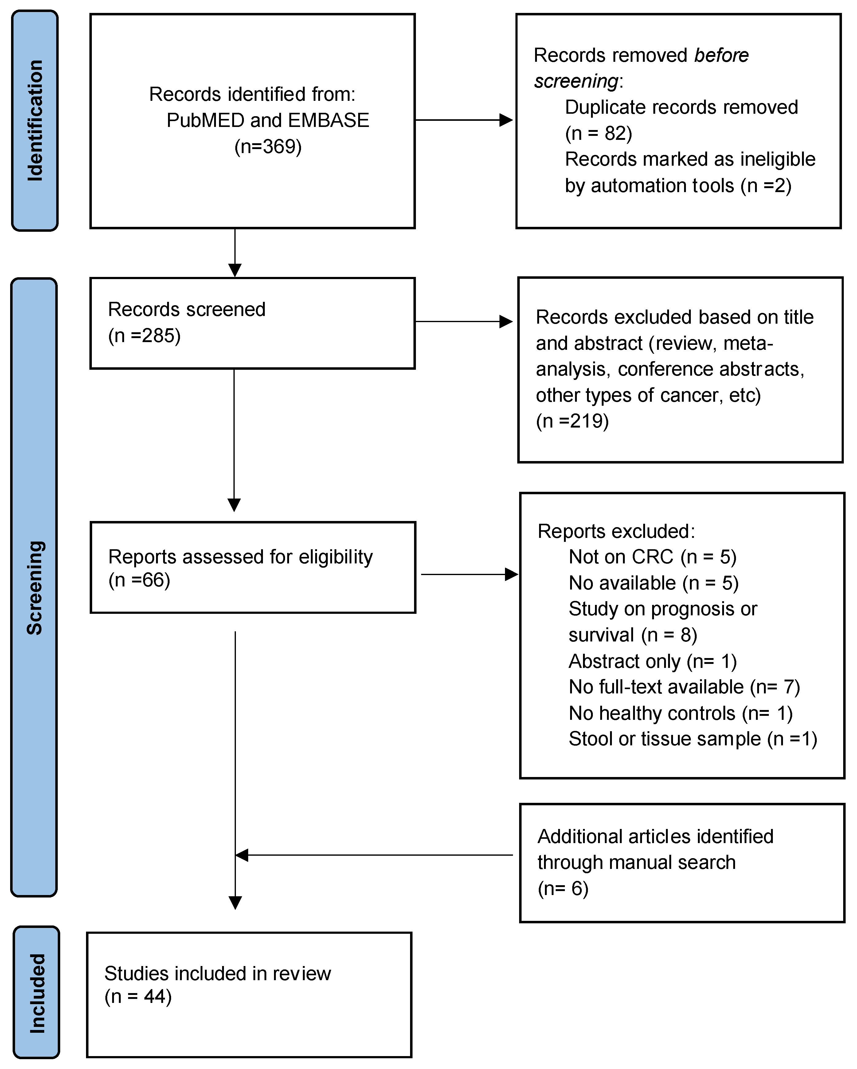
3.1. Systematic Review

| First Author, Year | Country | Cases (Mean Age + SD/ Median + Range) | Controls (Mean Age + SD/ Median + Range) | Biological Sample | miRNA | miRNA Source | Expression |
|---|---|---|---|---|---|---|---|
| Silva, 2021 [13] | Brazil | 41 - | 68 - | plasma | miR-106a-5p | discovery | up |
| miR-542-5p | discovery | up | |||||
| let-7e-5p | discovery | up | |||||
| miR-28-3p | discovery | up | |||||
| Han, 2021 [14] | China | 123 (51.60 ± 11.4) | 150 (52.30 ± 11.25) | serum | miR-15b | candidate | up |
| miR-16 | candidate | up | |||||
| miR-21 | candidate | up | |||||
| miR-31 | candidate | up | |||||
| Panel miRNA-15, miRNA-21, MiRNA-32 | candidate | ||||||
| Peng, 2020 [15] | China | 80 (61.08 ± 12.69) | 88 (60.93 ± 10.89) | serum | miR-30e-3p | discovery | up |
| miR-31-5p | discovery | up | |||||
| miR-34b-3p | discovery | up | |||||
| miR-146a-5p | discovery | up | |||||
| miR-148a-3p | discovery | down | |||||
| miR-192-5p | discovery | down | |||||
| Liu, 2020 [16] | China | 80 - | 23 - | plasma | miRNA-139-3p | candidate | down |
| Pastor-navarro, 2020 [17] | Spain | 27 (70; 43–86) | 45 (56.5; 50–73) | serum | miR-21 | candidate | up |
| Li, 2020 [18] | China | 597 (62.89) | 585 (57.17) | plasma | miR-20b-5p | discovery | up |
| miR-329-3p | discovery | up | |||||
| miR-374b-5p | discovery | up | |||||
| miR-503-5p | discovery | up | |||||
| Bader El Din, 2020 [19] | Egypt | 60 (45.9 ± 9.8) | 30 (42.1 ± 10.8) | serum | let-7c | discovery | up |
| miR-21 | discovery | up | |||||
| miR-26a | discovery | up | |||||
| miR-146a | discovery | up | |||||
| Zhao, 2019 [20] | China | 169 - | 155 - | serum | mir-150-5p | discovery | down |
| miR-99b-5p | discovery | down | |||||
| Sabry, 2018 [21] | Egypt | 35 (48.47 ± 15.16) | 101 (49.59 ± 13.99) | blood | miR-210 | candidate | up |
| miR-21 | candidate | up | |||||
| miR-126 | candidate | down | |||||
| Marcuello, 2019 [22] | Spain | 59 (62.05) | 80 (62.02) | serum | miR-29a-3p, miR-15b-5p, miR-18a-5p, miR-19a-3p, miR-19b-3p, miR-335-5p | candidate | - |
| Liu, 2019 [23] | China | 80 (63.7 ± 9.2) | 30 (59.4 ± 10.3) | plasma | miR-1290 | discovery | up |
| miR-320d | discovery | down | |||||
| Karimi, 2018 [24] | Iran | 25 (58.7) | 13 | serum | miR-301a | discovery | up |
| miR-23a | discovery | up | |||||
| Villanueva, 2018 [25] | Spain | 96 (72.0) | 100 (60.3) | plasma | (miRNA19a, miRNA19b, miRNA15b, miRNA29a, miRNA335, miRNA18a) | candidate | - |
| Yang, 2018 [26] | China | 46 (60.67 ± 12.49) | 33 (42.39 ± 13.13) | serum | miR-20a | candidate | down |
| miR-486 | candidate | down | |||||
| Liu, 2018 [27] | China | 40 (52.8 ± 6.2) | 40 (51.4 ± 5.8) | plasma | miR-27a | discovery | up |
| miR-130a | discovery | up | |||||
| Bilegsaikhan, 2018 [28] | China | 80 (59.2 ± 11.1) | 80 (60.6 ± 11.3) | serum | miR-338-5p | candidate | up |
| Wikberg, 2018 [29] | Sweden | 67 - | 134 - | plasma | miR-21 | candidate | up |
| miR-18a | candidate | up | |||||
| miR-25 | candidate | up | |||||
| miR-22 | candidate | up | |||||
| Yan, 2017 [30] | China | 77 - | 20 - | serum | miR-18a | candidate | up |
| miR-25 | candidate | up | |||||
| miR-22 | candidate | up | |||||
| miR-6869-5p | discovery | down | |||||
| miR-548c-5p | discovery | down | |||||
| miR-486-5p | discovery | up | |||||
| miR-3180-5p | discovery | up | |||||
| Wang, 2017 [31] | China | 50 - | 44 - | serum | miR-31 | discovery | up |
| miR-141 | discovery | up | |||||
| miR-224-3p | discovery | up | |||||
| miR-576-5p | discovery | up | |||||
| miR-4669 | discovery | down | |||||
| Wang, 2016 [32] | China | 268 (58; 49–66) | 102 (56; 48–66) | serum | miR-210 | candidate | up |
| Wang, 2017 [33] | China | 50 (62.3) | 50 - | plasma | miR-125a-3p | discovery | up |
| miR-320c | discovery | up | |||||
| Ng, 2017 [34] | Hong Kong | 117 - | 90 - | serum | miR-139-3p | candidate | up |
| Zhang, 2017 [35] | China | 20 - | 20 - | serum | miR-4463 | discovery | up |
| miR-5704 | discovery | up | |||||
| miR-371b-3p | discovery | down | |||||
| miR-1247-5p | discovery | down | |||||
| miR-1293 | discovery | down | |||||
| miR-548at-5p | discovery | down | |||||
| miR-107 | discovery | down | |||||
| miR-139-3p | discovery | down | |||||
| Pan, 2017 [36] | China | 80 (63.75 ± 12.34) | 80 (62.25 ± 8.24) | plasma | miR-15b | discovery | up |
| miR-17 | discovery | up | |||||
| miR-21 | discovery | up | |||||
| miR-26b | discovery | up | |||||
| miR-145 | discovery | up | |||||
| Bastaminejad, 2017 [37] | Iran | 40 - | 40 - | serum | miR-21 | candidate | up |
| Sazanov, 2016 [38] | Russia | 31 - | 34 - | plasma | miR-21 | candidate | up |
| Zekri, 2016 [39] | Egypt | 100 (46.7 ± 14.5) | 24 (43.7 ± 14.2) | serum | miR-223 | candidate | up |
| miR-17 | candidate | up | |||||
| miR-19a | candidate | up | |||||
| miR-20a | candidate | up | |||||
| Vychytilova-Faltejskova, 2016 [40] | Czech Republic | 203 - | 100 - | serum | miR-142-5p | discovery | up |
| miR-23a-3p | discovery | up | |||||
| miR-27a-3p | discovery | up | |||||
| miR-376c-3p | discovery | up | |||||
| Chen, 2016 [41] | USA | 31 (63.71) | 52 (59.06) | plasma | miR-21 | candidate | up |
| miR-152 | candidate | up | |||||
| Sarlinova, 2016 [42] | Slovakia | 71 - | 80 - | blood | miR-21 | candidate | up |
| miR-221 | candidate | up | |||||
| miR-150 | candidate | down | |||||
| Basati, 2015 [43] | Iran | 55 (58.52 ± 10.02) | 55 (57.87 ± 10.15) | serum | miR-194 | candidate | down |
| miR-29b | candidate | down | |||||
| Yamada, 2015 [44] | Japan | 136 (68) | 52 (58) | serum | miR-21 | discovery | up |
| miR-29a | discovery | up | |||||
| miR-125b | discovery | up | |||||
| Nonaka, 2015 [45] | Japan | 84 - | 32 - | serum | miR-103 | discovery | up |
| miR-720 | discovery | up | |||||
| miR-21 | candidate | up | |||||
| Ghanbari, 2014 [46] | Iran | 61 (64.13 ± 8.673) | 24 (61.96 ± 8.67) | plasma | miR-142-3p | discovery | down |
| miR-26a-5p | discovery | down | |||||
| Fang, 2015 [47] | China | 111 (60) | 43 - | plasma | miR-24 | candidate | down |
| miR-320a | candidate | down | |||||
| miR-423-5p | candidate | down | |||||
| Chen, 2015 [48] | China | 100 - | 79 - | plasma | miR-106a | candidate | up |
| miR-20a | candidate | up | |||||
| Li, 2015 [49] | China | 200 (66.3 + 11.8) | 400 (65.5 + 10.8) | plasma | miR-29b | candidate | down |
| Wang, 2014 [50] | China | 83 (57 ± 10.4) | 59 (55 ± 7.6) | serum | miR-21 | discovery | up |
| Let-7g | discovery | up | |||||
| miR-31 | discovery | down | |||||
| miR-92a | discovery | down | |||||
| miR-181b | discovery | down | |||||
| miR-203 | discovery | down | |||||
| Nonaka, 2014 [51] | Japan | 114 - | 32 - | serum | miR-199a-3p | discovery | up |
| miR-21 | candidate | up | |||||
| Basati, 2014 [52] | Iran | 40 (55.35 ± 10.13) | 40 (55.00 ± 10.35) | serum | miR-21 | Candidate | up |
| Giraldez, 2013 [53] | Spain | 53 - | 42 - | plasma | miR-19b | discovery | up |
| miR-15b | discovery | up | |||||
| miR-29a | discovery | up | |||||
| miR-335 | discovery | up | |||||
| Luo, 2013 [54] | Germany | 80 (68.0± 10.4) | 144 (62.5 ± 7.5) | plasma | Panel: miR-29a, -106b, -133a, -342-3p, -532-3p-miR-18a, -20a, -21, -92a, -143, -145, -181b | Discovery and candidate | |
| Wang, 2012 [55] | China | 90 (62 ± 11) | 58 (58 ± 12) | plasma | miR-601 | discovery | down |
| miR-760 | discovery | down | |||||
| Wang, 2012 [56] | China | 32 (63; 45–80) | 39 - | serum | miR-21 | candidate | up |
3.2. Quality Assessment
3.3. Meta-Analysis
4. Discussion
5. Conclusions
Supplementary Materials
Author Contributions
Funding
Institutional Review Board Statement
Informed Consent Statement
Data Availability Statement
Conflicts of Interest
References
- Sung, H.; Ferlay, J.; Siegel, R.L.; Laversanne, M.; Soerjomataram, I.; Jemal, A.; Bray, F. Global Cancer Statistics 2020: GLOBOCAN Estimates of Incidence and Mortality Worldwide for 36 Cancers in 185 Countries. CA Cancer J. Clin. 2021, 71, 209–249. [Google Scholar] [CrossRef]
- Arnold, M.; Abnet, C.C.; Neale, R.E.; Vignat, J.; Giovannucci, E.L.; McGlynn, K.A.; Bray, F. Global Burden of 5 Major Types of Gastrointestinal Cancer. Gastroenterology 2020, 159, 335–349.e15. [Google Scholar] [CrossRef] [PubMed]
- Siegel, R.L.; Miller, K.D.; Goding Sauer, A.; Fedewa, S.A.; Butterly, L.F.; Anderson, J.C.; Cercek, A.; Smith, R.A.; Jemal, A. Colorectal Cancer Statistics, 2020. CA Cancer J. Clin. 2020, 70, 145–164. [Google Scholar] [CrossRef]
- US Preventive Services Task Force Screening for Colorectal Cancer: US Preventive Services Task Force Recommendation Statement. JAMA 2021, 325, 1965–1977. [CrossRef]
- He, L.; Hannon, G.J. MicroRNAs: Small RNAs with a Big Role in Gene Regulation. Nat. Rev. Genet. 2004, 5, 522–531. [Google Scholar] [CrossRef]
- Kosaka, N.; Iguchi, H.; Ochiya, T. Circulating MicroRNA in Body Fluid: A New Potential Biomarker for Cancer Diagnosis and Prognosis. Cancer Sci. 2010, 101, 2087–2092. [Google Scholar] [CrossRef] [PubMed]
- Sassen, S.; Miska, E.A.; Caldas, C. MicroRNA—Implications for Cancer. Virchows Arch. 2008, 452, 1–10. [Google Scholar] [CrossRef] [PubMed]
- Carter, J.V.; Galbraith, N.J.; Yang, D.; Burton, J.F.; Walker, S.P.; Galandiuk, S. Blood-Based MicroRNAs as Biomarkers for the Diagnosis of Colorectal Cancer: A Systematic Review and Meta-Analysis. Br. J. Cancer 2017, 116, 762–774. [Google Scholar] [CrossRef] [PubMed]
- Karkeni, E.; Astier, J.; Tourniaire, F.; El Abed, M.; Romier, B.; Gouranton, E.; Wan, L.; Borel, P.; Salles, J.; Walrand, S.; et al. Obesity-Associated Inflammation Induces MicroRNA-155 Expression in Adipocytes and Adipose Tissue: Outcome on Adipocyte Function. J. Clin. Endocrinol. Metab. 2016, 101, 1615–1626. [Google Scholar] [CrossRef] [PubMed]
- Karkeni, E.; Bonnet, L.; Marcotorchino, J.; Tourniaire, F.; Astier, J.; Ye, J.; Landrier, J.-F. Vitamin D Limits Inflammation-Linked MicroRNA Expression in Adipocytes in Vitro and in Vivo: A New Mechanism for the Regulation of Inflammation by Vitamin D. Epigenetics 2018, 13, 156–162. [Google Scholar] [CrossRef] [Green Version]
- Page, M.J.; McKenzie, J.E.; Bossuyt, P.M.; Boutron, I.; Hoffmann, T.C.; Mulrow, C.D.; Shamseer, L.; Tetzlaff, J.M.; Akl, E.A.; Brennan, S.E.; et al. The PRISMA 2020 Statement: An Updated Guideline for Reporting Systematic Reviews. BMJ 2021, 372, n71. [Google Scholar] [CrossRef] [PubMed]
- Whiting, P.F.; Rutjes, A.W.S.; Westwood, M.E.; Mallett, S.; Deeks, J.J.; Reitsma, J.B.; Leeflang, M.M.G.; Sterne, J.A.C.; Bossuyt, P.M.M.; QUADAS-2 Group. QUADAS-2: A Revised Tool for the Quality Assessment of Diagnostic Accuracy Studies. Ann. Intern. Med. 2011, 155, 529–536. [Google Scholar] [CrossRef] [PubMed]
- Silva, C.M.S.; Barros-Filho, M.C.; Wong, D.V.T.; Mello, J.B.H.; Nobre, L.M.S.; Wanderley, C.W.S.; Lucetti, L.T.; Muniz, H.A.; Paiva, I.K.D.; Kuasne, H.; et al. Circulating Let-7e-5p, Mir-106a-5p, Mir-28-3p, and Mir-542-5p as a Promising Microrna Signature for the Detection of Colorectal Cancer. Cancers 2021, 13, 1493. [Google Scholar] [CrossRef] [PubMed]
- Han, L.; Shi, W.-J.; Xie, Y.-B.; Zhang, Z.-G. Diagnostic Value of Four Serum Exosome MicroRNAs Panel for the Detection of Colorectal Cancer. World J. Gastrointest. Oncol. 2021, 13, 970–979. [Google Scholar] [CrossRef] [PubMed]
- Peng, X.; Wang, J.; Zhang, C.; Liu, K.; Zhao, L.; Chen, X.; Huang, G.; Lai, Y. A Three-MiRNA Panel in Serum as a Noninvasive Biomarker for Colorectal Cancer Detection. Int. J. Biol. Markers 2020, 35, 74–82. [Google Scholar] [CrossRef] [PubMed]
- Liu, W.; Yang, D.; Chen, L.; Liu, Q.; Wang, W.; Yang, Z.; Shang, A.; Quan, W.; Li, D. Plasma Exosomal MiRNA-139-3p Is a Novel Biomarker of Colorectal Cancer. J. Cancer 2020, 11, 4899–4906. [Google Scholar] [CrossRef] [PubMed]
- Pastor-Navarro, B.; García-Flores, M.; Fernández-Serra, A.; Blanch-Tormo, S.; Martínez de Juan, F.; Martínez-Lapiedra, C.; Maia de Alcantara, F.; Peñalver, J.C.; Cervera-Deval, J.; Rubio-Briones, J.; et al. A Tetra-Panel of Serum Circulating MiRNAs for the Diagnosis of the Four Most Prevalent Tumor Types. Int. J. Mol. Sci. 2020, 21, 2783. [Google Scholar] [CrossRef] [PubMed]
- Li, J.; Feng, Y.; Heng, D.; Chen, R.; Wang, Y.; Xu, Z.; Zhang, D.; Zhang, C.; Zhang, Y.; Ji, D.; et al. Circulating non-coding RNA cluster predicted the tumorigenesis and development of colorectal carcinoma. Aging 2020, 12, 23047–23066. [Google Scholar] [CrossRef]
- Bader El Din, N.G.; Ibrahim, M.K.; El-Shenawy, R.; Salum, G.M.; Farouk, S.; Zayed, N.; Khairy, A.; El Awady, M. MicroRNAs Expression Profiling in Egyptian Colorectal Cancer Patients. IUBMB Life 2020, 72, 275–284. [Google Scholar] [CrossRef] [PubMed]
- Zhao, Y.J.; Song, X.; Niu, L.; Tang, Y.; Song, X.; Xie, L. Circulating Exosomal Mir-150-5p and Mir-99b-5p as Diagnostic Biomarkers for Colorectal Cancer. Front. Oncol. 2019, 9, 1129. [Google Scholar] [CrossRef] [Green Version]
- Sabry, D.; El-Deek, S.E.M.; Maher, M.; El-Baz, M.A.H.; El-Bader, H.M.; Amer, E.; Hassan, E.A.; Fathy, W.; El-Deek, H.E.M. Role of MiRNA-210, MiRNA-21 and MiRNA-126 as Diagnostic Biomarkers in Colorectal Carcinoma: Impact of HIF-1α-VEGF Signaling Pathway. Mol. Cell. Biochem. 2019, 454, 177–189. [Google Scholar] [CrossRef]
- Marcuello, M.; Duran-Sanchon, S.; Moreno, L.; Lozano, J.J.; Bujanda, L.; Castells, A.; Gironella, M. Analysis of a 6-Mirna Signature in Serum from Colorectal Cancer Screening Participants as Non-Invasive Biomarkers for Advanced Adenoma and Colorectal Cancer Detection. Cancers 2019, 11, 1542. [Google Scholar] [CrossRef]
- Liu, X.; Xu, X.; Pan, B.; He, B.; Chen, X.; Zeng, K.; Xu, M.; Pan, Y.; Sun, H.; Xu, T.; et al. Circulating MiR-1290 and MiR-320d as Novel Diagnostic Biomarkers of Human Colorectal Cancer. J. Cancer 2019, 10, 43–50. [Google Scholar] [CrossRef] [PubMed]
- Karimi, N.; Feizi, M.A.H.; Safaralizadeh, R.; Hashemzadeh, S.; Baradaran, B.; Shokouhi, B.; Teimourian, S. Serum Overexpression of MiR-301a and MiR-23a in Patients with Colorectal Cancer. J. Chin. Med. Assoc. 2019, 82, 215–220. [Google Scholar] [CrossRef] [PubMed]
- Herreros-Villanueva, M.; Duran-Sanchon, S.; Martín, A.C.; Pérez-Palacios, R.; Vila-Navarro, E.; Marcuello, M.; Diaz-Centeno, M.; Cubiella, J.; Diez, M.S.; Bujanda, L.; et al. Plasma MicroRNA Signature Validation for Early Detection of Colorectal Cancer. Clin. Transl. Gastroenterol. 2019, 10, e00003. [Google Scholar] [CrossRef] [PubMed]
- Yang, Q.; Wang, S.; Huang, J.; Xia, C.; Jin, H.; Fan, Y. Serum MiR-20a and MiR-486 Are Potential Biomarkers for Discriminating Colorectal Neoplasia: A Pilot Study. J. Cancer Res. Ther. 2018, 14, 1572. [Google Scholar] [CrossRef]
- Liu, X.; Pan, B.; Sun, L.; Chen, X.; Zeng, K.; Hu, X.; Xu, T.; Xu, M.; Wang, S. Circulating Exosomal MiR-27a and MiR-130a Act as Novel Diagnostic and Prognostic Biomarkers of Colorectal Cancer. Cancer Epidemiol. Biomark. Prev. 2018, 27, 746–754. [Google Scholar] [CrossRef]
- Bilegsaikhan, E.; Liu, H.N.; Shen, X.Z.; Liu, T.T. Circulating MiR-338-5p Is a Potential Diagnostic Biomarker in Colorectal Cancer. J. Dig. Dis. 2018, 19, 404–410. [Google Scholar] [CrossRef] [PubMed]
- Wikberg, M.L.; Myte, R.; Palmqvist, R.; van Guelpen, B.; Ljuslinder, I. Plasma MiRNA Can Detect Colorectal Cancer, but How Early? Cancer Med. 2018, 7, 1697–1705. [Google Scholar] [CrossRef] [PubMed]
- Yan, S.; Han, B.; Gao, S.; Wang, X.; Wang, Z.; Wang, F.; Zhang, J.; Xu, D.; Sun, B. Exosome-Encapsulated MicroRNAs as Circulating Biomarkers for Colorectal Cancer. Oncotarget 2017, 8, 60149–60158. [Google Scholar] [CrossRef] [PubMed] [Green Version]
- Wang, Y.-N.; Chen, Z.-H.; Chen, W.-C. Novel Circulating MicroRNAs Expression Profile in Colon Cancer: A Pilot Study. Eur. J. Med. Res. 2017, 22, 51. [Google Scholar] [CrossRef]
- Wang, J.; Yan, F.; Zhao, Q.; Zhan, F.; Wang, R.; Wang, L.; Zhang, Y.; Huang, X. Circulating Exosomal MiR-125a-3p as a Novel Biomarker for Early-Stage Colon Cancer. Sci. Rep. 2017, 7, 4150. [Google Scholar] [CrossRef]
- Wang, W.; Qu, A.; Liu, W.; Liu, Y.; Zheng, G.; Du, L.; Zhang, X.; Yang, Y.; Wang, C.; Chen, X. Circulating MiR-210 as a Diagnostic and Prognostic Biomarker for Colorectal Cancer. Eur. J. Cancer Care 2017, 26, e12448. [Google Scholar] [CrossRef] [PubMed]
- Ng, L.; Wan, T.M.-H.; Man, J.H.-W.; Chow, A.K.-M.; Iyer, D.; Chen, G.; Yau, T.C.-C.; Lo, O.S.-H.; Foo, D.C.-C.; Poon, J.T.-C.; et al. Identification of Serum MiR-139-3p as a Non-Invasive Biomarker for Colorectal Cancer. Oncotarget 2017, 8, 27393–27400. [Google Scholar] [CrossRef]
- Zhang, Y.; Li, M.; Ding, Y.; Fan, Z.; Zhang, J.; Zhang, H.; Jiang, B.; Zhu, Y. Serum MicroRNA Profile in Patients with Colon Adenomas or Cancer. BMC Med. Genomics 2017, 10, 23. [Google Scholar] [CrossRef] [PubMed]
- Pan, C.; Yan, X.; Li, H.; Huang, L.; Yin, M.; Yang, Y.; Gao, R.; Hong, L.; Ma, Y.; Shi, C.; et al. Systematic Literature Review and Clinical Validation of Circulating MicroRNAs as Diagnostic Biomarkers for Colorectal Cancer. Oncotarget 2017, 8, 68317–68328. [Google Scholar] [CrossRef]
- Bastaminejad, S.; Taherikalani, M.; Ghanbari, R.; Akbari, A.; Shabab, N.; Saidijam, M. Investigation of MicroRNA-21 Expression Levels in Serum and Stool as a Potential Non-Invasive Biomarker for Diagnosis of Colorectal Cancer. Iran. Biomed. J. 2017, 21, 106–113. [Google Scholar] [CrossRef]
- Sazanov, A.A.; Kiselyova, E.V.; Zakharenko, A.A.; Romanov, M.N.; Zaraysky, M.I. Plasma and Saliva MiR-21 Expression in Colorectal Cancer Patients. J. Appl. Genet. 2017, 58, 231–237. [Google Scholar] [CrossRef]
- Zekri, A.-R.N.; Youssef, A.S.E.-D.; Lotfy, M.M.; Gabr, R.; Ahmed, O.S.; Nassar, A.; Hussein, N.; Omran, D.; Medhat, E.; Eid, S.; et al. Circulating Serum MiRNAs as Diagnostic Markers for Colorectal Cancer. PLoS ONE 2016, 11, e0154130. [Google Scholar] [CrossRef] [PubMed]
- Vychytilova-Faltejskova, P.; Radova, L.; Sachlova, M.; Kosarova, Z.; Slaba, K.; Fabian, P.; Grolich, T.; Prochazka, V.; Kala, Z.; Svoboda, M.; et al. Serum-Based MicroRNA Signatures in Early Diagnosis and Prognosis Prediction of Colon Cancer. Carcinogenesis 2016, 37, 941–950. [Google Scholar] [CrossRef] [PubMed] [Green Version]
- Chen, H.; Liu, H.; Zou, H.; Chen, R.; Dou, Y.; Sheng, S.; Dai, S.; Ai, J.; Melson, J.; Kittles, R.A.; et al. Evaluation of Plasma MiR-21 and MiR-152 as Diagnostic Biomarkers for Common Types of Human Cancers. J. Cancer 2016, 7, 490–499. [Google Scholar] [CrossRef] [PubMed]
- Sarlinova, M.; Halasa, M.; Mistuna, D.; Musak, L.; Iliev, R.; Slaby, O.; Mazuchova, J.; Valentova, V.; Plank, L.; Halasova, E. MiR-21, MiR-221 and MiR-150 Are Deregulated in Peripheral Blood of Patients with Colorectal Cancer. Anticancer Res. 2016, 36, 5449–5454. [Google Scholar] [CrossRef] [PubMed]
- Basati, G.; Razavi, A.E.; Pakzad, I.; Malayeri, F.A. Circulating Levels of the MiRNAs, MiR-194, and MiR-29b, as Clinically Useful Biomarkers for Colorectal Cancer. Tumour Biol. J. Int. Soc. Oncodevelopmental Biol. Med. 2016, 37, 1781–1788. [Google Scholar] [CrossRef]
- Yamada, A.; Horimatsu, T.; Okugawa, Y.; Nishida, N.; Honjo, H.; Ida, H.; Kou, T.; Kusaka, T.; Sasaki, Y.; Yagi, M.; et al. Serum MiR-21, MiR-29a, and MiR-125b Are Promising Biomarkers for the Early Detection of Colorectal Neoplasia. Clin. Cancer Res. Off. J. Am. Assoc. Cancer Res. 2015, 21, 4234–4242. [Google Scholar] [CrossRef] [PubMed]
- Nonaka, R.; Miyake, Y.; Hata, T.; Kagawa, Y.; Kato, T.; Osawa, H.; Nishimura, J.; Ikenaga, M.; Murata, K.; Uemura, M.; et al. Circulating MiR-103 and MiR-720 as Novel Serum Biomarkers for Patients with Colorectal Cancer. Int. J. Oncol. 2015, 47, 1097–1102. [Google Scholar] [CrossRef] [PubMed]
- Ghanbari, R.; Mosakhani, N.; Asadi, J.; Nouraee, N.; Mowla, S.J.; Yazdani, Y.; Mohamadkhani, A.; Poustchi, H.; Knuutila, S.; Malekzadeh, R. Downregulation of Plasma MiR-142-3p and MiR-26a-5p in Patients with Colorectal Carcinoma. Int. J. Cancer Manag. 2015, 8. [Google Scholar] [CrossRef] [PubMed]
- Fang, Z.; Tang, J.; Bai, Y.; Lin, H.; You, H.; Jin, H.; Lin, L.; You, P.; Li, J.; Dai, Z.; et al. Plasma Levels of MicroRNA-24, MicroRNA-320a, and MicroRNA-423-5p Are Potential Biomarkers for Colorectal Carcinoma. J. Exp. Clin. Cancer Res. CR 2015, 34, 86. [Google Scholar] [CrossRef]
- Chen, W.-Y.; Zhao, X.-J.; Yu, Z.-F.; Hu, F.-L.; Liu, Y.-P.; Cui, B.-B.; Dong, X.-S.; Zhao, Y.-S. The Potential of Plasma MiRNAs for Diagnosis and Risk Estimation of Colorectal Cancer. Int. J. Clin. Exp. Pathol. 2015, 8, 7092–7101. [Google Scholar] [PubMed]
- Li, L.; Guo, Y.; Chen, Y.; Wang, J.; Zhen, L.; Guo, X.; Liu, J.; Jing, C. The Diagnostic Efficacy and Biological Effects of MicroRNA-29b for Colon Cancer. Technol. Cancer Res. Treat. 2016, 15, 772–779. [Google Scholar] [CrossRef]
- Wang, J.; Huang, S.; Zhao, M.; Yang, M.; Zhong, J.; Gu, Y.; Peng, H.; Che, Y.; Huang, C. Identification of a Circulating MicroRNA Signature for Colorectal Cancer Detection. PLoS ONE 2014, 9, e87451. [Google Scholar] [CrossRef]
- Nonaka, R.; Nishimura, J.; Kagawa, Y.; Osawa, H.; Hasegawa, J.; Murata, K.; Okamura, S.; Ota, H.; Uemura, M.; Hata, T.; et al. Circulating MiR-199a-3p as a Novel Serum Biomarker for Colorectal Cancer. Oncol. Rep. 2014, 32, 2354–2358. [Google Scholar] [CrossRef]
- Basati, G.; Emami Razavi, A.; Abdi, S.; Mirzaei, A. Elevated Level of MicroRNA-21 in the Serum of Patients with Colorectal Cancer. Med. Oncol. Northwood Lond. Engl. 2014, 31, 205. [Google Scholar] [CrossRef] [PubMed]
- Giráldez, M.D.; Lozano, J.J.; Ramírez, G.; Hijona, E.; Bujanda, L.; Castells, A.; Gironella, M. Circulating MicroRNAs as Biomarkers of Colorectal Cancer: Results from a Genome-Wide Profiling and Validation Study. Clin. Gastroenterol. Hepatol. Off. Clin. Pract. J. Am. Gastroenterol. Assoc. 2013, 11, 681–688.e3. [Google Scholar] [CrossRef] [PubMed]
- Luo, X.; Stock, C.; Burwinkel, B.; Brenner, H. Identification and Evaluation of Plasma MicroRNAs for Early Detection of Colorectal Cancer. PLoS ONE 2013, 8, e62880. [Google Scholar] [CrossRef]
- Wang, Q.; Huang, Z.; Ni, S.; Xiao, X.; Xu, Q.; Wang, L.; Huang, D.; Tan, C.; Sheng, W.; Du, X. Plasma MiR-601 and MiR-760 Are Novel Biomarkers for the Early Detection of Colorectal Cancer. PLoS ONE 2012, 7, e44398. [Google Scholar] [CrossRef] [PubMed]
- Wang, B.; Zhang, Q. The Expression and Clinical Significance of Circulating MicroRNA-21 in Serum of Five Solid Tumors. J. Cancer Res. Clin. Oncol. 2012, 138, 1659–1666. [Google Scholar] [CrossRef] [PubMed]
- Page, M.J.; Higgins, J.P.T.; Sterne, J.A.C. Chapter 13: Assessing risk of bias due to missing results in a synthesis. In Cochrane Handbook for Systematic Reviews of Interventions; Version 6.3 (updated February 2022); Higgins, J.P.T., Thomas, J., Chandler, J., Cumpston, M., Li, T., Page, M.J., Welch, V.A., Eds.; Cochrane: London, UK, 2022; Available online: https://www.training.cochrane.org/handbook (accessed on 27 July 2022).
- Wang, K.; Yuan, Y.; Cho, J.-H.; McClarty, S.; Baxter, D.; Galas, D.J. Comparing the MicroRNA Spectrum between Serum and Plasma. PLoS ONE 2012, 7, e41561. [Google Scholar] [CrossRef] [PubMed]
- Feng, Y.-H.; Tsao, C.-J. Emerging Role of MicroRNA-21 in Cancer. Biomed. Rep. 2016, 5, 395–402. [Google Scholar] [CrossRef]
- Li, C.; Zhao, L.; Chen, Y.; He, T.; Chen, X.; Mao, J.; Li, C.; Lyu, J.; Meng, Q.H. MicroRNA-21 Promotes Proliferation, Migration, and Invasion of Colorectal Cancer, and Tumor Growth Associated with down-Regulation of Sec23a Expression. BMC Cancer 2016, 16, 605. [Google Scholar] [CrossRef] [PubMed]
- Wu, Y.; Song, Y.; Xiong, Y.; Wang, X.; Xu, K.; Han, B.; Bai, Y.; Li, L.; Zhang, Y.; Zhou, L. MicroRNA-21 (Mir-21) Promotes Cell Growth and Invasion by Repressing Tumor Suppressor PTEN in Colorectal Cancer. Cell. Physiol. Biochem. 2017, 43, 945–958. [Google Scholar] [CrossRef] [PubMed]
- Chadeau-Hyam, M.; Athersuch, T.J.; Keun, H.C.; De Iorio, M.; Ebbels, T.M.D.; Jenab, M.; Sacerdote, C.; Bruce, S.J.; Holmes, E.; Vineis, P. Meeting-in-the-Middle Using Metabolic Profiling—A Strategy for the Identification of Intermediate Biomarkers in Cohort Studies. Biomarkers 2011, 16, 83–88. [Google Scholar] [CrossRef] [PubMed]


Publisher’s Note: MDPI stays neutral with regard to jurisdictional claims in published maps and institutional affiliations. |
© 2022 by the authors. Licensee MDPI, Basel, Switzerland. This article is an open access article distributed under the terms and conditions of the Creative Commons Attribution (CC BY) license (https://creativecommons.org/licenses/by/4.0/).
Share and Cite
Dansero, L.; Ricceri, F.; De Marco, L.; Fiano, V.; Nesi, G.; Padroni, L.; Milani, L.; Caini, S.; Masala, G.; Agnoli, C.; et al. Investigating the Role of Circulating miRNAs as Biomarkers in Colorectal Cancer: An Epidemiological Systematic Review. Biomedicines 2022, 10, 2224. https://doi.org/10.3390/biomedicines10092224
Dansero L, Ricceri F, De Marco L, Fiano V, Nesi G, Padroni L, Milani L, Caini S, Masala G, Agnoli C, et al. Investigating the Role of Circulating miRNAs as Biomarkers in Colorectal Cancer: An Epidemiological Systematic Review. Biomedicines. 2022; 10(9):2224. https://doi.org/10.3390/biomedicines10092224
Chicago/Turabian StyleDansero, Lucia, Fulvio Ricceri, Laura De Marco, Valentina Fiano, Ginevra Nesi, Lisa Padroni, Lorenzo Milani, Saverio Caini, Giovanna Masala, Claudia Agnoli, and et al. 2022. "Investigating the Role of Circulating miRNAs as Biomarkers in Colorectal Cancer: An Epidemiological Systematic Review" Biomedicines 10, no. 9: 2224. https://doi.org/10.3390/biomedicines10092224