
Exploring beyond Common Cell Death Pathways in Oral Cancer: A Systematic Review
 , , ,
, , ,
Abstract
:Simple Summary
Abstract
1. Introduction
2. Materials and Methods
2.1. Review Approach
2.2. Search Strategy
2.3. Eligibility Criteria
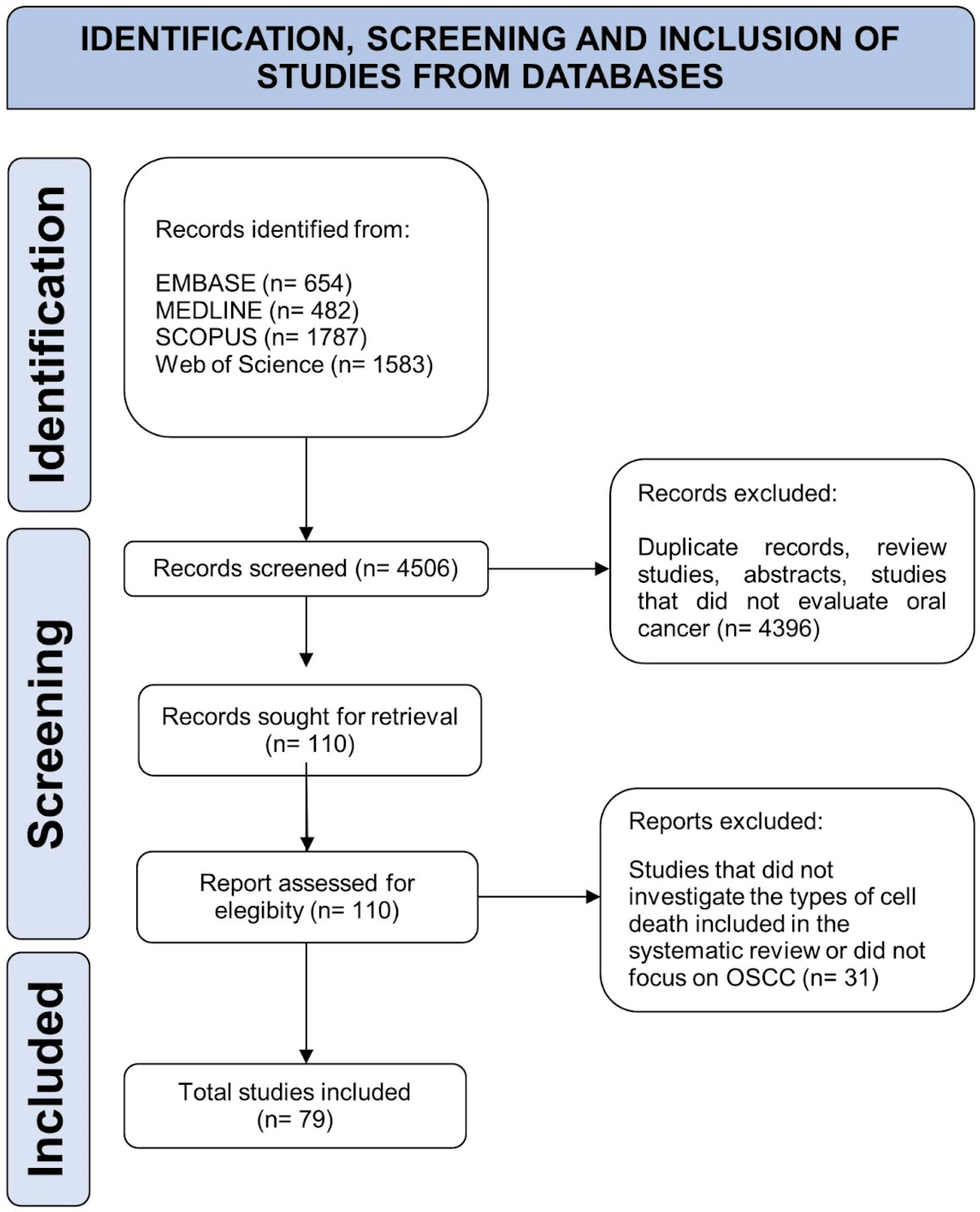
2.4. Study Selection
2.5. Data Extraction and Data Synthesis
2.6. Quality Assessment
3. Results
4. Discussion
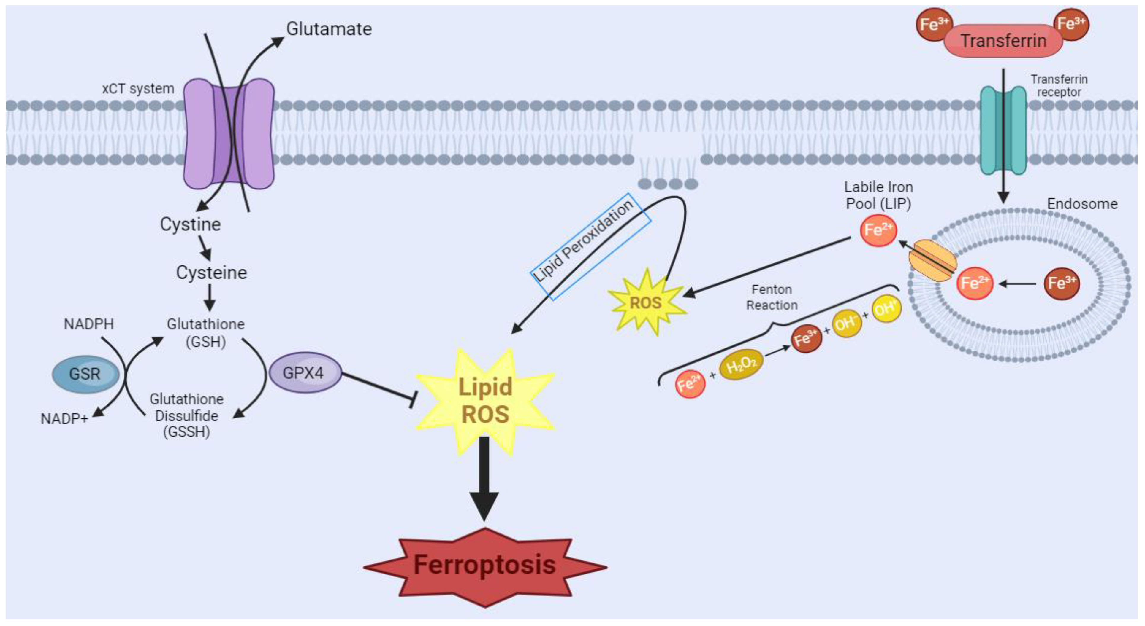
4.1. Ferroptosis
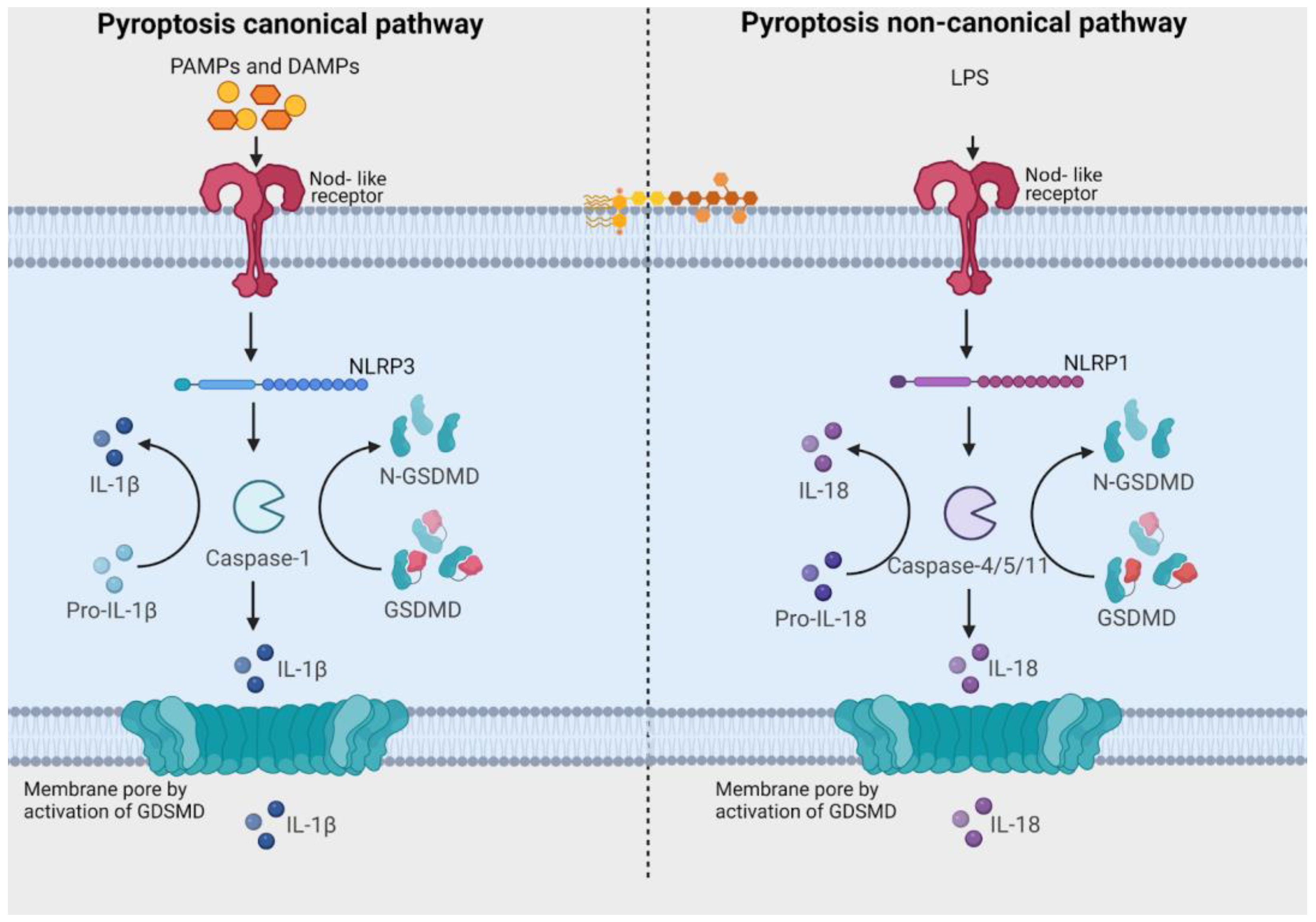
4.2. Pyroptosis
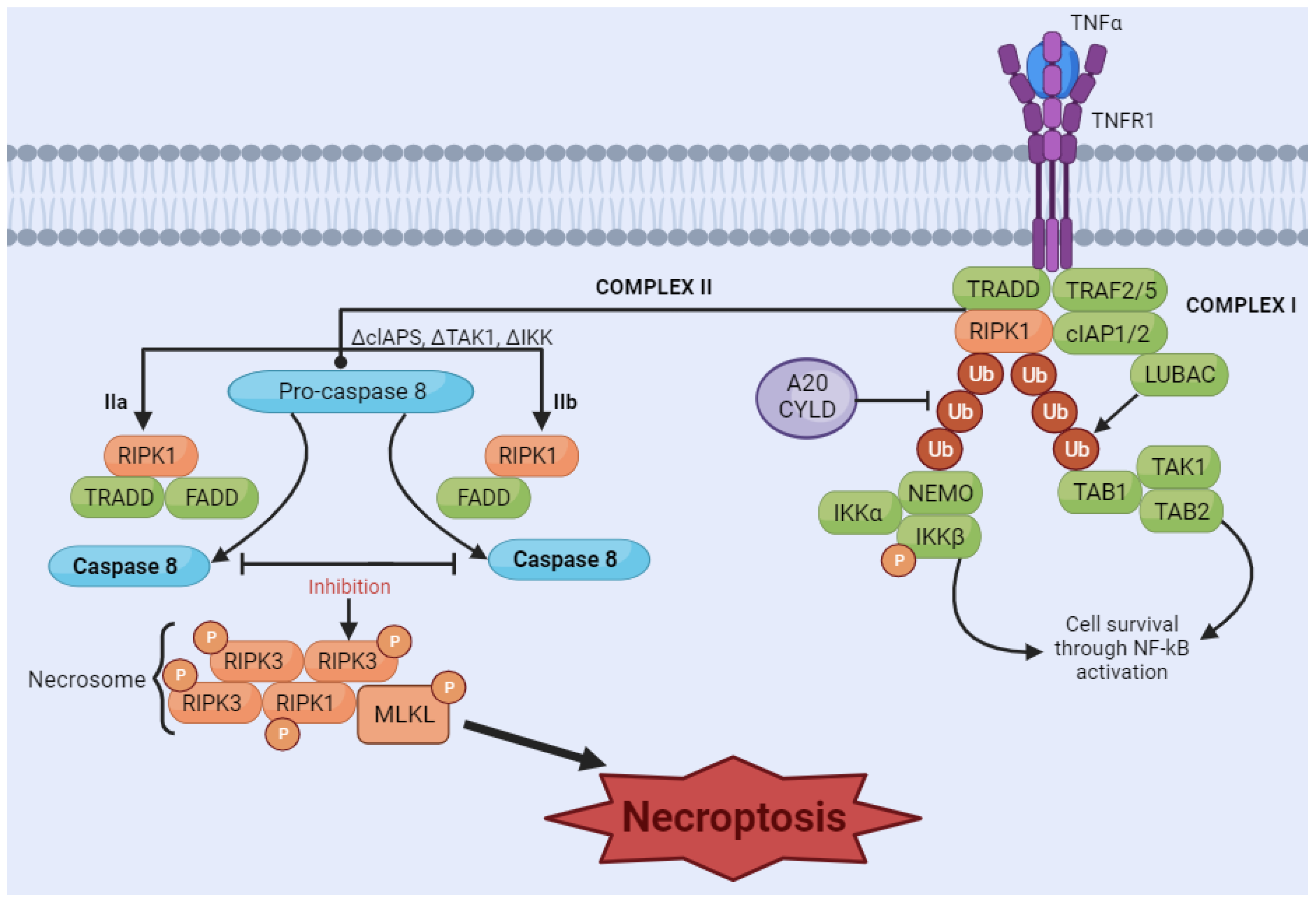
4.3. Necroptosis
4.4. Other Emerging Types of Cell Death
5. Conclusions
Author Contributions
Funding
Institutional Review Board Statement
Informed Consent Statement
Data Availability Statement
Conflicts of Interest
Appendix A. Search Strategies with Key Words and MeSH Terms
- EMBASE
- Search 1: ENTOSIS (Results: 7)
- Search 2: FERROPTOSIS (Results: 265)
- Search 3: PYROPTOSIS (Results: 124)
- Search 4: NETOSIS (Results: 7)
- Search 5: NECROPTOSIS (Results: 211)
- Search 6: PARTHANATOS (Results: 0)
- Search 7: MITOPTOSIS (Results: 7)
- Search 8: PARAPTOSIS (Results: 21)
- Search 9: METHUOSIS (Results: 12)
- PUBMED
- Search 1: ENTOSIS (Results: 3)
- Search 2: FERROPTOSIS (Results: 256)
- Search 3: PYROPTOSIS (Results: 77)
- Search 4: NETOSIS (Results: 2)
- Search 5: NECROPTOSIS (Results: 124)
- Search 6: PARTHANATOS (Results: 4)
- Search 7: MITOPTOSIS (Results: 2)
- Search 8: PARAPTOSIS (Results: 11)
- Search 9: METHUOSIS (Results: 3)
- SCOPUS
- Search 1: ENTOSIS (Results: 6)
- Search 2: FERROPTOSIS (Results: 218)
- Search 3: PYROPTOSIS (Results: 1.431)
- Search 4: NETOSIS (Results: 8)
- Search 5: NECROPTOSIS (Results: 108)
- Search 6: PARTHANATOS (Results: 7)
- Search 7: MITOPTOSIS (Results: 4)
- Search 8: PARAPTOSIS (Results: 2)
- Search 9: METHUOSIS (Results: 3)
- WEB OF SCIENCE
- Search 1: ENTOSIS (Results: 5)
- Search 2: FERROPTOSIS (Results: 151)
- Search 3: PYROPTOSIS (Results: 1090)
- Search 4: NETOSIS (Results: 11)
- Search 5: NECROPTOSIS (Results: 298)
- Search 6: PARTHANATOS (Results: 12)
- Search 7: MITOPTOSIS (Results: 9)
- Search 8: PARAPTOSIS (Results: 5)
- Search 9: METHUOSIS (Results: 2)
Appendix B. List of Excluded Studies along with Reasons for Exclusion
| Study (Year) | DOI | Reason for Exclusion * |
| Almangush et al. (2020) | https://doi.org/10.1186/s12885-020-07342-x | Did not evaluate non-apoptotic programmed cell death |
| Deng et al. (2022) | https://doi.org/10.1002/jcla.24292 | Study not specific to OSCC |
| Fan et al. (2021) | https://doi.org/10.3389/fgene.2021.732211 | Study not specific to OSCC |
| Fukuda et al. (2021) | https://doi.org/10.21873/anticanres.14944 | Unable to access the full article |
| Gohara et al. (2022) | https://doi.org/10.1016/j.omto.2022.10.001 | Study not specific to OSCC |
| Goreger et al. (2017) | https://doi.org/10.1186/s12865-016-0185-5 | Study not specific to OSCC |
| He et al. (2021) | https://doi.org/10.1016/j.intimp.2021.107789 | Study not specific to OSCC |
| Huang et al. (2022) | https://doi.org/10.1016/j.intimp.2021.108431 | Study not specific to OSCC |
| Kosim et al. (2023) | https://doi.org/10.3389/fphar.2022.988335 | Did not evaluate non-apoptotic programmed cell death |
| Li et al. (2022) | https://doi.org/10.1002/cam4.4825 | Study not specific to OSCC |
| Li et al. (2023) | https://doi.org/10.1186/s13027-023-00507-w | Study not specific to OSCC |
| Liu et al. (2023) | https://doi.org/10.3389/fgene.2022.988606 | Study not specific to OSCC |
| Lu et al. (2021) | https://doi.org/10.3389/fgene.2021.755486 | Study not specific to OSCC |
| Lu et al. (2022) | https://doi.org/10.1155/2022/1539659 | Study not specific to OSCC |
| Qian et al. (2021) | https://doi.org/10.2147/IJGM.S337089 | Study not specific to OSCC |
| Roh et al. (2017) | https://doi.org/10.1016/j.redox.2016.12.010 | Study not specific to OSCC |
| Savic et al. (2023) | https://doi.org/10.3390/cells12020336 | Study not specific to OSCC |
| Shin et al. (2018) | https://doi.org/10.1016/j.freeradbiomed.2018.10.426 | Study not specific to OSCC |
| Takasu et al. (2016) | https://doi.org/10.1038/cgt.2016.8 | Study not specific to OSCC |
| Tian et al. (2022) | https://doi.org/10.4103/2221-1691.357743 | Study not specific to OSCC |
| Wang et al. (2022) | https://doi.org/10.1016/j.csbj.2022.06.046 | Mixed together tumors from several locations |
| Wei et al. (2020) | https://doi.org/10.21037/atm.2020.02.36 | Study not specific to OSCC |
| Wei et al. (2022) | https://doi.org/10.3892/etm.2022.11449 | Study not specific to OSCC |
| Wu et al. (2022) | https://doi.org/10.1155/2022/7602482 | Study not specific to OSCC |
| Wu et al. (2022) | https://doi.org/10.3389/fcell.2022.702224 | Study not specific to OSCC |
| Xu et al. (2021) | https://doi.org/10.1155/2021/5759927 | Study not specific to OSCC |
| Yaghmaei et al. (2017) | https://doi.org/10.2174/1871520616666160725110844 | Unable to access the full article |
| Yu et al. (2020) | https://doi.org/10.3390/cancers12061670 | Study not specific to OSCC |
| Yu et al. (2022) | https://doi.org/10.1155/2022/3713929 | Unable to access the full article |
| Zhou et al. (2022) | https://doi.org/10.1002/cam4.4819 | Study not specific to OSCC |
| Zhu et al. (2021) | https://doi.org/10.1016/j.intimp.2021.108268 | Study not specific to OSCC |
| * Abbreviation: OSCC: oral squamous cell carcinoma. | ||
Appendix C. Risk of Bias Assessment of In Vitro Studies
| Experimental Conditions | Blinding * | Complete Outcome | Exposure Characterization | Outcome Assessment | Reporting | Other | Overall Risk | |
| Ruggieri et al. [92] | Low | NI | Low | Low | Low | Low | Low | Low Risk |
| Sulkshane and Teni [84] | High | NI | Low | Low | Low | Low | High | Moderate Risk |
| Feng et al. [64] | Low | NI | Low | Low | Low | Low | Low | Low Risk |
| Okazaki et al. [18] | Low | NI | Low | Low | Low | Low | Low | Low Risk |
| Garley et al. [93] | Low | NI | Low | High | Low | Low | Low | Moderate Risk |
| Yue et al. [65] | Low | NI | Low | Low | Low | Low | Low | Low Risk |
| Huang et al. [19] | Low | NI | Low | Low | Low | Low | Low | Low Risk |
| Zhu et al. [21] | Low | NI | Low | Low | Low | Low | Low | Low Risk |
| Sato et al. [20] | Low | NI | Low | Low | Low | Low | Low | Low Risk |
| Huang et al. [66] | Low | NI | Low | Low | Low | Low | Low | Low Risk |
| Lee et al. [23] | Low | NI | Low | Low | Low | Low | Low | Low Risk |
| Lin et al. [24] | Low | NI | Low | Low | Low | Low | Low | Low Risk |
| Hémon et al. [22] | Low | NI | Low | Low | Low | Low | Low | Low Risk |
| Uzunparmak et al. [86] | Low | NI | Low | Low | Low | Low | Low | Low Risk |
| Li et al. [96] | Low | NI | Low | Low | Low | Low | Low | Low Risk |
| Garley et al. [94] | Low | NI | Low | High | Low | Low | Low | Moderate Risk |
| Chen et al. [95] | Low | NI | Low | Low | Low | Low | Low | Low Risk |
| Yang et al. [69] | Low | NI | Low | Low | Low | Low | Low | Low Risk |
| Wang et al. [28] | Low | NI | Low | Low | Low | Low | Low | Low Risk |
| Yao et al. [70] | Low | NI | Low | Low | Low | Low | Low | Low Risk |
| Luo et al. [68] | Low | NI | Low | Low | Low | Low | Low | Low Risk |
| Jiang et al. [67] | Low | NI | Low | Low | Low | Low | Low | Low Risk |
| Yang et al. [29] | Low | NI | Low | Low | Low | Low | Low | Low Risk |
| Tomita et al. [27] | Low | NI | Low | NI | Low | Low | Low | Low Risk |
| You et al. [30] | Low | NI | Low | Low | Low | Low | Low | Low Risk |
| You et al. [31] | Low | NI | Low | Low | Low | Low | Low | Low Risk |
| Zhang et al. [32] | Low | NI | Low | Low | Low | Low | Low | Low Risk |
| Huang et al. [87] | Low | NI | Low | Low | Low | Low | Low | Low Risk |
| Li et al. [35] | Low | NI | Low | Low | Low | Low | Low | Low Risk |
| Rioja-Blanco et al. [71] | Low | NI | Low | Low | Low | Low | Low | Low Risk |
| Zhu et al. [77] | Low | NI | Low | Low | Low | Low | Low | Low Risk |
| Wang et al. [73] | Low | NI | Low | Low | Low | Low | Low | Low Risk |
| Shen et al. [72] | Low | NI | Low | Low | Low | Low | Low | Low Risk |
| Zhu et al. [78] | Low | NI | Low | Low | Low | Low | Low | Low Risk |
| Bhuyan et al. [33] | Low | NI | Low | Low | Low | Low | Low | Low Risk |
| Lu et al. [40] | Low | NI | Low | High | Low | Low | Low | Moderate Risk |
| Liu et al. [39] | Low | NI | Low | Low | Low | Low | Low | Low Risk |
| Xu C. et al. [44] | Low | NI | Low | Low | Low | Low | Low | Low Risk |
| Liu et al. [38] | Low | NI | Low | NI | Low | Low | Low | Low Risk |
| Han and Wu [34] | Low | NI | Low | High | Low | Low | Low | Moderate Risk |
| Sun et al. [42] | Low | NI | Low | Low | Low | Low | Low | Low Risk |
| Wang et al. [58] | Low | NI | Low | High | Low | Low | Low | Moderate Risk |
| Wang et al. [43] | Low | NI | Low | Low | Low | Low | Low | Low Risk |
| Li J, Tang and Ma [35] | Low | NI | Low | Low | Low | Low | Low | Low Risk |
| Yang et al. [46] | Low | NI | Low | NI | Low | Low | High | Moderate Risk |
| Kaokaen et al. [88] | Low | NI | Low | Low | Low | Low | Low | Low Risk |
| Liu et al. [80] | Low | NI | Low | Low | Low | Low | Low | Low Risk |
| Wang et al. [82] | Low | NI | Low | Low | Low | Low | Low | Low Risk |
| Nan et al. [81] | Low | NI | Low | Low | Low | Low | Low | Low Risk |
| Zi et al. [79] | Low | NI | Low | Low | Low | Low | Low | Low Risk |
| Yan et al. [83] | Low | NI | Low | Low | Low | Low | Low | Low Risk |
| Wu et al. [59] | Low | NI | Low | Low | Low | Low | Low | Low Risk |
| Pan et al. [55] | Low | NI | Low | Low | Low | Low | Low | Low Risk |
| Jehl et al. [52] | Low | NI | Low | Low | Low | Low | Low | Low Risk |
| Xie et al. [60] | Low | NI | Low | Low | Low | Low | Low | Low Risk |
| Zhang et al. [62] | Low | NI | Low | Low | Low | Low | Low | Low Risk |
| Yu et al. [61] | Low | NI | Low | Low | Low | Low | Low | Low Risk |
| Wang et al. [56] | Low | NI | Low | Low | Low | Low | Low | Low Risk |
| Li et al. [54] | Low | NI | Low | Low | Low | Low | Low | Low Risk |
| Wang et al. [57] | Low | NI | Low | High | Low | Low | Low | Moderate Risk |
| Zhao and Zhu [63] | Low | NI | Low | High | Low | Low | Low | Moderate Risk |
| Lee and Roh [53] | Low | NI | Low | High | Low | Low | Low | Moderate Risk |
| Gupta et al. [89] | Low | NI | Low | Low | Low | Low | Low | Low Risk |
| Yun et al. [91] | Low | NI | Low | Low | Low | Low | Low | Low Risk |
| Huang et al. [90] | Low | NI | Low | Low | Low | Low | Low | Low Risk |
| Zhou et al. [76] | Low | NI | Low | Low | Low | Low | Low | Low Risk |
| * Abbreviation: NI: not informed. | ||||||||
Appendix D. Risk of Bias of In Vivo Studies Based on Animal Models Using the SYRCLE Risk of Bias Tool
| Sequence Generation (Selection Bias) | Baseline Characteristics (Selection Bias) | Allocation Concealment (Selection Bias) | Random Housing (Performance Bias) | Blinding (Performance Bias) | Random Outcome Assessment (Detection Bias) | Blinding (Detection Bias) | Incomplete Outcome Data (Attrition Bias) | Selective Outcome Reporting (Reporting Bias) | Other Sources of Bias | |
| Feng et al. [64] | Low | Unclear | Unclear | Unclear | Unclear | Unclear | High | High | Low | High |
| Sulkshane and Teni [84] | Low | Unclear | Unclear | Unclear | Unclear | Unclear | High | High | Low | Low |
| Okazaki et al. [18] | Unclear | Unclear | Unclear | Unclear | Unclear | Unclear | Unclear | High | Unclear | High |
| Huang et al. [19] | Low | Unclear | Unclear | Unclear | Unclear | Unclear | High | High | Low | Low |
| Huang et al. [66] | Unclear | Unclear | Unclear | Unclear | Unclear | Unclear | High | High | Low | Low |
| Lee et al. [23] | Unclear | Unclear | Unclear | Unclear | Unclear | Unclear | High | High | Low | High |
| Uzunparmak et al. [86] | Low | Unclear | Unclear | Unclear | Unclear | Unclear | High | High | Low | Low |
| Yang et al. [29] | Unclear | Unclear | Unclear | Unclear | Unclear | Unclear | High | High | Low | Low |
| Wang et al. [28] | Low | Low | Unclear | Unclear | Unclear | Unclear | High | High | Low | Low |
| Yao et al. [70] | Low | Unclear | Unclear | Unclear | Unclear | Unclear | High | High | Low | Low |
| Luo et al. [68] | Low | Unclear | Unclear | Unclear | Unclear | Unclear | High | High | Low | Low |
| Yang et al. [69] | Unclear | Unclear | Unclear | Unclear | Unclear | Unclear | High | High | Low | Low |
| You et al. [30] | Unclear | Unclear | Unclear | Unclear | Unclear | Unclear | High | High | Low | Low |
| Zhang et al. [32] | Low | Unclear | Unclear | Unclear | Unclear | Unclear | High | High | Low | Low |
| Li et al. [96] | Low | Low | Unclear | Unclear | Unclear | Unclear | High | High | Low | Low |
| Rioja-Blanco et al. [71] | Low | Low | Unclear | Unclear | Unclear | Unclear | High | High | Low | Low |
| Shen et al. [72] | Low | Low | Unclear | Unclear | Unclear | Unclear | High | High | Low | High |
| Zhu et al. [77] | Low | Unclear | Unclear | Unclear | Unclear | Unclear | Unclear | High | Low | High |
| Xu et al. [44] | Unclear | Unclear | Unclear | Unclear | Unclear | Unclear | High | High | Low | Low |
| Wang et al. [57] | Low | Unclear | Unclear | Unclear | Unclear | Unclear | High | High | Low | Low |
| Yang et al. [46] | Low | Low | Unclear | Unclear | Unclear | Unclear | High | High | Low | Low |
| Liu et al. [44] | Low | Low | Unclear | Unclear | Unclear | Unclear | High | High | Low | Low |
| Wang et al. [82] | Low | Low | Unclear | Unclear | Unclear | Unclear | High | High | Low | Low |
| Zi et al. [79] | Low | Unclear | Unclear | Unclear | Unclear | Unclear | High | High | Low | High |
| Wu et al. [59] | Low | Low | Unclear | Unclear | Unclear | Unclear | High | High | Low | Low |
| Chung et al. [50] | Unclear | Unclear | Unclear | Unclear | Unclear | Unclear | High | High | Low | Low |
| Wang et al. [56] | Low | Unclear | Unclear | Unclear | Unclear | Unclear | High | High | Low | Low |
| Wang et al. [43] | Low | Unclear | Unclear | Unclear | Unclear | Unclear | High | High | Low | Low |
| Zhao and Zhu [63] | Unclear | Unclear | Unclear | Unclear | Unclear | Unclear | High | High | Unclear | Low |
| Zhou et al. [76] | Low | Unclear | Unclear | Unclear | Unclear | Unclear | High | High | Low | Low |
| Zhou et al. [48] | Unclear | Unclear | Unclear | Unclear | Unclear | Unclear | High | High | Low | Low |
References
- Safa, A.R. Resistance to Cell Death and Its Modulation in Cancer Stem Cells. Crit. Rev. Oncog. 2016, 21, 203. [Google Scholar] [CrossRef] [PubMed]
- Galluzzi, L.; Vitale, I.; Aaronson, S.A.; Abrams, J.M.; Adam, D.; Agostinis, P.; Alnemri, E.S.; Altucci, L.; Amelio, I.; Andrews, D.W.; et al. Molecular mechanisms of cell death: Recommendations of the Nomenclature Committee on Cell Death 2018. Cell Death Differ. 2018, 25, 486–541. [Google Scholar] [CrossRef] [PubMed]
- Yan, G.; Elbadawi, M.; Efferth, T. Multiple cell death modalities and their key features (Review). World Acad. Sci. J. 2020, 2, 39–48. [Google Scholar] [CrossRef]
- Peng, F.; Liao, M.; Qin, R.; Zhu, S.; Peng, C.; Fu, L.; Chen, Y.; Han, B. Regulated cell death (RCD) in cancer: Key pathways and targeted therapies. Signal Transduct. Target. Ther. 2022, 7, 286. [Google Scholar] [CrossRef]
- Tong, X.; Tang, R.; Xiao, M.; Xu, J.; Wang, W.; Zhang, B.; Liu, J.; Yu, X.; Shi, S. Targeting cell death pathways for cancer therapy: Recent developments in necroptosis, pyroptosis, ferroptosis, and cuproptosis research. J. Hematol. Oncol. 2022, 15, 174. [Google Scholar] [CrossRef] [PubMed]
- Wang, X.; Hua, P.; He, C.; Chen, M. Non-apoptotic cell death-based cancer therapy: Molecular mechanism, pharmacological modulators, and nanomedicine. Acta Pharm. Sin. B 2022, 12, 3567–3593. [Google Scholar] [CrossRef]
- Xi, Y.; Gao, L.; Li, S.; Sun, K.; Chen, P.; Cai, Z.; Ren, W.; Zhi, K. The role of novel programmed cell death in head and neck squamous cell carcinoma: From mechanisms to potential therapies. Front. Pharmacol. 2023, 14, 1228985. [Google Scholar] [CrossRef] [PubMed]
- Bedoui, S.; Herold, M.J.; Strasser, A. Emerging connectivity of programmed cell death pathways and its physiological implications. Nat. Rev. Mol. Cell Biol. 2020, 21, 678–695. [Google Scholar] [CrossRef]
- Li, J.; He, D.; Li, S.; Xiao, J.; Zhu, Z. Ferroptosis: The emerging player in remodeling triple-negative breast cancer. Front. Immunol. 2023, 14, 1284057. [Google Scholar] [CrossRef]
- Jia, Y.-J.; Zhang, Y.; Ma, X.-B.; Wang, Y.; Tian, Y.-Q.; He, P.-X.; Xu, Y.-C. Ferroptosis: Opportunities and Challenges in Cancer. J. Explor. Res. Pharmacol. 2023, 8, 243–253. [Google Scholar] [CrossRef]
- Frank, D.; Vince, J.E. Pyroptosis versus necroptosis: Similarities, differences, and crosstalk. Cell Death Differ. 2019, 26, 99–114. [Google Scholar] [CrossRef]
- Bertheloot, D.; Latz, E.; Franklin, B.S. Necroptosis, pyroptosis and apoptosis: An intricate game of cell death. Cell. Mol. Immunol. 2021, 18, 1106–1121. [Google Scholar] [CrossRef] [PubMed]
- Kayagaki, N.; Webster, J.D.; Newton, K. Control of Cell Death in Health and Disease. Annu. Rev. Pathol. Mech. Dis. 2023, 19, 157–180. [Google Scholar] [CrossRef]
- Gulia, S.; Chandra, P.; Das, A. The Prognosis of Cancer Depends on the Interplay of Autophagy, Apoptosis, and Anoikis within the Tumor Microenvironment. Cell Biochem. Biophys. 2023, 81, 621–658. [Google Scholar] [CrossRef] [PubMed]
- Page, M.J.; McKenzie, J.E.; Bossuyt, P.M.; Boutron, I.; Hoffmann, T.C.; Mulrow, C.D.; Shamseer, L.; Tetzlaff, J.M.; Akl, E.A.; Brennan, S.E.; et al. The PRISMA 2020 statement: An updated guideline for reporting systematic reviews. BMJ 2021, 372, 71. [Google Scholar] [CrossRef] [PubMed]
- Hooijmans, C.R.; Rovers, M.M.; de Vries, R.B.M.; Leenaars, M.; Ritskes-Hoitinga, M.; Langendam, M.W. SYRCLE’s risk of bias tool for animal studies. BMC Med. Res. Methodol. 2014, 14, 43. [Google Scholar] [CrossRef] [PubMed]
- Bezemer, J.M.; van der Ende, J.; Limpens, J.; de Vries, H.J.C.; Schallig, H.D.F.H. Safety and efficacy of allylamines in the treatment of cutaneous and mucocutaneous leishmaniasis: A systematic review. PLoS ONE 2021, 16, e0249628. [Google Scholar] [CrossRef]
- Okazaki, S.; Shintani, S.; Hirata, Y.; Suina, K.; Semba, T.; Yamasaki, J.; Umene, K.; Ishikawa, M.; Saya, H.; Nagano, O. Synthetic lethality of the ALDH3A1 inhibitor dyclonine and xCT inhibitors in glutathione deficiency-resistant cancer cells. Oncotarget 2018, 9, 33832–33843. [Google Scholar] [CrossRef]
- Huang, K.-J.; Wei, Y.-H.; Chiu, Y.-C.; Wu, S.-R.; Shieh, D.-B. Assessment of zero-valent iron-based nanotherapeutics for ferroptosis induction and resensitization strategy in cancer cells. Biomater. Sci. 2019, 7, 1311–1322. [Google Scholar] [CrossRef]
- Sato, K.; Shi, L.; Ito, F.; Ohara, Y.; Motooka, Y.; Tanaka, H.; Mizuno, M.; Hori, M.; Hirayama, T.; Hibi, H.; et al. Non-thermal plasma specifically kills oral squamous cell carcinoma cells in a catalytic Fe(II)-dependent manner. J. Clin. Biochem. Nutr. 2019, 65, 8–15. [Google Scholar] [CrossRef] [PubMed]
- Zhu, T.; Shi, L.; Yu, C.; Dong, Y.; Qiu, F.; Shen, L.; Qian, Q.; Zhou, G.; Zhu, X. Ferroptosis Promotes Photodynamic Therapy: Supramolecular Photosensitizer-Inducer Nanodrug for Enhanced Cancer Treatment. Theranostics 2019, 9, 3293–3307. [Google Scholar] [CrossRef] [PubMed]
- Hémon, A.; Louandre, C.; Lailler, C.; Godin, C.; Bottelin, M.; Morel, V.; François, C.; Galmiche, A.; Saidak, Z. SLC7A11 as a biomarker and therapeutic target in HPV-positive head and neck Squamous Cell Carcinoma. Biochem. Biophys. Res. Commun. 2020, 533, 1083–1087. [Google Scholar] [CrossRef] [PubMed]
- Lee, J.; You, J.H.; Shin, D.; Roh, J.-L. Inhibition of Glutaredoxin 5 predisposes Cisplatin-resistant Head and Neck Cancer Cells to Ferroptosis. Theranostics 2020, 10, 7775–7786. [Google Scholar] [CrossRef] [PubMed]
- Lin, Y.H.; Chiu, V.; Huang, C.Y.; Tzeng, I.S.; Hsieh, P.C.; Kuo, C.Y. Promotion of Ferroptosis in Oral Cancer Cell Lines by Chrysophanol. Curr. Top. Nutraceutical Res. 2020, 18, 273–276. [Google Scholar] [CrossRef]
- Gu, W.; Kim, M.; Wang, L.; Yang, Z.; Nakajima, T.; Tsushima, Y. Multi-omics Analysis of Ferroptosis Regulation Patterns and Characterization of Tumor Microenvironment in Patients with Oral Squamous Cell Carcinoma. Int. J. Biol. Sci. 2021, 17, 3476–3492. [Google Scholar] [CrossRef] [PubMed]
- Li, H.; Zhang, X.; Yi, C.; He, Y.; Chen, X.; Zhao, W.; Yu, D. Ferroptosis-related gene signature predicts the prognosis in Oral squamous cell carcinoma patients. BMC Cancer 2021, 21, 835. [Google Scholar] [CrossRef] [PubMed]
- Tomita, K.; Nagasawa, T.; Kuwahara, Y.; Torii, S.; Igarashi, K.; Roudkenar, M.H.; Roushandeh, A.M.; Kurimasa, A.; Sato, T. MiR-7-5p Is Involved in Ferroptosis Signaling and Radioresistance Thru the Generation of ROS in Radioresistant HeLa and SAS Cell Lines. Int. J. Mol. Sci. 2021, 22, 8300. [Google Scholar] [CrossRef]
- Wang, X.; Liu, K.; Gong, H.; Li, D.; Chu, W.; Zhao, D.; Wang, X.; Xu, D. Death by histone deacetylase inhibitor quisinostat in tongue squamous cell carcinoma via apoptosis, pyroptosis, and ferroptosis. Toxicol. Appl. Pharmacol. 2020, 410, 115363. [Google Scholar] [CrossRef]
- Yang, J.; Cao, X.-H.; Luan, K.-F.; Huang, Y.-D. Circular RNA FNDC3B Protects Oral Squamous Cell Carcinoma Cells From Ferroptosis and Contributes to the Malignant Progression by Regulating miR-520d-5p/SLC7A11 Axis. Front. Oncol. 2021, 11, 672724. [Google Scholar] [CrossRef]
- You, J.H.; Lee, J.; Roh, J.-L. Mitochondrial pyruvate carrier 1 regulates ferroptosis in drug-tolerant persister head and neck cancer cells via epithelial-mesenchymal transition. Cancer Lett. 2021, 507, 40–54. [Google Scholar] [CrossRef]
- You, J.H.; Lee, J.; Roh, J.-L. PGRMC1-dependent lipophagy promotes ferroptosis in paclitaxel-tolerant persister cancer cells. J. Exp. Clin. Cancer Res. 2021, 40, 1–18. [Google Scholar] [CrossRef]
- Zhang, X.; Yang, S.; Wang, Q.; Ye, W.; Liu, S.; Wang, X.; Zhang, Z.; Cao, L.; Jiang, X. Tailored theranostic nanoparticles cause efficient ferroptosis in head and neck squamous cell carcinoma through a reactive oxygen species “butterfly effect”. Chem. Eng. J. 2021, 423, 130083. [Google Scholar] [CrossRef]
- Bhuyan, S.; Pal, B.; Pathak, L.; Saikia, P.J.; Mitra, S.; Gayan, S.; Mokhtari, R.B.; Li, H.; Ramana, C.V.; Baishya, D.; et al. Targeting hypoxia-induced tumor stemness by activating pathogen-induced stem cell niche defense. Front. Immunol. 2022, 13, 933329. [Google Scholar] [CrossRef]
- Han, L.; Li, L.; Wu, G. Induction of ferroptosis by carnosic acid-mediated inactivation of Nrf2/HO-1 potentiates cisplatin responsiveness in OSCC cells. Mol. Cell. Probes 2022, 64, 101821. [Google Scholar] [CrossRef]
- Li, J.; Tang, L.-L.; Ma, J. Survival-related indicators ALOX12B and SPRR1A are associated with DNA damage repair and tumor microenvironment status in HPV 16-negative head and neck squamous cell carcinoma patients. BMC Cancer 2022, 22, 714. [Google Scholar] [CrossRef]
- Li, T.; Wang, Y.; Xiang, X.; Chen, C. Development and Validation of a Ferroptosis-Related lncRNAs Prognosis Model in Oral Squamous Cell Carcinoma. Front. Genet. 2022, 13, 847940. [Google Scholar] [CrossRef]
- Liu, F.; Tang, L.; Li, Q.; Chen, L.; Pan, Y.; Yin, Z.; He, J.; Tian, J. Single-cell transcriptomics uncover the key ferroptosis regulators contribute to cancer progression in head and neck squamous cell carcinoma. Front. Mol. Biosci. 2022, 9, 962742. [Google Scholar] [CrossRef]
- Liu, J.; An, W.; Zhao, Q.; Liu, Z.; Jiang, Y.; Li, H.; Wang, D. Hyperbaric oxygen enhances X-ray induced ferroptosis in oral squamous cell carcinoma cells. Oral Dis. 2022. online ahead of print. [Google Scholar] [CrossRef]
- Liu, S.; Yan, S.; Zhu, J.; Lu, R.; Kang, C.; Tang, K.; Zeng, J.; Ding, M.; Guo, Z.; Lai, X.; et al. Combination RSL3 Treatment Sensitizes Ferroptosis- and EGFR-Inhibition-Resistant HNSCCs to Cetuximab. Int. J. Mol. Sci. 2022, 23, 9014. [Google Scholar] [CrossRef]
- Lu, T.; Zhang, Z.; Pan, X.; Zhang, J.; Wang, X.; Wang, M.; Li, H.; Yan, M.; Chen, W. Caveolin-1 promotes cancer progression via inhibiting ferroptosis in head and neck squamous cell carcinoma. J. Oral Pathol. Med. 2022, 51, 52–62. [Google Scholar] [CrossRef]
- Qiu, L.; Tao, A.; Liu, F.; Ge, X.; Li, C. Potential prognostic value of a eight ferroptosis-related lncRNAs model and the correlative immune activity in oral squamous cell carcinoma. BMC Genet. 2022, 23, 80. [Google Scholar] [CrossRef] [PubMed]
- Sun, K.; Ren, W.; Li, S.; Zheng, J.; Huang, Y.; Zhi, K.; Gao, L. MiR-34c-3p upregulates erastin-induced ferroptosis to inhibit proliferation in oral squamous cell carcinomas by targeting SLC7A11. Pathol. Res. Pract. 2022, 231, 153778. [Google Scholar] [CrossRef] [PubMed]
- Wang, M.; Li, F.; Lu, T.; Wu, R.; Yang, S.; Chen, W. Photodynamic and ferroptotic Ce6@ZIF-8@ssPDA for head and neck cancer treatment. Mater. Des. 2022, 224, 111403. [Google Scholar] [CrossRef]
- Xu, C.; Shen, Y.; Shi, Y.; Zhang, M.; Zhou, L. Eukaryotic translation initiation factor 3 subunit B promotes head and neck cancer via CEBPB translation. Cancer Cell Int. 2022, 22, 161. [Google Scholar] [CrossRef] [PubMed]
- Xu, Y.; Hong, M.; Kong, D.; Deng, J.; Zhong, Z.; Liang, J. Ferroptosis-associated DNA methylation signature predicts overall survival in patients with head and neck squamous cell carcinoma. BMC Genom. 2022, 23, 63. [Google Scholar] [CrossRef] [PubMed]
- Yang, Y.; Tang, H.; Zheng, J.; Yang, K. The PER1/HIF-1alpha negative feedback loop promotes ferroptosis and inhibits tumor progression in oral squamous cell carcinoma. Transl. Oncol. 2022, 18, 101360. [Google Scholar] [CrossRef]
- Yin, J.; Fu, J.; Zhao, Y.; Xu, J.; Chen, C.; Zheng, L.; Wang, B. Comprehensive Analysis of the Significance of Ferroptosis-Related Genes in the Prognosis and Immunotherapy of Oral Squamous Cell Carcinoma. Bioinform. Biol. Insights 2022, 16, 1–13. [Google Scholar] [CrossRef]
- Zhou, Q.; Wang, X.; Zhang, Y.; Wang, L.; Chen, Z. Inhibition of AEBP1 predisposes cisplatin-resistant oral cancer cells to ferroptosis. BMC Oral Health 2022, 22, 478. [Google Scholar] [CrossRef]
- Zhu, H.; Tao, Y.; Huang, Q.; Chen, Z.; Jiang, L.; Yan, H.; Zhong, J.; Liang, L. Identification of ferroptosis-related genes as potential biomarkers of tongue squamous cell carcinoma using an integrated bioinformatics approach. FEBS Open Bio 2022, 12, 412–429. [Google Scholar] [CrossRef]
- Chung, C.; Lin, C.; Chen, C.; Hsueh, C.; Chang, Y.; Wang, C.; Chu, P.; Tai, S.; Yang, M. Ferroptosis Signature Shapes the Immune Profiles to Enhance the Response to Immune Checkpoint Inhibitors in Head and Neck Cancer. Adv. Sci. 2023, 10, 2204514. [Google Scholar] [CrossRef] [PubMed]
- Fan, X.; Zhong, Y.; Yuan, F.; Zhang, L.; Cai, Y.; Liao, L. A ferroptosis-related prognostic model with excellent clinical performance based on the exploration of the mechanism of oral squamous cell carcinoma progression. Sci. Rep. 2023, 13, 1461. [Google Scholar] [CrossRef]
- Jehl, A.; Conrad, O.; Burgy, M.; Foppolo, S.; Vauchelles, R.; Ronzani, C.; Etienne-Selloum, N.; Chenard, M.-P.; Danic, A.; Dourlhes, T.; et al. Blocking EREG/GPX4 Sensitizes Head and Neck Cancer to Cetuximab through Ferroptosis Induction. Cells 2023, 12, 733. [Google Scholar] [CrossRef] [PubMed]
- Lee, J.; Roh, J.-L. Promotion of ferroptosis in head and neck cancer with divalent metal transporter 1 inhibition or salinomycin. Hum. Cell 2023, 36, 1090–1098. [Google Scholar] [CrossRef] [PubMed]
- Li, J.; Xiao, W.; Wei, W.; Wu, M.; Xiong, K.; Lyu, J.; Li, Y. HSPA5, as a ferroptosis regulator, may serve as a potential therapeutic for head and neck squamous cell carcinoma. Mol. Immunol. 2023, 158, 79–90. [Google Scholar] [CrossRef] [PubMed]
- Pan, X.; Xu, X.; Wang, L.; Zhang, S.; Chen, Y.; Yang, R.; Chen, X.; Cheng, B.; Xia, J.; Ren, X. BASP1 is a prognostic biomarker associated with immunotherapeutic response in head and neck squamous cell carcinoma. Front. Oncol. 2023, 13, 1021262. [Google Scholar] [CrossRef]
- Wang, Z.; Han, J.; Guo, Z.; Wu, H.; Liu, Y.; Wang, W.; Zhang, C.; Liu, J. Ginseng-based carbon dots inhibit the growth of squamous cancer cells by increasing ferroptosis. Front. Oncol. 2023, 13, 1097692. [Google Scholar] [CrossRef] [PubMed]
- Wang, L.; Wang, C.; Li, X.; Tao, Z.; Zhu, W.; Su, Y.; Choi, W.S. Melatonin and erastin emerge synergistic anti-tumor effects on oral squamous cell carcinoma by inducing apoptosis, ferroptosis, and inhibiting autophagy through promoting ROS. Cell. Mol. Biol. Lett. 2023, 28, 36. [Google Scholar] [CrossRef] [PubMed]
- Wang, Z.-Q.; Li, Y.-Q.; Wang, D.-Y.; Shen, Y.-Q. Natural product piperlongumine inhibits proliferation of oral squamous carcinoma cells by inducing ferroptosis and inhibiting intracellular antioxidant capacity. Transl. Cancer Res. 2022, 12, 2911–2922. [Google Scholar] [CrossRef] [PubMed]
- Wu, X.; Wu, Q.; Wang, Y.; Liu, Y.; Li, Z.; Liu, Q.; Huang, Z.; Li, M.; Zhang, B.; Zhan, Q. Aqueous-soluble components of sporoderm-removed Ganoderma lucidum spore powder promote ferroptosis in oral squamous cell carcinoma. Chin. J. Cancer Res. 2023, 35, 176–190. [Google Scholar] [CrossRef]
- Xie, J.; Lan, T.; Zheng, D.-L.; Ding, L.-C.; Lu, Y.-G. CDH4 inhibits ferroptosis in oral squamous cell carcinoma cells. BMC Oral Health 2023, 23, 329. [Google Scholar] [CrossRef]
- Yu, Y.; MohamedAl-Sharani, H.; Zhang, B. EZH2-mediated SLC7A11 upregulation via miR-125b-5p represses ferroptosis of TSCC. Oral Dis. 2021, 29, 880–891. [Google Scholar] [CrossRef]
- Zhang, P.; Cui, Y.; Wang, J.; Cheng, J.; Zhu, L.; Liu, C.; Yue, S.; Pang, R.; Guan, J.; Xie, B.; et al. Dual-stimuli responsive smart nanoprobe for precise diagnosis and synergistic multi-modalities therapy of superficial squamous cell carcinoma. J. Nanobiotechnology 2023, 21, 4. [Google Scholar] [CrossRef]
- Zhao, Y.; Zhu, S. Nrf2/HO-1 Alleviates Disulfiram/Copper-Induced Ferroptosis in Oral Squamous Cell Carcinoma. Biochem. Genet. 2023. online ahead of print. [Google Scholar] [CrossRef] [PubMed]
- Feng, X.; Luo, Q.; Zhang, H.; Wang, H.; Chen, W.; Meng, G.; Chen, F. The role of NLRP3 inflammasome in 5-fluorouracil resistance of oral squamous cell carcinoma. J. Exp. Clin. Cancer Res. 2017, 36, 81. [Google Scholar] [CrossRef] [PubMed]
- Yue, E.; Tuguzbaeva, G.; Chen, X.; Qin, Y.; Li, A.; Sun, X.; Dong, C.; Liu, Y.; Yu, Y.; Zahra, S.M.; et al. Anthocyanin is involved in the activation of pyroptosis in oral squamous cell carcinoma. Phytomedicine 2019, 56, 286–294. [Google Scholar] [CrossRef] [PubMed]
- Huang, Z.; Zhang, Q.; Wang, Y.; Chen, R.; Wang, Y.; Huang, Z.; Zhou, G.; Li, H.; Rui, X.; Jin, T.; et al. Inhibition of caspase-3-mediated GSDME-derived pyroptosis aids in noncancerous tissue protection of squamous cell carcinoma patients during cisplatin-based chemotherapy. Am. J. Cancer Res. 2020, 10, 4287. [Google Scholar] [PubMed]
- Jiang, L.; Ge, W.; Cui, Y.; Wang, X. The regulation of long non-coding RNA 00958 (LINC00958) for oral squamous cell carcinoma (OSCC) cells death through absent in melanoma 2 (AIM2) depending on microRNA-4306 and Sirtuin1 (SIRT1) in vitro. Bioengineered 2021, 12, 5085–5098. [Google Scholar] [CrossRef] [PubMed]
- Luo, Q.; Li, X.; Gan, G.; Yang, M.; Chen, X.; Chen, F. PPT1 Reduction Contributes to Erianin-Induced Growth Inhibition in Oral Squamous Carcinoma Cells. Front. Cell Dev. Biol. 2021, 9, 764263. [Google Scholar] [CrossRef] [PubMed]
- Yang, M.; Luo, Q.; Chen, X.; Chen, F. Bitter melon derived extracellular vesicles enhance the therapeutic effects and reduce the drug resistance of 5-fluorouracil on oral squamous cell carcinoma. J. Nanobiotechnol. 2021, 19, 259. [Google Scholar] [CrossRef] [PubMed]
- Yao, Y.; Shen, X.; Zhou, M.; Tang, B. Periodontal Pathogens Promote Oral Squamous Cell Carcinoma by Regulating ATR and NLRP3 Inflammasome. Front. Oncol. 2021, 11, 722797. [Google Scholar] [CrossRef]
- Rioja-Blanco, E.; Arroyo-Solera, I.; Álamo, P.; Casanova, I.; Gallardo, A.; Unzueta, U.; Serna, N.; Sánchez-García, L.; Quer, M.; Villaverde, A.; et al. CXCR4-targeted nanotoxins induce GSDME-dependent pyroptosis in head and neck squamous cell carcinoma. J. Exp. Clin. Cancer Res. 2022, 41, 49. [Google Scholar] [CrossRef]
- Shen, X.; Zhang, B.; Hu, X.; Li, J.; Wu, M.; Yan, C.; Yang, Y.; Li, Y. Neisseria sicca and Corynebacterium matruchotii inhibited oral squamous cell carcinomas by regulating genome stability. Bioengineered 2022, 13, 14094–14106. [Google Scholar] [CrossRef]
- Wang, S.; Zhang, M.; Wu, Z.; Zhu, S.; Wan, S.; Zhang, B.; Yang, Q.; Xiao, Y.; Chen, L.; Sun, Z. GSDME Is Related to Prognosis and Response to Chemotherapy in Oral Cancer. J. Dent. Res. 2022, 101, 848–858. [Google Scholar] [CrossRef]
- Xin, Y.; Zhang, J.; Jiang, Q.; Qiu, J. Construction of prognostic signature of patients with oral squamous cell carcinoma based on pyroptosis-related long non-coding RNAs. Front. Surg. 2022, 9, 935765. [Google Scholar] [CrossRef]
- Zeng, D.; Wang, X.; Zhang, S.; Zheng, A.; Huang, Q.; Cao, L. Pyroptosis-related gene-based prognostic signature for predicting the overall survival of oral squamous cell carcinoma patients. Front. Surg. 2022, 9, 903271. [Google Scholar] [CrossRef]
- Zhou, J.-Y.; Wang, W.-J.; Zhang, C.-Y.; Ling, Y.-Y.; Hong, X.-J.; Su, Q.; Li, W.-G.; Mao, Z.-W.; Cheng, B.; Tan, C.-P.; et al. Ru(II)-modified TiO2 nanoparticles for hypoxia-adaptive photo-immunotherapy of oral squamous cell carcinoma. Biomaterials 2022, 289, 121757. [Google Scholar] [CrossRef]
- Zhu, S.-W.; Ye, M.; Ma, X.; Wu, Z.-Z.; Wan, S.-C.; Yang, S.-C.; Li, H.; Xu, Z.; Sun, Z.-J. pH-responsive nanoprodrugs combining a Src inhibitor and chemotherapy to potentiate antitumor immunity via pyroptosis in head and neck cancer. Acta Biomater. 2022, 154, 497–509. [Google Scholar] [CrossRef] [PubMed]
- Zhu, W.; Zhang, J.; Wang, M.; Zhai, R.; Xu, Y.; Wang, J.; Wang, M.; Zhang, H.; Liu, L. Development of a prognostic pyroptosis-related gene signature for head and neck squamous cell carcinoma patient. Cancer Cell Int. 2022, 22, 62. [Google Scholar] [CrossRef] [PubMed]
- Zi, M.; Xingyu, C.; Yang, C.; Xiaodong, S.; Shixian, L.; Shicheng, W. Improved antitumor immunity of chemotherapy in OSCC treatment by Gasdermin-E mediated pyroptosis. Apoptosis 2022, 28, 348–361. [Google Scholar] [CrossRef] [PubMed]
- Liu, X.; Zhan, W.; Gao, G.; Jiang, Q.; Zhang, X.; Zhang, H.; Sun, X.; Han, W.; Wu, F.-G.; Liang, G. Apoptosis-Amplified Assembly of Porphyrin Nanofiber Enhances Photodynamic Therapy of Oral Tumor. J. Am. Chem. Soc. 2023, 145, 7918–7930. [Google Scholar] [CrossRef] [PubMed]
- Nan, Z.; Dou, Y.; Chen, A.; Wang, K.; Sun, J.; Meng, Z.; Neckenig, M.; Ai, D.; Liu, S.; Dong, Z.; et al. Identification and validation of a prognostic signature of autophagy, apoptosis and pyroptosis-related genes for head and neck squamous cell carcinoma: To imply therapeutic choices of HPV negative patients. Front. Immunol. 2023, 13, 1100417. [Google Scholar] [CrossRef]
- Wang, S.; Wu, Z.-Z.; Zhu, S.-W.; Wan, S.-C.; Zhang, M.-J.; Zhang, B.-X.; Yang, Q.-C.; Xiao, Y.; Li, H.; Mao, L.; et al. CTLA-4 blockade induces tumor pyroptosis via CD8+ T cells in head and neck squamous cell carcinoma. Mol. Ther. 2023, 31, 2154–2168. [Google Scholar] [CrossRef]
- Yan, L.; Sun, Y.; Guo, J.; Jia, R. PD-L1 Exon 3 Is a Hidden Switch of Its Expression and Function in Oral Cancer Cells. Int. J. Mol. Sci. 2023, 24, 8193. [Google Scholar] [CrossRef] [PubMed]
- Sulkshane, P.; Teni, T. BH3 mimetic Obatoclax (GX15-070) mediates mitochondrial stress predominantly via MCL-1 inhibition and induces autophagy-dependent necroptosis in human oral cancer cells. Oncotarget 2016, 8, 60060–60079. [Google Scholar] [CrossRef] [PubMed]
- Li, J.; Huang, S.; Zeng, L.; Li, K.; Yang, L.; Gao, S.; Guan, C.; Zhang, S.; Lao, X.; Liao, G.; et al. Necroptosis in head and neck squamous cell carcinoma: Characterization of clinicopathological relevance and in vitro cell model. Cell Death Dis. 2020, 11, 391. [Google Scholar] [CrossRef] [PubMed]
- Uzunparmak, B.; Gao, M.; Lindemann, A.; Erikson, K.; Wang, L.; Lin, E.; Frank, S.J.; Gleber-Netto, F.O.; Zhao, M.; Skinner, H.D.; et al. Caspase-8 loss radiosensitizes head and neck squamous cell carcinoma to SMAC mimetic–induced necroptosis. J. Clin. Investig. 2020, 5, 139837. [Google Scholar] [CrossRef]
- Huang, Y.-C.; Yuan, T.-M.; Liu, B.-H.; Liu, K.-L.; Wung, C.-H.; Chuang, S.-M. Capsaicin Potentiates Anticancer Drug Efficacy Through Autophagy-Mediated Ribophorin II Downregulation and Necroptosis in Oral Squamous Cell Carcinoma Cells. Front. Pharmacol. 2021, 12, 676813. [Google Scholar] [CrossRef] [PubMed]
- Kaokaen, P.; Chaicharoenaudomrung, N.; Kunhorm, P.; Mesil, K.; Binlateh, T.; Noisa, P.; Jitprasertwong, P. Nanoencapsulation of Cordycepin Induces Switching from Necroptosis to Apoptosis in Human Oral Cancer Cells (HSC-4) Through Inhibition of Receptor-Interacting Serine/Threonine-Protein Kinase 3 (RIPK3) and Autophagy Modulation. Nat. Prod. Commun. 2022, 17, 1934578X221074838. [Google Scholar] [CrossRef]
- Gupta, P.; Singh, A.; Verma, A.K.; Kant, S.; Pandey, A.K.; Mishra, A.; Khare, P.; Prakash, V. Nanoencapsulation of Docetaxel Induces Concurrent Apoptosis and Necroptosis in Human Oral Cancer Cells (SCC-9) via TNF-α/RIP1/RIP3 Pathway. Indian J. Clin. Biochem. 2022, 38, 351–360. [Google Scholar] [CrossRef] [PubMed]
- Huang, K.; Gu, X.; Xu, H.; Li, H.; Shi, M.; Wei, D.; Wang, S.; Li, Y.; Liu, B.; Li, Y. Prognostic Value of Necroptosis-Related Genes Signature in Oral Squamous Cell Carcinoma. Cancers 2023, 15, 4539. [Google Scholar] [CrossRef]
- Yun, H.-M.; Kwon, Y.-J.; Kim, E.; Chung, H.-J.; Park, K.-R. Machilin D Promotes Apoptosis and Autophagy, and Inhibits Necroptosis in Human Oral Squamous Cell Carcinoma Cells. Int. J. Mol. Sci. 2023, 24, 4576. [Google Scholar] [CrossRef] [PubMed]
- Ruggieri, V.; Agriesti, F.; Scrima, R.; Laurenzana, I.; Perrone, D.; Tataranni, T.; Mazzoccoli, C.; Muzio, L.L.; Capitanio, N.; Piccoli, C. Dichloroacetate, a selective mitochondria-targeting drug for oral squamous cell carcinoma: A metabolic perspective of treatment. Oncotarget 2014, 6, 1217–1230. [Google Scholar] [CrossRef] [PubMed]
- Garley, M.; Dziemiańczyk-Pakieła, D.; Grubczak, K.; Surażyński, A.; Dąbrowska, D.; Ratajczak-Wrona, W.; Sawicka-Powierza, J.; Borys, J.; Moniuszko, M.; Pałka, J.A.; et al. Differences and similarities in the phenomenon of NETs formation in oral inflammation and in oral squamous cell carcinoma. J. Cancer 2018, 9, 1958–1965. [Google Scholar] [CrossRef]
- Garley, M.; Jabłońska, E.; Miltyk, W.; Grubczak, K.; Surażyński, A.; Ratajczak-Wrona, W.; Grudzińska, M.; Nowacka, K.H.; Moniuszko, M.; Pałka, J.A.; et al. Cancers Cells in Traps? The Pathways of NETs Formation in Response to OSCC in Humans—A Pilot Study. Cancer Control. 2020, 27, 1073274820960473. [Google Scholar] [CrossRef]
- Chen, Q.; Song, S.; Wang, Z.; Shen, Y.; Xie, L.; Li, J.; Jiang, L.; Zhao, H.; Feng, X.; Zhou, Y.; et al. Isorhamnetin induces the paraptotic cell death through ROS and the ERK/MAPK pathway in OSCC cells. Oral Dis. 2020, 27, 240–250. [Google Scholar] [CrossRef] [PubMed]
- Li, D.; Kou, Y.; Gao, Y.; Liu, S.; Yang, P.; Hasegawa, T.; Su, R.; Guo, J.; Li, M. Oxaliplatin induces the PARP1-mediated parthanatos in oral squamous cell carcinoma by increasing production of ROS. Aging 2021, 13, 4242–4257. [Google Scholar] [CrossRef] [PubMed]
- Li, J.; Cao, F.; Yin, H.; Huang, Z.; Lin, Z.; Mao, N.; Sun, B.; Wang, G. Ferroptosis: Past, present and future. Cell Death Dis. 2020, 11, 88. [Google Scholar] [CrossRef]
- Stockwell, B.R.; Angeli, J.P.F.; Bayir, H.; Bush, A.I.; Conrad, M.; Dixon, S.J.; Fulda, S.; Gascón, S.; Hatzios, S.K.; Kagan, V.E.; et al. Ferroptosis: A Regulated Cell Death Nexus Linking Metabolism, Redox Biology, and Disease. Cell 2017, 171, 273–285. [Google Scholar] [CrossRef]
- Zhang, C.; Liu, X.; Jin, S.; Chen, Y.; Guo, R. Ferroptosis in cancer therapy: A novel approach to reversing drug resistance. Mol. Cancer 2022, 21, 47. [Google Scholar] [CrossRef]
- Hou, W.; Xie, Y.; Song, X.; Sun, X.; Lotze, M.T.; Zeh, H.J., 3rd; Kang, R.; Tang, D. Autophagy promotes ferroptosis by degradation of ferritin. Autophagy 2016, 12, 1425–1428. [Google Scholar] [CrossRef] [PubMed]
- Chen, Y.; Fan, Z.; Hu, S.; Lu, C.; Xiang, Y.; Liao, S. Ferroptosis: A New Strategy for Cancer Therapy. Front. Oncol. 2022, 12, 830561. [Google Scholar] [CrossRef]
- Tang, D.; Chen, X.; Kang, R.; Kroemer, G. Ferroptosis: Molecular mechanisms and health implications. Cell Res. 2021, 31, 107–125. [Google Scholar] [CrossRef] [PubMed]
- Cao, J.Y.; Dixon, S.J. Mechanisms of ferroptosis. Cell. Mol. Life Sci. 2016, 73, 2195–2209. [Google Scholar] [CrossRef] [PubMed]
- Ying, J.F.; Lu, Z.B.; Fu, L.Q.; Tong, Y.; Wang, Z.; Li, W.F.; Mou, X.Z. The role of iron homeostasis and iron-mediated ROS in cancer. Am. J. Cancer Res. 2021, 11, 1895. [Google Scholar] [PubMed]
- Seibt, T.M.; Proneth, B.; Conrad, M. Role of GPX4 in ferroptosis and its pharmacological implication. Free. Radic. Biol. Med. 2018, 133, 144–152. [Google Scholar] [CrossRef]
- Dodson, M.; Castro-Portuguez, R.; Zhang, D.D. NRF2 plays a critical role in mitigating lipid peroxidation and ferroptosis. Redox Biol. 2019, 23, 101107. [Google Scholar] [CrossRef]
- Jiang, L.; Kon, N.; Li, T.; Wang, S.-J.; Su, T.; Hibshoosh, H.; Baer, R.; Gu, W. Ferroptosis as a p53-mediated activity during tumour suppression. Nature 2015, 520, 57–62. [Google Scholar] [CrossRef]
- Hassannia, B.; Vandenabeele, P.; Berghe, T.V. Targeting Ferroptosis to Iron Out Cancer. Cancer Cell 2019, 35, 830–849. [Google Scholar] [CrossRef]
- Hsieh, C.-H.; Hsieh, H.-C.; Shih, F.-H.; Wang, P.-W.; Yang, L.-X.; Shieh, D.-B.; Wang, Y.-C. An innovative NRF2 nano-modulator induces lung cancer ferroptosis and elicits an immunostimulatory tumor microenvironment. Theranostics 2021, 11, 7072–7091. [Google Scholar] [CrossRef]
- Ma, Q. Role of Nrf2 in Oxidative Stress and Toxicity. Annu. Rev. Pharmacol. Toxicol. 2013, 53, 401–426. [Google Scholar] [CrossRef] [PubMed]
- Sun, X.; Ou, Z.; Chen, R.; Niu, X.; Chen, D.; Kang, R.; Tang, D. Activation of the p62-Keap1-NRF2 pathway protects against ferroptosis in hepatocellular carcinoma cells. Hepatology 2016, 63, 173–184. [Google Scholar] [CrossRef] [PubMed]
- Zheng, S.; Mo, J.; Zhang, J.; Chen, Y. HIF-1α inhibits ferroptosis and promotes malignant progression in non-small cell lung cancer by activating the Hippo-YAP signalling pathway. Oncol. Lett. 2023, 25, 1–9. [Google Scholar] [CrossRef] [PubMed]
- Jun, J.C.; Rathore, A.; Younas, H.; Gilkes, D.; Polotsky, V.Y. Hypoxia-Inducible Factors and Cancer. Curr. Sleep Med. Rep. 2017, 3, 1–10. [Google Scholar] [CrossRef]
- Wu, J.; Minikes, A.M.; Gao, M.; Bian, H.; Li, Y.; Stockwell, B.R.; Chen, Z.-N.; Jiang, X. Intercellular interaction dictates cancer cell ferroptosis via NF2–YAP signalling. Nature 2019, 572, 402–406. [Google Scholar] [CrossRef] [PubMed]
- Wang, W.; Green, M.; Choi, J.E.; Gijón, M.; Kennedy, P.D.; Johnson, J.K.; Liao, P.; Lang, X.; Kryczek, I.; Sell, A.; et al. CD8+ T cells regulate tumour ferroptosis during cancer immunotherapy. Nature 2019, 569, 270–274. [Google Scholar] [CrossRef] [PubMed]
- Sun, S.; Shen, J.; Jiang, J.; Wang, F.; Min, J. Targeting ferroptosis opens new avenues for the development of novel therapeutics. Signal Transduct. Target. Ther. 2023, 8, 372. [Google Scholar] [CrossRef]
- Lei, G.; Mao, C.; Yan, Y.; Zhuang, L.; Gan, B. Ferroptosis, radiotherapy, and combination therapeutic strategies. Protein Cell 2021, 12, 836–857. [Google Scholar] [CrossRef]
- Lei, G.; Zhuang, L.; Gan, B. Targeting ferroptosis as a vulnerability in cancer. Nat. Rev. Cancer 2022, 22, 381–396. [Google Scholar] [CrossRef]
- Luo, L.; Wang, H.; Tian, W.; Zeng, J.; Huang, Y.; Luo, H. Targeting ferroptosis for cancer therapy: Iron metabolism and anticancer immunity. Am. J. Cancer Res. 2021, 11, 5508–5525. [Google Scholar]
- Cosialls, E.; El Hage, R.; Dos Santos, L.; Gong, C.; Mehrpour, M.; Hamaï, A. Ferroptosis: Cancer Stem Cells Rely on Iron until “to Die for” It. Cells 2021, 10, 2981. [Google Scholar] [CrossRef] [PubMed]
- Taylor, W.R.; Fedorka, S.R.; Gad, I.; Shah, R.; Alqahtani, H.D.; Koranne, R.; Kuganesan, N.; Dlamini, S.; Rogers, T.; Al-Hamashi, A.; et al. Small-Molecule Ferroptotic Agents with Potential to Selectively Target Cancer Stem Cells. Sci. Rep. 2019, 9, 5926. [Google Scholar] [CrossRef] [PubMed]
- Wang, Y.; Yu, L.; Ding, J.; Chen, Y. Iron Metabolism in Cancer. Int. J. Mol. Sci. 2019, 20, 95. [Google Scholar] [CrossRef] [PubMed]
- Wang, H.; Zhang, Z.; Ruan, S.; Yan, Q.; Chen, Y.; Cui, J.; Wang, X.; Huang, S.; Hou, B. Regulation of iron metabolism and ferroptosis in cancer stem cells. Front. Oncol. 2023, 13, 1251561. [Google Scholar] [CrossRef] [PubMed]
- Gnanapradeepan, K.; Basu, S.; Barnoud, T.; Budina-Kolomets, A.; Kung, C.-P.; Murphy, M.E. The p53 Tumor Suppressor in the Control of Metabolism and Ferroptosis. Front. Endocrinol. 2018, 9, 124. [Google Scholar] [CrossRef]
- Liu, Y.; Gu, W. p53 in ferroptosis regulation: The new weapon for the old guardian. Cell Death Differ. 2022, 29, 895–910. [Google Scholar] [CrossRef] [PubMed]
- Poltorack, C.D.; Dixon, S.J. Understanding the role of cysteine in ferroptosis: Progress & paradoxes. FEBS J. 2021, 289, 374–385. [Google Scholar] [CrossRef] [PubMed]
- Liu, M.-Z.; Kong, N.; Zhang, G.-Y.; Xu, Q.; Xu, Y.; Ke, P.; Liu, C. The critical role of ferritinophagy in human disease. Front. Pharmacol. 2022, 13, 933732. [Google Scholar] [CrossRef]
- Li, J.; Jia, Y.-C.; Ding, Y.-X.; Bai, J.; Cao, F.; Li, F. The crosstalk between ferroptosis and mitochondrial dynamic regulatory networks. Int. J. Biol. Sci. 2023, 19, 2756–2771. [Google Scholar] [CrossRef]
- Bartolacci, C.; Andreani, C.; El-Gammal, Y.; Scaglioni, P.P. Lipid Metabolism Regulates Oxidative Stress and Ferroptosis in RAS-Driven Cancers: A Perspective on Cancer Progression and Therapy. Front. Mol. Biosci. 2021, 8, 706650. [Google Scholar] [CrossRef]
- Hirata, Y.; Cai, R.; Volchuk, A.; Steinberg, B.E.; Saito, Y.; Matsuzawa, A.; Grinstein, S.; Freeman, S.A. Lipid peroxidation increases membrane tension, Piezo1 gating, and cation permeability to execute ferroptosis. Curr. Biol. 2023, 33, 1282–1294. [Google Scholar] [CrossRef]
- Yang, J.; Mo, J.; Dai, J.; Ye, C.; Cen, W.; Zheng, X.; Jiang, L.; Ye, L. Cetuximab promotes RSL3-induced ferroptosis by suppressing the Nrf2/HO-1 signalling pathway in KRAS mutant colorectal cancer. Cell Death Dis. 2021, 12, 1079. [Google Scholar] [CrossRef] [PubMed]
- Zhang, C.; Wang, C.; Yang, Z.; Bai, Y.; Shukuya, T.; Poh, M.-E.; Ekman, S.; Li, J.; Xu, Y.; Deng, S. Identification of GPX4 as a therapeutic target for lung adenocarcinoma after EGFR-TKI resistance. Transl. Lung Cancer Res. 2022, 11, 786–801. [Google Scholar] [CrossRef] [PubMed]
- Stępień, K.; Ostrowski, R.P.; Matyja, E. Hyperbaric oxygen as an adjunctive therapy in treatment of malignancies, including brain tumours. Med. Oncol. 2016, 33, 101. [Google Scholar] [CrossRef] [PubMed]
- Xu, Z.; Xie, Y.; Mao, Y.; Huang, J.; Mei, X.; Song, J.; Sun, Y.; Yao, Z.; Shi, W. Ferroptosis-Related Gene Signature Predicts the Prognosis of Skin Cutaneous Melanoma and Response to Immunotherapy. Front. Genet. 2021, 12, 758981. [Google Scholar] [CrossRef] [PubMed]
- Qi, X.; Wang, R.; Lin, Y.; Yan, D.; Zuo, J.; Chen, J.; Shen, B. A Ferroptosis-Related Gene Signature Identified as a Novel Prognostic Biomarker for Colon Cancer. Front. Genet. 2021, 12, 692426. [Google Scholar] [CrossRef] [PubMed]
- Jin, J.; Liu, C.; Yu, S.; Cai, L.; Sitrakiniaina, A.; Gu, R.; Li, W.; Wu, F.; Xue, X. A novel ferroptosis-related gene signature for prognostic prediction of patients with lung adenocarcinoma. Aging 2021, 13, 16144–16164. [Google Scholar] [CrossRef] [PubMed]
- Qian, Y.; Daza, J.; Itzel, T.; Betge, J.; Zhan, T.; Marmé, F.; Teufel, A. Prognostic Cancer Gene Expression Signatures: Current Status and Challenges. Cells 2021, 10, 648. [Google Scholar] [CrossRef] [PubMed]
- Shimizu, S.; Hiratsuka, H.; Koike, K.; Tsuchihashi, K.; Sonoda, T.; Ogi, K.; Miyakawa, A.; Kobayashi, J.; Kaneko, T.; Igarashi, T.; et al. Tumor-infiltrating CD8+ T-cell density is an independent prognostic marker for oral squamous cell carcinoma. Cancer Med. 2019, 8, 80–93. [Google Scholar] [CrossRef]
- Foy, J.-P.; Bertolus, C.; Michallet, M.-C.; Deneuve, S.; Incitti, R.; Bendriss-Vermare, N.; Albaret, M.-A.; Ortiz-Cuaran, S.; Thomas, E.; Colombe, A.; et al. The immune microenvironment of HPV-negative oral squamous cell carcinoma from never-smokers and never-drinkers patients suggests higher clinical benefit of IDO1 and PD1/PD-L1 blockade. Ann. Oncol. 2017, 28, 1934–1941. [Google Scholar] [CrossRef]
- Wang, H.; Mao, L.; Zhang, T.; Zhang, L.; Wu, Y.; Guo, W.; Hu, J.; Ju, H.; Ren, G. Altered expression of TIM-3, LAG-3, IDO, PD-L1, and CTLA-4 during nimotuzumab therapy correlates with responses and prognosis of oral squamous cell carcinoma patients. J. Oral Pathol. Med. 2019, 48, 669–676. [Google Scholar] [CrossRef]
- Yu, P.; Zhang, X.; Liu, N.; Tang, L.; Peng, C.; Chen, X. Pyroptosis: Mechanisms and diseases. Signal Transduct. Target. Ther. 2021, 6, 128. [Google Scholar] [CrossRef] [PubMed]
- Fang, Y.; Tian, S.; Pan, Y.; Li, W.; Wang, Q.; Tang, Y.; Yu, T.; Wu, X.; Shi, Y.; Ma, P.; et al. Pyroptosis: A new frontier in cancer. Biomed. Pharmacother. 2020, 121, 109595. [Google Scholar] [CrossRef] [PubMed]
- Kelley, N.; Jeltema, D.; Duan, Y.; He, Y. The NLRP3 Inflammasome: An Overview of Mechanisms of Activation and Regulation. Int. J. Mol. Sci. 2019, 20, 3328. [Google Scholar] [CrossRef]
- Jiang, M.; Qi, L.; Li, L.; Li, Y. The caspase-3/GSDME signal pathway as a switch between apoptosis and pyroptosis in cancer. Cell Death Discov. 2020, 6, 112. [Google Scholar] [CrossRef]
- Bhat, A.A.; Thapa, R.; Afzal, O.; Agrawal, N.; Almalki, W.H.; Kazmi, I.; Alzarea, S.I.; Altamimi, A.S.A.; Prasher, P.; Singh, S.K.; et al. The pyroptotic role of Caspase-3/GSDME signalling pathway among various cancer: A Review. Int. J. Biol. Macromol. 2023, 242, 124832. [Google Scholar] [CrossRef]
- Zhang, Z.; Li, X.; Wang, Y.; Wei, Y.; Wei, X. Involvement of inflammasomes in tumor microenvironment and tumor therapies. J. Hematol. Oncol. 2023, 16, 24. [Google Scholar] [CrossRef]
- Vyas, D.; Laput, G.; Vyas, A. Chemotherapy-enhanced inflammation may lead to the failure of therapy and metastasis. OncoTargets Ther. 2014, 7, 1015–1023. [Google Scholar] [CrossRef] [PubMed]
- Moossavi, M.; Parsamanesh, N.; Bahrami, A.; Atkin, S.L.; Sahebkar, A. Role of the NLRP3 inflammasome in cancer. Mol. Cancer 2018, 17, 158. [Google Scholar] [CrossRef]
- Sharma, B.R.; Kanneganti, T.-D. NLRP3 inflammasome in cancer and metabolic diseases. Nat. Immunol. 2021, 22, 550–559. [Google Scholar] [CrossRef]
- Liu, X.; Yin, L.; Shen, S.; Hou, Y. Inflammation and cancer: Paradoxical roles in tumorigenesis and implications in immunotherapies. Genes Dis. 2023, 10, 151–164. [Google Scholar] [CrossRef]
- Lu, L.; Zhang, Y.; Tan, X.; Merkher, Y.; Leonov, S.; Zhu, L.; Deng, Y.; Zhang, H.; Zhu, D.; Tan, Y.; et al. Emerging mechanisms of pyroptosis and its therapeutic strategy in cancer. Cell Death Discov. 2022, 8, 338. [Google Scholar] [CrossRef]
- Lu, X.; Guo, T.; Zhang, X. Pyroptosis in Cancer: Friend or Foe? Cancers 2021, 13, 3620. [Google Scholar] [CrossRef]
- Feng, X.; Luo, Q.; Wang, H.; Zhang, H.; Chen, F. MicroRNA-22 suppresses cell proliferation, migration and invasion in oral squamous cell carcinoma by targeting NLRP3. J. Cell. Physiol. 2018, 233, 6705–6713. [Google Scholar] [CrossRef]
- Wei, X.; Xie, F.; Zhou, X.; Wu, Y.; Yan, H.; Liu, T.; Huang, J.; Wang, F.; Zhou, F.; Zhang, L. Role of pyroptosis in inflammation and cancer. Cell. Mol. Immunol. 2022, 19, 971–992. [Google Scholar] [CrossRef]
- Dhuriya, Y.K.; Sharma, D. Necroptosis: A regulated inflammatory mode of cell death. J. Neuroinflammation 2018, 15, 199. [Google Scholar] [CrossRef] [PubMed]
- Nicolè, L.; Sanavia, T.; Cappellesso, R.; Maffeis, V.; Akiba, J.; Kawahara, A.; Naito, Y.; Radu, C.M.; Simioni, P.; Serafin, D.; et al. Necroptosis-driving genes RIPK1, RIPK3 and MLKL-p are associated with intratumoral CD3+ and CD8+ T cell density and predict prognosis in hepatocellular carcinoma. J. Immunother. Cancer 2022, 10, e004031. [Google Scholar] [CrossRef] [PubMed]
- Grootjans, S.; Vanden Berghe, T.; Vandenabeele, P. Initiation and execution mechanisms of necroptosis: An overview. Cell Death Differ. 2017, 24, 1184–1195. [Google Scholar] [CrossRef] [PubMed]
- Pinci, F.; Gaidt, M.M.; Jung, C.; Nagl, D.; Kuut, G.; Hornung, V. Tumor necrosis factor is a necroptosis-associated alarmin. Front. Immunol. 2022, 13, 1074440. [Google Scholar] [CrossRef] [PubMed]
- Seo, J.; Nam, Y.W.; Kim, S.; Oh, D.-B.; Song, J. Necroptosis molecular mechanisms: Recent findings regarding novel necroptosis regulators. Exp. Mol. Med. 2021, 53, 1007–1017. [Google Scholar] [CrossRef]
- Deragon, M.A.; McCaig, W.D.; Truong, P.V.; Metz, K.R.; Carron, K.A.; Hughes, K.J.; Knapp, A.R.; Dougherty, M.J.; LaRocca, T.J. Mitochondrial Trafficking of MLKL, Bak/Bax, and Drp1 Is Mediated by RIP1 and ROS which Leads to Decreased Mitochondrial Membrane Integrity during the Hyperglycemic Shift to Necroptosis. Int. J. Mol. Sci. 2023, 24, 8609. [Google Scholar] [CrossRef]
- Juan, C.A.; de la Lastra, J.M.P.; Plou, F.J.; Pérez-Lebeña, E. The Chemistry of Reactive Oxygen Species (ROS) Revisited: Outlining Their Role in Biological Macromolecules (DNA, Lipids and Proteins) and Induced Pathologies. Int. J. Mol. Sci. 2021, 22, 4642. [Google Scholar] [CrossRef]
- Choi, M.E.; Price, D.R.; Ryter, S.W.; Choi, A.M.K. Necroptosis: A crucial pathogenic mediator of human disease. J. Clin. Investig. 2019, 4, 128834. [Google Scholar] [CrossRef]
- Galluzzi, L.; Kepp, O.; Chan, F.K.-M.; Kroemer, G. Necroptosis: Mechanisms and Relevance to Disease. Annu. Rev. Pathol. Mech. Dis. 2017, 12, 103–130. [Google Scholar] [CrossRef]
- Pistritto, G.; Trisciuoglio, D.; Ceci, C.; Garufi, A.; D’Orazi, G. Apoptosis as anticancer mechanism: Function and dysfunction of its modulators and targeted therapeutic strategies. Aging 2016, 8, 603–619. [Google Scholar] [CrossRef]
- Gong, Y.; Fan, Z.; Luo, G.; Yang, C.; Huang, Q.; Fan, K.; Cheng, H.; Jin, K.; Ni, Q.; Yu, X.; et al. The role of necroptosis in cancer biology and therapy. Mol. Cancer 2019, 18, 100. [Google Scholar] [CrossRef] [PubMed]
- Sprooten, J.; De Wijngaert, P.; Vanmeerbeek, I.; Martin, S.; Vangheluwe, P.; Schlenner, S.; Krysko, D.V.; Parys, J.B.; Bultynck, G.; Vandenabeele, P.; et al. Necroptosis in Immuno-Oncology and Cancer Immunotherapy. Cells 2020, 9, 1823. [Google Scholar] [CrossRef] [PubMed]
- Yan, J.; Wan, P.; Choksi, S.; Liu, Z.-G. Necroptosis and tumor progression. Trends Cancer 2022, 8, 21–27. [Google Scholar] [CrossRef]
- Khamseh, M.E.; Sheikhi, A.; Shahsavari, Z.; Ghorbani, M.; Akbari, H.; Imani, M.; Panahi, M.; Alimohammadi, A.; Ameri, M.; Nazem, S.; et al. Evaluation of the expression of necroptosis pathway mediators and its association with tumor characteristics in functional and non-functional pituitary adenomas. BMC Endocr. Disord. 2022, 22, 1. [Google Scholar] [CrossRef] [PubMed]
- Zhang, T.; Wang, Y.; Inuzuka, H.; Wei, W. Necroptosis pathways in tumorigenesis. Semin. Cancer Biol. 2022, 86, 32–40. [Google Scholar] [CrossRef] [PubMed]
- Wu, Y.; Dong, G.; Sheng, C. Targeting necroptosis in anticancer therapy: Mechanisms and modulators. Acta Pharm. Sin. B 2020, 10, 1601–1618. [Google Scholar] [CrossRef] [PubMed]
- Murphy, J.M.; Rodriguez, Y.A.R.; Jeong, K.; Ahn, E.-Y.E.; Lim, S.-T.S. Targeting focal adhesion kinase in cancer cells and the tumor microenvironment. Exp. Mol. Med. 2020, 52, 877–886. [Google Scholar] [CrossRef]
- Su, Z.; Yang, Z.; Xie, L.; DeWitt, J.P.; Chen, Y. Cancer therapy in the necroptosis era. Cell Death Differ. 2016, 23, 748–756. [Google Scholar] [CrossRef]
- Basit, F.; Cristofanon, S.; Fulda, S. Obatoclax (GX15-070) triggers necroptosis by promoting the assembly of the necrosome on autophagosomal membranes. Cell Death Differ. 2013, 20, 1161–1173. [Google Scholar] [CrossRef]
- Yuan, T.-M.; Liang, R.-Y.; Chueh, P.J.; Chuang, S.-M. Role of ribophorin II in the response to anticancer drugs in gastric cancer cell lines. Oncol. Lett. 2015, 9, 1861–1868. [Google Scholar] [CrossRef] [PubMed]
- Bhosale, P.G.; Kennedy, R.A.; Watt, F.M. Caspase activation in tumour-infiltrating lymphocytes is associated with lymph node metastasis in oral squamous cell carcinoma. J. Pathol. 2023, 261, 43–54. [Google Scholar] [CrossRef] [PubMed]
- Vorobjeva, N.V.; Chernyak, B.V. NETosis: Molecular Mechanisms, Role in Physiology and Pathology. Biochemistry 2020, 85, 1178–1190. [Google Scholar] [CrossRef] [PubMed]
- Ronchetti, L.; Boubaker, N.S.; Barba, M.; Vici, P.; Gurtner, A.; Piaggio, G. Neutrophil extracellular traps in cancer: Not only catching microbes. J. Exp. Clin. Cancer Res. 2021, 40, 231. [Google Scholar] [CrossRef] [PubMed]
- Jaboury, S.; Wang, K.; O’sullivan, K.M.; Ooi, J.D.; Ho, G.Y. NETosis as an oncologic therapeutic target: A mini review. Front. Immunol. 2023, 14, 1170603. [Google Scholar] [CrossRef] [PubMed]
- Garley, M. Unobvious Neutrophil Extracellular Traps Signification in the Course of Oral Squamous Cell Carcinoma: Current Understanding and Future Perspectives. Cancer Control. 2023, 30, 1–8. [Google Scholar] [CrossRef] [PubMed]
- Huang, P.; Chen, G.; Jin, W.; Mao, K.; Wan, H.; He, Y. Molecular Mechanisms of Parthanatos and Its Role in Diverse Diseases. Int. J. Mol. Sci. 2022, 23, 7292. [Google Scholar] [CrossRef] [PubMed]
- Galia, A.; Calogero, A.; Condorelli, R.; Fraggetta, F.; La Corte, A.; Ridolfo, F.; Bosco, P.; Castiglione, R.; Salemi, M. PARP-1 protein expression in glioblastoma multiforme. Eur. J. Histochem. 2012, 56, 9. [Google Scholar] [CrossRef] [PubMed]
- Jangamreddy, J.R.; Los, M.J. Mitoptosis, a Novel Mitochondrial Death Mechanism Leading Predominantly to Activation of Autophagy. Hepat. Mon. 2012, 12, e6159. [Google Scholar] [CrossRef] [PubMed]
- Pimenova, E.A.; Reunova, Y.A.; Menchinskaya, E.S.; Reunov, A.A.; Aminin, D.L. An Unusual Pathway of Mitoptosis Found in Ehrlich Carcinoma Cells. Dokl. Biol. Sci. 2020, 494, 240–243. [Google Scholar] [CrossRef] [PubMed]
- Hanson, S.; Dharan, A.; PV, J.; Pal, S.; Nair, B.G.; Kar, R.; Mishra, N. Paraptosis: A unique cell death mode for targeting cancer. Front. Pharmacol. 2023, 14, 1159409. [Google Scholar] [CrossRef]


| Author | Country | Year | Cell Death | Type of Study | Cell Lines | In Silico Model * | In Vivo Model | Main Finding |
|---|---|---|---|---|---|---|---|---|
| Ruggieri et al. [92] | Italy | 2015 | Mitoptosis | In vitro | HSC-2, HSC-3, PE15 | NA | NA | Dichloroacetate (DCA) promoted mitochondria fragmentation and enhanced production of reactive oxygen species |
| Sulkshane and Teni [84] | India | 2016 | Necroptosis | In vitro and in vivo | AW8507, AW13516, SCC029B | NA | Xenograft murine model | Obatoclax induced members of the BCL-2 (B-cell lymphoma 2) family, specifically antagonizing the myeloid cell leukemia sequence 1 (MCL-1) protein, which induces necroptosis through extensive stress and mitochondrial dysfunction |
| Feng et al. [64] | China | 2017 | Pyroptosis | In vitro and in vivo | CAL-27 | NA | Xenograft murine model | Activation of Nod-like receptor family, pyrin domain containing 3 (NLRP3) inflammasome was slightly increased in OSCC tissues from patients who received 5-fluorouracil (5-FU), and 5-FU increased the expression and activation of NLRP3 xenograft model |
| Garley et al. [93] | Poland | 2018 | NETosis | In vitro | Primary culture of neutrophils from OSCC patients | NA | NA | Cells stimulated with lipopolysaccharides (LPS) and interleukin 17 (IL-17) produced more neutrophil extracellular traps (NETs) compared to unstimulated cells |
| Okazaki et al. [18] | Japan | 2018 | Ferroptosis | In vitro and in vivo | OSC19, HSC-2, HSC-3, HSC-4 | NA | Xenograft murine model | xCT (also called SLC7A11, solute carrier family 7 member 11) regulated lipid peroxidation, reactive oxygen species (ROS) and ferroptosis, while aldehyde dehydrogenase 3 family member A1 (ALDH3A1) mediated the detoxification of 4-hydroxynonenal (4-HNE) derived from lipid peroxides |
| Huang et al. [19] | Taiwan | 2019 | Ferroptosis | In vitro and in vivo | OEC-M1, SCC-9, HSC-3, SAS, OC-3 | NA | Xenograft murine model | Zero-valent iron nanoparticles promoted ferroptosis |
| Sato et al. [20] | Japan/Australia | 2019 | Ferroptosis | In vitro | SAS, HSC-3, HSC-4, Ca9-22, Sa3, HSC-2, Ho-1-u-1 | NA | NA | Non-thermal plasma induced ferroptosis of cancer cells |
| Yue et al. [65] | China | 2019 | Pyroptosis | In vitro | SCC-15 | NA | NA | Anthocyanin induced NLRP3 inflammasome, Gasdermin-D (GSDMD), caspase-1 and interleukin-1β (IL-1β) |
| Zhu et al. [21] | China | 2019 | Ferroptosis | In vitro | CAL-27 | NA | NA | The photosensitizer chlorin e6 inhibited SLC7A11 and promoted ROS accumulation, increasing ferroptosis |
| Garley et al. [94] | Poland | 2020 | NETosis | in vitro | CAL-27 | NA | NA | The enhanced process of NET formation was accompanied by changes in the expression of proteins from the phosphatidylinositol 3-kinase (PI3K)/protein kinase B (AKT) pathway |
| Hémon et al. [22] | France | 2020 | Ferroptosis | In vitro | PECAPJ41 | NA | NA | Defect in cystine transport sensibilized cells to ferroptosis |
| Huang et al. [66] | China | 2020 | Pyroptosis | In vitro and in vivo | CAL-27, SCC-9 | NA | Xenograft murine model and human tumors | Vitamin D inhibited caspase-3-mediated Gasdermin-E (GSDME) cleavage, reducing pyroptosis |
| Lee et al. [23] | Republic of Korea | 2020 | Ferroptosis | In vitro and in vivo | HN-4 | NA | Xenograft murine model | Sulfasalazine, cysteine deprivation and glutaredoxin 5 (GLRX5) silencing resulted in ferroptosis |
| Li et al. [85] | China | 2020 | Necroptosis | In vitro and in vivo | SCC-9, SCC-25, CAL-27, SCC-15, HSC-3, HSC-4, CALL-33, HSC6, SCC1 | NA | Human tumors | OSCC cells displayed a high tendency to necroptosis, and necroptosis was an independent risk factor for poor overall survival and progression-free survival |
| Lin et al. [24] | Taiwan | 2020 | Ferroptosis | In vitro | SAS | NA | NA | The natural compound Chrysophanol promoted ferroptosis by decreasing glutathione peroxidase 4 (GPX4) and increasing lipocalin-2 (LCN2) |
| Uzunparmak et al. [86] | United States | 2020 | Necroptosis | In vitro and in vivo | MOC-1, TR146, UMSCC-1 | NA | Xenograft murine model | Caspase-8 knockdown enhanced the radiosensitizing effects of birinapant and Z-VAD-FMK through the induction of necroptosis |
| Chen et al. [95] | China | 2021 | Paraptosis | in vitro | HSC-3, HSC-4 | NA | NA | Isorhamnetin induced paraptosis cell death, which was mediated by ROS and extracellular regulated MAP kinase (ERK) signal pathway |
| Gu et al. [25] | Japan/China | 2021 | Ferroptosis | In silico | NA | mRNA expression data from TCGA and GEO | NA | Developed a ferroptosis score, which was associated with prognosis and treatment of OSCC patients |
| Huang et al. [66] | Taiwan | 2021 | Necroptosis | In vitro | HSC-3, SAS | NA | NA | Coadministration of capsaicin with conventional anticancer agents increased levels of the necroptosis markers phospho-MLKL (mixed lineage kinase domain like pseudokinase) and phospho-RIPK3 (receptor interacting protein kinase 3) |
| Jiang et al. [67] | China | 2021 | Pyroptosis | In vitro | HSC-4, CAL-27 | NA | NA | Knockdown of the LINC00958 reduced GSDMD, inflammasome-mediated proteins and pyroptosis |
| Li et al. [26] | China | 2021a | Ferroptosis | In silico | NA | mRNA expression data from TCGA | NA | Established a 10-ferroptosis-related gene signature and nomogram that were associated with OSCC prognosis |
| Li et al. [96] | China | 2021b | Parthanatos | in vitro and in vivo | CAL-27, SCC-25 | NA | Xenograft murine model | Oxaliplatin inhibited poly(ADP-ribose) polymerase 1 (PARP1)-mediated parthanatos through increasing ROS production |
| Luo et al. [68] | China | 2021 | Pyroptosis | In vitro and in vivo | WSU-HN4, CAL-27, SCC-9 | NA | Xenograft murine model | GSDME was cleaved by activated caspase-3 to generate the GSDME-N fragment, which promoted pyroptosis. Although Erianine increased GSDME-N fragment, it was unable to induce pyroptosis |
| Tomita et al. [27] | Japan | 2021 | Ferroptosis | In vitro | SAS | NA | NA | miR-7-5p control radioresistance via ROS generation that leads to ferroptosis. |
| Wang et al. [28] | China | 2021 | Ferroptosis and Pyroptosis | In vitro and in vivo | CAL-27, TCA-8113 | NA | Xenograft murine model | Quisinostat, a histone inhibitor, induced both pyroptosis and ferroptosis |
| Yao et al. [70] | China | 2021 | Pyroptosis | In vitro and in vivo | HSC-3, SCC-7 | NA | Xenograft murine model | The periodontitis-related bacteria (P. gingivalis and F. nucleatum) overexpressed NLRP3 inflammasome, activating upstream signal molecules of ataxia telangiectasia and Rad3-related (ATR) checkpoint kinase 1 (CHK1) pathway and inhibiting CHK1 |
| Yang et al. [29] | China | 2021a | Ferroptosis | In vitro and in vivo | CAL-27, SCC-15 | NA | Xenograft murine model | Silencing of circFNDC3B/miR-520d-5p/SLC7A11 axis inhibited GPX4 and SLC7A11 and increased ROS and Fe2+, attenuating ferroptosis |
| Yang et al. [69] | China | 2021b | Pyroptosis | In vitro and in vivo | Cal-27, WSU-HN-6 | NA | Xenograft murine model | Bitter melon-derived extracellular vesicles decreased NLRP3 inflammasome |
| You et al. [30] | Republic of Korea | 2021a | Ferroptosis | In vitro and in vivo | AMC-HN4 | NA | Xenograft murine model | Lysine demethylase 5A (KDM5A)/mitochondrial pyruvate carrier 1 (MPC-1) axis promoted cancer ferroptosis |
| You et al. [31] | Republic of Korea | 2021b | Ferroptosis | In vitro | HN-4 | NA | NA | Silencing of progesterone receptor membrane component 1 (PGRMC1) caused ferroptosis by xCT inhibitors |
| Zhang et al. [32] | China | 2021 | Ferroptosis | In vitro and in vivo | HN-6, CAL-27 | NA | Xenograft murine model | The pH-sensitive Zif-8 particles led to ferroptosis |
| Bhuyan et al. [33] | India/United States | 2022 | Ferroptosis and Pyroptosis | In vitro | SCC-25 and Cancer stem cells derived from SCC-25 | NA | NA | Ferrostatin-1, a ferroptosis inhibitor, prevented the effect of conditioned media from Bacillus Calmette Guerin (BCG) and induced loss of cell viability; infected cancer stem cells exhibited significant upregulation of caspase-3, caspase-1 and Gasdermin D (GSDMD), well-known markers of pyroptosis |
| Han et al. [34] | China | 2022 | Ferroptosis | In vitro | CAL-27, SCC-9 | NA | NA | The carnosic acid sensitized cisplatin-resistant cells to cisplatin by evoking ferroptosis, which involves the inactivation of nuclear erythroid factor 2-related factor 2 (Nrf2)/heme oxygenase 1 (HO-1)/xCT pathway |
| Kaokaen et al. [88] | Thailand | 2022 | Necroptosis | In vitro | HSC-4 | NA | NA | Nanoencapsulation of cordycepin induced a switch from necroptosis to apoptosis |
| Li et al. [35] | China | 2022a | Ferroptosis | In silico and in vitro | HN-6, CAL-27 | mRNA expression data from TCGA and GEO | NA | Arachidonate 12-lipoxygenase, 12R type (ALOX12B) and small proline rich protein 1A (SPRR1A) were associated with overall survival and correlated with the number of cancer-associated fibroblasts |
| Li et al. [36] | China | 2022b | Ferroptosis | In silico | NA | mRNA expression data from TCGA | NA | The combination of ferroptosis-related non-coding RNA was associated with prognosis |
| Liu et al. [37] | China | 2022a | Ferroptosis | In silico and in vivo | NA | mRNA expression data from GEO single cells | Human tumors | The acyl-CoA synthetase long chain family member 1 (ACSL1), solute carrier family 39 member 14 (SLC39A14), transferrin receptor (TFRC) and prion protein (PRNP) expressions were closely associated with ferroptosis-related development and tumor progression |
| Liu et al. [38] | China | 2022b | Ferroptosis | In vitro and in vivo | SCC-15 | NA | Human tumors | Co-exposure of hyperbaric oxygen and X-ray radiation promoted ferroptosis by regulating GPX4 |
| Liu et al. [39] | China | 2022c | Ferroptosis | In vitro | CAL-33 | NA | NA | Erastin and ferroptosis-inducer agent RSL3 promoted ferroptosis independently of GPX4 or HO-1, and RSL3, together with epidermal growth factor receptor inhibitor Gefitinib or monoclonal antibody Cetuximab, significantly reduced the viability of the tumor cells |
| Lu et al. [40] | China | 2022 | Ferroptosis | In vitro | HN-6, CAL-27 | NA | NA | Overexpression of caveolin 1 (CAV1) inhibited ferroptosis |
| Qiu et al. [41] | China | 2022 | Ferroptosis | In silico | NA | mRNA expression data from TCGA | NA | Eight ferroptosis-related lncRNAs (FIRRE, LINC01305, AC099850.3, AL512274.1, AC090246.1, MIAT, AC079921.2 and LINC00524) were associated with prognosis |
| Rioja-Blanco et al. [71] | Spain | 2022 | Pyroptosis | In vitro and in vivo | UM-SCC-74B | NA | Xenograft murine model | Nanotoxins T22-PE24-H6 and T22-DITOX-H6 triggered caspase-3/GSDME-mediated pyroptosis |
| Shen et al. [72] | China | 2022 | Pyroptosis | In vitro and in vivo | HSC-3, SCC-7 | NA | Xenograft murine model | The natural compounds N. sicca and C. matruchotii promoted pyroptosis |
| Sun et al. [42] | China | 2022 | Ferroptosis | In vitro | SCC-25 | NA | NA | miR-34c-3p negatively regulated SLC7A11 expression, promoting ferroptosis |
| Wang et al. [43] | China | 2022a | Ferroptosis | In vitro and in vivo | HN-6 | NA | Xenograft murine model | ZIF-8@ssPDA as a drug carrier for the chlorin e6 photosensitizer induced ferroptosis |
| Wang et al. [73] | China | 2022b | Pyroptosis | In vitro and in vivo | SCC-7 | NA | Human tumors | GSDME expression in OSCC was related to better prognosis, and knockdown of GSDME attenuated the antitumor effect induced by cisplatin |
| Xin et al. [74] | China | 2022 | Pyroptosis | In silico and in vivo | NA | mRNA expression data from TCGA | Human tumors | The expression levels of pyroptosis-related lncRNA genes ZJPX, ZFAS1, TNFRSF10A-AS1, LINC00847, AC099850.3 and IER3-AS1 were associated with the prognosis of OSCC patients |
| Xu et al. [44] | China | 2022a | Ferroptosis | In vitro and in vivo | CAL-27 | NA | Xenograft murine model | Ferroptosis was pointed out as a top activated pathway after eukaryotic translation initiation factor 3 subunit B (EIF3B) knockdown |
| Xu et al. [45] | China | 2022b | Ferroptosis | In silico | NA | mRNA expression data from TCGA and GEO | NA | Established a ferroptosis-related 16-DNA methylation signature with potential to predict prognosis outcome |
| Yang et al. [46] | China | 2022 | Ferroptosis | In vitro and in vivo | TSCCA, SCC-15, CAL-27 | NA | Xenograft murine model | Period 1 promoted ferroptosis in a hypoxia inducible factor 1 subunit alpha (HIF-1α)-dependent manner |
| Yin et al. [47] | China | 2022 | Ferroptosis | In silico | NA | mRNA expression data from TCGA | NA | A gene expression signature containing 4 ferroptosis-related genes was associated with prognosis and response to immunotherapy of OSCC patients |
| Zeng et al. [75] | China | 2022 | Pyroptosis | In silico | NA | mRNA expression data from TCGA | NA | Established a pyroptosis-related prognostic signature associated with OSCC prognosis |
| Zhou et al. [48] | China | 2022a | Ferroptosis | In vitro and in vivo | CAL-27, SCC15 | NA | Xenograft murine model | Silencing of the adipocyte enhancer-binding protein 1 (AEBP1) predisposed cisplatin-resistant oral cancer cells to ferroptosis via the JNK/p38/ERK pathway |
| Zhou et al. [76] | China | 2022b | Pyroptosis | In vivo | NA | NA | Patient-derived xenograft model | TiO2@Ru@siRNA induced pyroptosis |
| Zhu et al. [49] | China | 2022a | Ferroptosis | In silico and in vivo | NA | mRNA expression data from TCGA and GEO | Human tumors | A model based on ferroptosis-related genes showed a good ability to predict overall survival |
| Zhu et al. [77] | China | 2022b | Pyroptosis | In vitro and in vivo | SCC-7, 4MOSC2 | NA | Xenograft murine model | GSDME-mediated pyroptosis could awaken potent antitumor immunity |
| Zhu et al. [78] | China | 2022c | Pyroptosis | In vitro | HN-6, Cal-27 | NA | NA | NLRP3 inflammasome inhibited the invasion and migration of OSCC cells |
| Zi et al. [79] | China | 2022 | Pyroptosis | In vitro and in vivo | HN-6, Cal-27 | NA | Xenograft murine model | GSDME expression improved the sensitivity of chemotherapeutics, activated pyroptosis and altered the tumor immune-suppressive microenvironment |
| Chung et al. [50] | Taiwan | 2023 | Ferroptosis | In vitro and in vivo | HSC-3, CAL-27 | NA | Xenograft murine model and Human tumors | Ferroptosis induced programmed cell death ligand 1 (PD-L1) expression through the membrane damage-independent (NF-κB pathway) and -dependent (calcium influx) mechanisms |
| Fan et al. [51] | China | 2023 | Ferroptosis | In silico | NA | mRNA expression data from TCGA, GEO and ICGC | NA | The model composed of 9 prognostic-related differently expressed ferroptosis-related genes (CISD2, DDIT4, CA9, ALOX15, ATG5, BECN1, BNIP3, PRDX5 and MAP1LC3A) was able to predict overall survival |
| Gupta et al. [89] | India | 2023 | Necroptosis | In vitro | SCC-9 | NA | NA | Docetaxel nanoformulation (PLGA-Dtx) induced both apoptosis and necroptosis via TNF-α/RIP1/RIP3 and caspase-dependent pathway |
| Huang et al. [90] | China | 2023 | Necroptosis | In silico and in vitro | SAS, SCC-9 | mRNA expression data from TCGA and GTEx | NA | The signature containing necroptosis-related genes (HPRT1, PGAM5, BID, SMN1, FADD, and KIAA1191) was associated with survival of OSCC patients, and hypoxanthine phosphoribosyltransferase 1 (HPRT1) was an independent prognostic factor |
| Jehl et al. [52] | France | 2023 | Ferroptosis | In vitro | CAL-27, CALL-33, SCC-9 | NA | NA | Silencing of epiregulin sensitized cells to Cetuximab with induction of ferroptosis |
| Lee and Roh [53] | Republic of Korea | 2023 | Ferroptosis | In vitro | HN3, HN-6, HN12 | NA | NA | Divalent metal transporter 1 silencing or salinomycin promoted ferroptosis |
| Li et al. [54] | China | 2023 | Ferroptosis | In silico and in vitro | CAL-27, HSC-3 | mRNA expression data from TCGA and GEO | NA | Heat shock protein family A (Hsp70) member 5 (HSPA5) is a ferroptosis regulator by reducing GPX4 and ferritin heavy chain 1 (FTH1) mRNA amounts and increasing acyl-CoA synthetase long chain family member 4 (ACSL4) expression |
| Liu et al. [80] | China | 2023 | Pyroptosis | In vitro and in vivo | SCC-7 | NA | Xenograft murine model | The porphyrin derivative Ac-Asp-Glu-Val-Asp-Asp-TPP (Ac-DEVDD-TPP) was able to induce pyroptosis and apoptosis |
| Nan et al. [81] | China | 2023 | Pyroptosis | In vitro | CAL-27, SCC-15 | NA | NA | An increase in the expression of pyroptosis markers was observed in cells treated with radiotherapy |
| Pan et al. [55] | China | 2023 | Ferroptosis | In vitro | HSC-3, HSC-4 | NA | NA | Brain abundant membrane attached signal protein 1 (BASP1) suppressed immunogenic ferroptosis |
| Wang et al. [57] | China | 2023a | Ferroptosis | In vitro and in vivo | SCC-15 | NA | Xenograft murine model | Melatonin combined with erastin exhibited synergistic anticancer effects by inducing ferroptosis |
| Wang et al. [56] | China | 2023b | Ferroptosis | In vitro and in vivo | CAL-27, SCC-25, SCC-7 | NA | Xenograft murine model | Ginseng-based carbon dots inhibited cancer invasion and migration in vitro and tumor growth in vivo by inducing ferroptosis |
| Wang et al. [58] | China | 2023c | Ferroptosis | In vitro | HSC-3, H400 | NA | NA | Piperlongumine (PL) induced ferroptosis, with synergic effects with CB-839 |
| Wang et al. [82] | China | 2023d | Pyroptosis | In vitro and in vivo | SCC4, SCC-9, SCC-25, CAL-27 | NA | Xenograft murine model | The blockade of cytotoxic T-lymphocyte associated protein 4 (CTLA-4) triggered pyroptosis via the release of interferon-gamma (IFN-γ) and tumor necrosis factor alpha (TNF-α) from activated CD8+ T cells |
| Wu et al. [59] | China | 2023 | Ferroptosis | In vitro and in vivo | SCC-15, SCC-25 | NA | Xenograft murine model | Aqueous-soluble sporoderm-removed Ganoderma lucidum spore powder promotes ferroptosis of OSCC cells |
| Xie et al. [60] | China | 2023 | Ferroptosis | In vitro | CAL-27, SCC-9 | NA | NA | Cadherin 4 (CDH4) positively correlated with ferroptosis suppressor genes |
| Yan et al. [83] | China | 2023 | Pyroptosis | In vitro | CAL-27 | NA | NA | The blockage of two exonic splicing enhancers in PD-L1 inhibited cell growth and induced pyroptosis |
| Yu et al. [61] | China | 2023 | Ferroptosis | In vitro and in vivo | CAL-27, SCC-9 | NA | Human tumors | Enhancer of zeste 2 polycomb repressive complex 2 subunit (EZH2) inhibited erastin-induced ferroptosis in tongue cancer cells via miR-125b-5p/SLC7A11 axis |
| Yun et al. [91] | Republic of Korea | 2023 | Necroptosis | In vitro | YD-8, YD-10B | NA | NA | Machilin D, a lignin from the roots of Saururus chinensis, promoted apoptosis and autophagy and inhibited necroptosis |
| Zhao and Zhu [63] | China | 2023 | Ferroptosis | In vitro and in vivo | SCC-9, CAL-27 | NA | Xenograft murine model | Disulfiram/copper complex induced ferroptosis, and inhibition of NRF2 or HO-1 enhanced the sensitivity of OSCC cells |
| Zhang et al. [62] | China | 2023 | Ferroptosis | In vitro | SCC-9 | NA | NA | pH/HAase dual-stimuli triggered smart nanoprobe FeIIITA@HA-induced ferroptosis |
Disclaimer/Publisher’s Note: The statements, opinions and data contained in all publications are solely those of the individual author(s) and contributor(s) and not of MDPI and/or the editor(s). MDPI and/or the editor(s) disclaim responsibility for any injury to people or property resulting from any ideas, methods, instructions or products referred to in the content. |
© 2024 by the authors. Licensee MDPI, Basel, Switzerland. This article is an open access article distributed under the terms and conditions of the Creative Commons Attribution (CC BY) license (https://creativecommons.org/licenses/by/4.0/).
Share and Cite
Siquara da Rocha, L.d.O.; de Morais, E.F.; de Oliveira, L.Q.R.; Barbosa, A.V.; Lambert, D.W.; Gurgel Rocha, C.A.; Coletta, R.D. Exploring beyond Common Cell Death Pathways in Oral Cancer: A Systematic Review. Biology 2024, 13, 103. https://doi.org/10.3390/biology13020103
Siquara da Rocha LdO, de Morais EF, de Oliveira LQR, Barbosa AV, Lambert DW, Gurgel Rocha CA, Coletta RD. Exploring beyond Common Cell Death Pathways in Oral Cancer: A Systematic Review. Biology. 2024; 13(2):103. https://doi.org/10.3390/biology13020103
Chicago/Turabian StyleSiquara da Rocha, Leonardo de Oliveira, Everton Freitas de Morais, Lilianny Querino Rocha de Oliveira, Andressa Vollono Barbosa, Daniel W. Lambert, Clarissa A. Gurgel Rocha, and Ricardo D. Coletta. 2024. "Exploring beyond Common Cell Death Pathways in Oral Cancer: A Systematic Review" Biology 13, no. 2: 103. https://doi.org/10.3390/biology13020103