Review

Therapeutic Potential of Rosmarinic Acid: A Comprehensive Review

1
Department of Environmental Sciences, Comsats University Islamabad, Vehari Campus, Vehari 61100, Pakistan
2
University Institute of Diet and Nutritional Sciences, Faculty of Allied Health Sciences, The University of Lahore, Lahore 54000, Pakistan
3
School of Exercise and Nutrition, Deakin University, Victoria 3221, Australia
4
Department of Food Science, Nutrition & Home Economics, Institute of Home and Food Sciences, Government College University Faisalabad, Faisalabad 38000, Pakistan
5
Department of Food Science and Technology, MNS-University of Agriculture, Multan 66000, Pakistan
6
Institute of Food and Nutritional Sciences, PMAS-Arid Agriculture University, Rawalpindi 46300, Pakistan
7
Department of Biosciences, COMSATS Institute of Information Technology, Sahiwal 57000, Pakistan
8
Department of Agricultural Engineering and Technology, Ghazi University, Dera Ghazi Khan 32200, Pakistan
9
Department of Clinical Laboratory Sciences, College of Applied Medical Sciences, Jouf University, Sakaka 72341, Saudi Arabia
10
Department of Physiology, Faculty of Life Sciences, Government College University, Faisalabad 38000, Pakistan
11
Student Research Committee, School of Medicine, Bam University of Medical Sciences, Bam 44340847, Iran
12
Medical Illustration, Kendall College of Art and Design, Ferris State University, Grand Rapids, MI 49503, USA
13
Department of Nutrition and Dietetics, Faculty of Pharmacy, University of Concepcion, Concepcion 4070386, Chile
14
Zabol Medicinal Plants Research Center, Zabol University of Medical Sciences, Zabol 61615-585, Iran
15
Department of Clinical Oncology, Queen Elizabeth Hospital, 30 Gascoigne Road, Hong Kong, China
16
Faculty of Medicine, University of Porto, Alameda Prof. Hernâni Monteiro, 4200-319 Porto, Portugal
17
Institute for Research and Innovation in Health (i3S), University of Porto, 4200-135 Porto, Portugal
*
Authors to whom correspondence should be addressed.
Appl. Sci. 2019, 9(15), 3139; https://doi.org/10.3390/app9153139
Submission received: 26 June 2019 / Revised: 25 July 2019 / Accepted: 29 July 2019 / Published: 2 August 2019
(This article belongs to the Special Issue Biological Activity and Applications of Natural Compounds)

Abstract

:
Naturally occurring food-derived active ingredients have received huge attention for their chemopreventive and chemotherapy capabilities in several diseases. Rosmarinic acid (RA) is a caffeic acid ester and a naturally-occurring phenolic compound in a number of plants belonging to the Lamiaceae family, such as Rosmarinus officinalis (rosemary) from which it was formerly isolated. RA intervenes in carcinogenesis through different ways, including in tumor cell proliferation, apoptosis, metastasis, and inflammation. On the other hand, it also exerts powerful antimicrobial, anti-inflammatory, antioxidant and even antidepressant, anti-aging effects. The present review aims to provide an overview on anticancer activities of RA and to deliberate its therapeutic potential against a wide variety of diseases. Given the current evidence, RA may be considered as part of the daily diet in the treatment of several diseases, with pre-determined doses avoiding cytotoxicity.

Graphical Abstract

1. Introduction

Rosmarinic acid (RA) is an ester of caffeic acid and 3,4-dihydroxyphenyl lactic acid that occurs in nature as phenolic compounds. Its molecular formula is C18H16O8 and is formally known as (R)-α-[[3-(3,4-dihydroxyphenyl)-1-oxo-2 E-propenyl]oxy]-3,4-dihydroxy-enzenepropanoic acid (Figure 1). The main sources of RA are plants belonging to the Boraginaceae family, subfamily Nepetoideae. It was isolated for the first time in 1958 from the rosemary plant (Rosmarinus officinalis L.), and recently it has been reported in Forsythia koreana (Rehder) Nakai, Hyptis pectinate (L.) Poit., Ocimum tenuiflorum L., Thymus mastichina (L.) L., and plants belonging to Lamiaceae family [1].
RA has remarkable biological effects, including antiviral, antibacterial, anticancer, antioxidant, anti-aging, antidiabetic, cardioprotective, hepatoprotective, nephroprotective, antidepressant, antiallergic, and anti-inflammatory activities (Figure 2). RA and some rosemary extract-isolated compounds, like carnosic and ursolic acids and carnosol have also shown to be able to reduce the likelihood of tumor development in several body organs, such as stomach, colon, liver, breast, and leukemia cells [2,3,4]. Thus, here we review the various therapeutic potentials of RA in this article.

2. Bioavailability of Rosmarinic Acid and Its Metabolic Changes in the Human Body

RA is partially metabolized to the coumaric acid and caffeic acid in the body of the rat [5], and the hypolipidemic effect of RA may also be a consequence of the action of its metabolites. For example, caffeic acid inhibited the synthesis of hepatic fatty acid synthase, 3-hydroxy-3-methylglutaryl CoA reductase and acyl-CoA:cholesterol acyltransferase activities and increased fatty acid β-oxidation activity in high-fat diet-induced obese mice [6]. Caffeic acid and sinapic acid increased serum estradiol concentrations in rats with estrogen deficiency, which may have contributed to the observed metabolic effects [7]. In the rat ovary ovulation, external ovarian tissues such as fatty tissue, skin, bones and brain are the source of estradiol. In these sites, C19 cannot be synthetized. Steroids C19 (androgens) can be converted to estrogens by aromatase. Therefore, it seems possible that RA or its metabolites increase the activity of aromatase. Caffeic acid increased estradiol and reduced total cholesterol concentrations only in rats that were fed standard food containing soy, and these effects were not observed in rats fed without soy with reduced phenolic acid contents [8]. It is therefore possible that at least some of the RA effects reported depend on the diet. RA showed similar beneficial effects on some lipid parameters and insulin resistance (HOMA-IR) as that demonstrated for sinapic acid in a parallel study [7]. Moreover, RA had positive effects on expression of hepatic genes or proteins involved in signaling insulin and glucose and lipid metabolism, such as insulin receptor substrate-1 (IRS-1), 5′ AMP-activated protein kinase (AMPK), phosphoenolpyruvate carboxykinase (PEPCK), glucose transporter 2 (GLUT2), forkhead box protein O1 (FOXO1), sterol regulatory element-binding protein 1 (SREBP1), and carnitine palmitoyltransferase 1 (CPT1) in diabetic rats [9]. The possible mechanism of action of RA on glucose and lipid metabolism may be mediated by peroxisome proliferator-activated receptor (PPAR) peroxidation; RA has been shown to activate these receptors. It should be noted that the lower RA dose (10 mg/kg) was sufficient to reduce the HOMA-IR index and the concentration of fructosamine, while a higher dose (50 mg/kg) was required to reduce the total cholesterol and triglyceride levels in rats with estrogen deficiency. Moreover, RA and its metabolites can directly neutralize reactive oxygen species (ROS) [10] and thereby reduce the formation of oxidative damage products. The antioxidant activity of RA directly derives from its structure, namely the presence of 4 hydrogens in the phenolic system and two catecholic moieties, which give this compound polar character. Electrochemical studies have shown that RA oxidizes in two steps. In the first step, the rest of the caffeic acid is oxidized and in the second step the residue of 3,4-dihydroxyphenyl lactic acid.
RA is therefore considered to be the strongest antioxidant of all hydroxycinnamic acid derivatives [11]. Inhibition of the production of advanced glycation end products under the influence of RA was previously presented in vitro and in vivo [12]. The use of RA in doses of 10 and 50 mg/kg in rats with estrogen deficiency did not affect the body mass. RA administered at a dose of 10 mg/kg of ovariectomized rats did not affect estradiol and progesterone concentrations compared with ovariectomy control rats, whereas RA at a dose of 50 mg/kg of estradiol showed a trend of growth. Orchids containing RA are often used in self-healing and daily diets, so it is possible to consume 5–10 g of these plants daily in the form of infusions and spices [13]. RA is water-soluble, and according to literature data, the efficacy of secretion of this compound in infusions is about 90% [14]. Accordingly, it is possible to consume approximately 110 mg RA daily, i.e., approximately 1.6 mg/kg for adult men weighing 70 kg. Increasing the concentration of reduced glutathione (GSH) in plasma due to the use of RA was previously described in various models of diabetes [15]. RA has been shown to stimulate the regulation of the catalytic subunits of the glutamate cysteine ligase (the enzyme involved in the biosynthesis of GSH) in the hematopoietic stem cells [16]. It can be assumed that the increase in the concentration of GSH, after the administration of RA, was previously the result of the intense biosynthesis of GSH rather than its recovery from the oxidized form. Moreover, it should be noted that the RA appears to be absorbed into the rat mainly as its metabolites [5]. It is possible that these metabolites also play a role in the observed increase in GSH concentration. Furthermore, serum GSH/oxidized glutathione (GSSG) was calculated, as it is known to be an important indicator of redox cell status as well as for the state of redox at the tissue and whole body [17]. The adventitious effect of RA on redox homeostasis has been shown to increase the ratio of GSH/GSSG in serum rats.

3. Health Benefits of Rosmarinic Acid

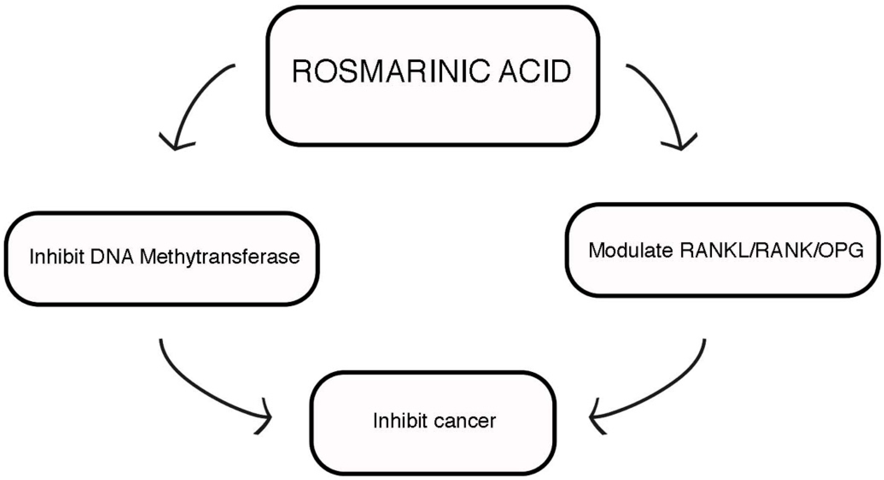
3.1. Anticancer Potential

Several mechanisms have been proposed for RA anticancer activity (Figure 3). For instance, in rats with colon cancer, RA at the concentration of 5 mg/kg body weight (b.w.) impaired tumor formation and development, reduced lipid peroxidation by-products and pro-apoptotic proteins expression, modulated xenobiotic enzymes, and increased apoptotic proteins expression [18]. In human liver cancer cell line, HepG2, transfected with plasmid containing ARE-luciferin gene, RA predominantly enhances ARE-luciferin activity and promotes nuclear factor E2-related factor-2 (Nrf2) translocation from cytoplasm to the nucleus and also increases MRP2 and P-gp efflux activity along with intercellular ATP level [19]. A study conducted by Wu et al. [20] reported that RA inhibited CCRF-CEM and CEM/ADR5000 cells in a dose-dependent pattern but caused less cytotoxicity towards normal lymphocytes. RA concurrently induced necrosis and apoptosis and stimulated MMP dysfunction activated PARP-cleavage and caspase-independent apoptosis. RA also blocked the translocation of p65 from the cytosol to the nucleus [20]. Moreover, it inhibits transcription factor hypoxia-inducible factor-1α (HIF-1α) expression, which affects the glycolytic pathway; meanwhile, it also suppressed glucose consumption and lactate production in colorectal cells [21]. RA also inhibits micro RNAs and pro-inflammatory cytokines and thus may suppress the Warburg effects through an inflammatory pathway involving activator of transcription-3 (STAT3) and signal transducer of interleukin (IL)-6 [22]. Furthermore, RA inhibits HL-60 promyelocytic leukemia cells’ growth and development and provides strong scavenging free radical effects, disturbing the balance of nuclear deoxyribonucleotide triphosphate (dNTP) levels without affecting protein levels of RR (R1, R2, p53R2) subunits, ultimately leading to apoptosis induction [23,24].
RA application, at a concentration of 5 mg/kg b.w. during 30 weeks in 1,2-dimethyldrazin stimulated colon carcinogenesis in the rat at 20 mg/kg b.w. and significantly stopped tumor formation and proliferation. RA supplementation also reduced tumor necrosis factor-α (TNF-α), cyclooxygenase-2 (COX-2) and IL-6 levels, and modulated p65 expression [25]. It is also able to inhibit the release of the highly mobile group box 1 (HMGB1) and to slow down HMGB1-dependent inflammatory responses in human endothelial cells, stopping HMBG1-mediated hyperpermeability and leukocytes migration in mice [26]. RA supplementation primarily decreases aberrant crypt foci (ACF) formation and multiplicity in rats [27].
RA inhibited APC10.1 cell growth that comes in Apc (Min) mouse model of colorectal carcinogenesis [28]. Through oral administration, RA totally prevented skin tumor cells formation in DMBA-induced mouse skin carcinogenesis and decreased lipid peroxidation byproducts levels [29]. It also inhibited human ovarian cancer A2780 cell line, disturbing the cell cycle at multiple phases and stimulating apoptosis by modifying multiple genes expression, involved in apoptosis regulation [30].
RA induced the cell cycle arrest and apoptosis in prostate cancer cell lines (PCa, PC-3, and DU145) [31]. These effects were mediated through modulation of histone deacetylases expression (HDACs), specifically HDAC2; the aberrant expression of theses enzymes is related with the onset of human cancer.
In rats with 1,2 dimethylhydrazine-induced colon carcinogenesis, RA administration at dose of 2.5, 5, and 10 mg/kg b.w. led to a decrease in the number of polyps (50%), reversed oxidative markers (21%), antioxidant status (38.6%), CYP450 contents (29.4%), and PNPH activities (21.9%) [32]. RA can also inhibit adhesion, invasion, and migration of Ls 174-T human colon carcinoma cells through enhancing GSH levels and decreasing ROS levels. Finally, RA may also inhibit colorectal carcinoma metastasis, by reducing the extracellular signal-regulated kinase pathway and the number and weight of lung tumors [33]. MDA-MB-231BO human bone homing breast cancer cells migrations are also inhibited by RA, whereas the number and size of mineralized nodules in ST-2 murine bone marrow stoma cells cultures raise [34].
RA also enhances chemosensitivity of human resistant gastric carcinoma SGC7901 cells [35]. The anticancer potential of RA analogues has also been tested. RA analogue-11 induces apoptosis of SGC7901 via the epidermal growth factor receptor (EGFR)/Akt/nuclear factor kappa B (NF-κB) pathway [36].

3.2. Antidiabetic Activity

RA supplementation increases the expression of mitochondrial biogenesis key genes, like sirtuin 1 (SIRT-1), peroxisome proliferator-activated receptor gamma coactivator 1-alpha (PGC-1α), and mitochondrial transcription factor A (TFAM) via activation of AMP-activated protein kinase (AMPK) in the skeletal muscle of insulin-resistant rats as well as in L6 myotubes. It also increased glucose faster and decreased serine IRS-1 phosphorylation, while increasing the glucose transporter type 4 (GLUT4) transfer [37].
In streptozotocin (STZ)-induced diabetic rats, RA exerted a noticeable hypoglycemic effect, whereas in high-fat diet (HFD) fed diabetic rats it increased glucose utilization and ameliorated insulin sensitivity. RA supplementation inverted the STZ- and HFD-induced increase in phosphoenolpyruvate carboxykinase (PEPCK) expression in the liver and the STZ- and HFD-induced decrease in GLUT4 expression in skeletal muscle. RA exerts hypoglycemic effects and improves insulin sensitivity, also increasing GLUT4 expression and decreasing PEPCK expression [38]. In addition, it also reverses memory and learning defects through improving cognition in healthy rats, inhibiting hyperglycemia, lipid peroxidation, and enhancing antioxidant defense system [39]. At a concentration of 10 mg/kg, RA decreased TBARS levels in kidney and liver of STZ-induced diabetic rats. This effect was mainly conferred by its ability to increase superoxide dismutase (SOD) and catalase (CAT) activity and to reverse the decrease in ascorbic acid and of non-protein thiol levels in diabetic rats [40]. RA administration also ameliorated oxidative stress markers in diabetic rats and water consumption and urination. Thus, it was proposed that RA mitigates STZ-induced diabetic manifestations by protecting rat’s tissues against free radicals’ damaging effects [15]. At 100 mg/kg, RA significantly increased insulin index sensitivity and reduced blood glucose, advanced glycation end-products, HbA1c, IL-1β, TNFα, IL-6, p-JNK, P38 mitogen-activated protein kinase (MAPK), and NF-κB levels. Moreover, it significantly reduced free fatty acids (FFA), triglycerides, serum cholesterol, AOPPs, lipid peroxides, and protein carbonyls levels in plasma and pancreas of diabetic rats. The reduced activities of CAT, SOD, glutathione S-transferases (GST), and glutathione peroxidase (GPx) and the reduced levels of vitamins C and E, ceruloplasmin, and GSH in plasma of diabetic rats were also significantly recovered by RA application. Furthermore, it protects pancreatic β-cells from oxidative stress in HFD-STZ-induced experimental diabetes [41]. The protective effects of RA (30 mg/kg) against hypoglycemia, hyperlipidemia, oxidative stress, and an imbalanced gut microbiota architecture was studied in diabetic rats. The treatment decreased the levels of fasting plasma glucose, total cholesterol, and triglyceride, exhibited an antioxidant and antiglycative effect, showed protective effects against tissue damage and inflammation in the abdominal aorta, increased the population of diabetes-resistant bacteria, and decreased the number of diabetes-sensitive bacteria [12].
RA also reduced diabetes occurrence and preserved normal insulin secretion, ROS, and reactive nitrogen species (RNS) by regulating antioxidant enzymes, and attenuating the pro-inflammatory T helper 2 and T regulatory cells levels [42]. At a concentration of 10 mg/kg, RA treatment significantly reduced lipid peroxidation levels in the hippocampus (28%), cortex (38%), and striatum (47%) of diabetic rats [43]. In Wistar rats, RA administrated orally at 50 mg/kg for 10 weeks in STZ-induced diabetes diminished endothelium-dependent relaxation accompanied by IL-1β, TNF-α, preproendothelin-1, and endothelin converting enzyme 1 overexpression. It also provided aortic endothelial function protection against diabetes-induced damage [44]. Finally, it significantly inhibited the carbohydrate-induced adaptive increase of sodium-dependent glucose cotransporter 1 (SGLT1) in the enterocyte brush border membrane [45].

3.3. Antimicrobial Activity

Regarding RA antimicrobial activity, it exerted antibacterial effects against Staphylococcus aureus strains, and the lowest blocking concentration was found to be 0.8 and 10 mg/mL against S. aureus and methicillin-resistant S. aureus (MRSA), respectively. Moreover, it displayed synergistic effects with amoxicillin, ofloxacin and vancomycin antibiotics against S. aureus, and only with vancomycin against MRSA. Time-kill analysis showed that using a combination of RA with antibiotics is more effective than using individual antibiotics. Microbial surface components recognizing adhesive matrix molecules (MSCRAMM) adhesion protein expression in MRSA and S. aureus was also significantly suppressed by using a combination of RA with vancomycin rather than RA alone [46].
On the other hand, RA administration reduced biofilm formation in a concentration- and time-dependent manner, suggesting that it could be used as an effective antimicrobial agent to kill the planktonic cells activity and to reduces the biofilm formation activity in early-stage development [47]. RA also exerts inhibitory effects against Escherichia coli K-12 and Staphylococcus carnosus LTH1502 growth, through decreasing cell counts and cell number [48]. Under acidic conditions, RA was reacted with nitrite ions to give 6,6-nitro and 6-dinitrorosmarinic acids. These compounds were active as HIV-1 integrase inhibitors at sub-molecular levels and inhibited viral replication in MT-4 cells. Without increasing cellular toxicity levels, RA nitration A strongly improved anti-integrase inhibition and antiviral effects [49]. Moreover, RA also exerted antimicrobial effects against Enterobacteriaceae spp., Pseudomonas spp., lactic acid bacteria, yeast and mold, and psychotropic counts, as well as fate Listeria monocytogenes inculcated in chicken meats [50]. Finally, RA also displayed inhibitory effects against S. aureus cocktail through inducing morphological changes and reducing viable cells counts and causing morphological changes in cheese and meat samples, such as cell shrinkage and appearance of blabbing-like structures in cell surfaces [51,52,53].

3.4. Cardioprotective Activity

RA at 25, 50, 10 mg/L had the capacity to maintain ATP levels in cells and inhibit the decrease in H/R-induced cell viability, lactate dehydrogenase (LDH) leakage, and excessive ROS. It also inhibited H/R-induced cardiomyocyte apoptosis and down-regulated p-AKt cleaved caspase expression [54].
The endothelial protein C receptor (EPCR) has a prominent role in inflammation and coagulation, whereas its activity is significantly changed by ectodomain cleavage and release as the soluble protein (sEPCR). RA has been found to be a strong anti-inflammatory agent. Monitoring RA effects in TNF-α, phorbol-12-myristate 13-acetate (PMA), IL-1β and in cecal ligation and puncture (CLP)-mediated EPCR shedding and underlying mechanisms, it was found that RA treatment led to a potent inhibition of PMA, TNF-α, and IL-induced EPCR shedding through TACE expression suppression. Furthermore, RA reduced extracellular regulated kinases (ERK) 1/2, PMA-stimulated p38, and c-Jun N-terminal kinase (JNK) phosphorylation. These results support the upcoming use of RA as an anti-sEPCR shedding reagent against IL-1β, TNF-α, PMA, and CLP-mediated EPCR shedding [55,56].
RA administration in fructose-fed rats (FFR) significantly enhanced insulin sensitivity, reduced lipid levels, oxidative damage, and a p22phox subunit of nicotinamide adenine dinucleotide phosphate reduced oxidase expression as well as prevented cardiac hypertrophy. RA lowered fructose-induced blood pressure through decreasing angiotensin-converting enzymes activity and endothelin-1 and increasing the level of nitric oxide (NO) [57]. RA also reduced fasting serum levels of vascular cell adhesion molecule 1 (VCAM-1), inter-cellular adhesion molecule 1 (ICAM-1), plasminogen-activator-inhibitor-1 (PAI-1), and increased GPX and SOD levels [58]. RA also intervenes in many important steps of angiogenesis, including adhesion, migration, proliferation, and tube formation of human umbilical vein endothelial cells (HUVEC) in a dose-dependent pattern. It also decreased IL-8 release from endothelial cells, H2O2-dependent vascular endothelial growth factor (VEGF) expression, and intracellular ROS levels [59]. Finally, in H9c2 cardiac muscle cells, RA inhibited apoptosis by decreasing intracellular ROS generation and recovering mitochondria membrane potential [60,61].

3.5. Antioxidant Activity

RA exhibited free radicals scavenging activity in hepatic stellate cells (HSCs) as a result of its antioxidant effects by boosting GSH synthesis and participating in NF-κB-dependent inhibition of MMP-2 activity. It also has the ability to reverse activated HSCs to quiescent cells and ultimately inhibits MMP-2 activity. RNA interference-imposed knockdown of NF-κB abolished MMP-2 down-regulation by RA. NF-κB inactivation mediated by RA could be blocked by the diphenyleneiodonium chloride, a potent inhibitor of NADH/NADPH oxidase. Moreover, transfection of dominant-negative (DN) mutant JNK1, p38α kinase, or extracellular signal-regulated kinases 2 (ERK2) had no such effect. At once, RA suppresses lipid peroxidation (LPO) and ROS generation, whereas in HSC-T6 cells it increases cellular GSH. Additionally, it significantly increases Nrf2 translocation and catalytic subunits from glutamate cysteine ligase (GCLc) expression but was not able to modulate GCL (GCLm) subunits and antioxidant response element (ARE)-mediated luciferase activity. GClc up-regulation mediated by RA is inhibited by shRNA-induced Nrf2 knockdown. The knocking down of Nrf2 abolished RA-mediated inhibition of ROS [16,62].
On the other hand, lycopene and RA administration reduced elevated blood urea nitrogen, renal malondialdehyde (MDA), proapoptotic protein (Bax) immuno-expression, serum creatinine, inducible nitric oxide synthase (iNOS), and autophagic marker protein (LC3/B) levels induced by gentamicin. This combination also increased the reduced SOD, an antiapoptotic protein (Bcl2) immuno-expression, GPx, and GSH levels and ameliorated gentamicin-induced histopathological changes. Moreover, it also evidenced a greater protective effect than corresponding monotherapy [63].
The in vivo antioxidant defense system consists of antioxidant enzymes including CAT, GPx, SOD, and nutritional antioxidants. Any disturbance in normal antioxidant defense system triggers several diseases, including diabetes, cancer, atherosclerosis, and degenerative diseases [64]. In experimental animals, carbon tetrachloride (CCl4) induced neurotoxicity, whereas CCl4 skin absorption, inhalation, and ingestion increased lipid peroxidation and reduced protein and antioxidant enzymes contents. This molecule produces free radicals in the lungs, heart, blood cells, and kidney. Under aerobic conditions, CCl4 is converted into highly reactive trichloromethyl radicals through the action of the cytochrome P450 system [65].
Brain amyloid-β (Aβ) accumulation is a hallmark of Alzheimer’s disease (AD) and has an important role in cognitive dysfunction [66]. At a dose of 0.25 mg/kg, RA significantly enhanced cognitive function and object discrimination and recognition test. Furthermore, RA decreased the time to reach the platform and increased the number of crossings over the removed platform, when compared with Aβ25-35-induced group in Morris water maze test; moreover, it reduced NO and MDA levels in kidney, brain, and liver [67]. RA also suppresses AD development by reducing amyloid β aggregation by increasing monoamine secretion in mice [68].
RA also prevented oxidative stress in C6 glial cells by increasing cell viability and inhibiting lipid peroxidation. It also decreased H2O2-induced COX-2 and iNOS expression at the transcriptional level and down-regulated COX-2 protein expression and iNOS in C6 glial cells treated with RA [67]. It significantly reduced oxidative stress and increased antioxidant status in Wistar rats post-spinal cord injury (SCI). RA also facilitated the inflammatory process through pro-inflammatory post-SCI and down-regulated NF-κB [69]. It also exerted a significant cytoprotective effect through covering the intercellular ROS in HaCat keratinocytes. RA also increased CAT, SOD, heme oxygenase-1 (HO-1), and transcription factor Nrf2 expression and activity, markedly reduced by UVB radiation [70].
On the other hand, in G93A-SOD1 transgenic mice with amyotrophic lateral sclerosis (ALS), RA application at a daily dose of 400 mg/kg significantly increased their survival by reliving function motor neurons deficits. These types of systematic changes were closely correlated with a decrease in neuronal loss and oxidative stress in ventral horns of G93A-SOD1 mice [71]. RA also increased SOD, GSH and GPx activities and decreased MDA levels in kidney and liver of sepsis-induced rats [72].
RA also reduced threshold shift, attenuated noise-induced hearing loss, and promoted hair cells survival. Moreover, it enhanced the endogenous antioxidant defense system by decreasing SOD production and up-regulation and decreased 4-HNE expression [73,74]. SOD, CAT, and GPx activities were increased through RA application at a dose of 50, 100, and 200 mg/kg. Furthermore, it induced structural changes in the kidney and liver at a dose of 200 mg/kg [75,76].

3.6. Hepatoprotective Activity

Sepsis, shock, and renal artery stenosis are the major clinical problems of acute renal failure, usually associated with high morbimortality rates. Ischemia-reperfusion (I-R) injury provokes cell damage, cell death, tissue necrosis, multiorgan dysfunction and increases vascular permeability. These types of physiopathological processes include RNS, ROS, neutrophils, cytokines, platelets, coagulation system, endothelium, and xanthine oxidoreductase enzyme system activation. During I-R injury, cell death occurs as a result of both apoptosis and necrosis [77,78,79]. RA modulates lipid peroxidation, production of ROS, peroxynitrite formation, complement factors and proinflammatory mediators, such as cytokines and chemokines. These processes are involved in hepatic diseases [80].
On the other hand, RA at dose of 150 mg/kg was able to treat rats with I-R, lowered lipid peroxidation and nitro tyrosine levels, enhanced GSH contents, and reduced neutrophil infiltration, hepatocellular damage, and all oxidative or nitrative stress markers. It also exerted anti-inflammatory and antioxidant effects in the ischemic liver, protecting hepatocytes from ischemic injury [81,82]. Moreover, it also conferred marked protection against oxidative stress through increasing CAT and GPx contents, thus preventing hepatic steatosis. Different RA derivatives have also been reported as having anti-secretory and antiulcer effects in epithelial tissues, being able to heal gastric ulceration [83]. Indeed, RA led to a significant reduction in hepatic toxicity, including alanine aminotransferase (ALT), aspartate aminotransferase (AST), lipid peroxidation, and oxidized glutathione levels and improved antioxidant effects of GPx, CAT, and SOD enzymes [26]. A marked improvement in liver serum markers and histology and inflammatory process decrease were also stated after RA administration at a dose of 10, 25, and 50 mg/kg by gavage once daily for two consecutive days against CCl4-induced hepatic necrosis. Furthermore, RA prevented α-smooth muscle actin (α-SMA) and transforming growth factor β1 (TGF-β1) expression, suggesting profibrotic response suppression [84].
Peroxisome proliferator-activated receptor γ (PPARγ) is required for HSCs differentiation, and its epigenetic repression carries on the HSCs activation. RA inhibits HSCs signaling and expression by canonical Wnts as well as suppresses liver fibrosis progression and activation [85,86,87]. In CCl4-induced rat liver fibrosis model, RA inhibited HSCs proliferation and TGF-βl, connective transforming growth factor (CTGF), and α-SMA expression. Much evidence has shown that RA can decrease fibrosis grade and ameliorate biochemical and histopathological morphology in CCl4-induced liver fibrosis [88].
In lipopolysaccharide (LPS)-activated RAW164.7 cells, RA concentration-dependently down-regulated IL-6, TNF-α, and high mobility group box1 protein levels. RA also inhibited I kappa B kinase pathway and modulated NF-κB. RA intravenous injection also decreased puncture-induced lethality and cecal ligation in rats. Additionally, RA down-regulated serum IL-6 and TNF-α levels, triggering receptor expressed on myeloid cells, high mobility group box1 protein, and endotoxin whilst up-regulating the serum IL-6 level. Moreover, post-RA injection, a marked decrease in serum enzyme activities was observed, along with amelioration of liver, lungs, and small intestine hemodynamics; this anti-inflammatory mechanism may be explained through inhibition of NF-κB pathway activation by inhibiting I kappa B kinase activity [89]. RA also decreased ROS production and protein and DNA synthesis inhibition in a dose-dependent manner [90].
In extrahepatic cholestasis rat model by bile-duct ligation, RA showed hepatoprotective effect via mechanisms involving resolution of oxidative burden and down-regulation of HMGB1/TLR4, NF-κB, AP-1, and TGF-β1/Smad signaling [91].

3.7. Antidepressant Potential

RA effects at a dose of 0, 3, 10, or 30 mg/kg were investigated in female C57BL/6 mice for 60 min before pilocarpine (300 mg/kg) or pentylenetetrazol (PTZ, 60 mg/kg) injection. Generalized seizure duration and myoclonic generalized tonic-clonic seizure latencies were analyzed by electroencephalographic (EEG) and behavioral methods. The effect of an acute RA dose on mice was also evaluated in behavior in the open field, rotarod, novel object recognition, and forced swim tests. In the PTZ model, RA dose-dependently increased generalized seizures and latency to myoclonic jerks and improved pilocarpine-induced myoclonic jerks’ latency. Additionally, RA (30 mg/kg) improved the time at the center of the open field, the crossings number and the immobility time in forced swim test [92].
Worldwide mood disorders are the most spreading forms of mental illness and, in main cases, morbidity. According to the World Health Organization (WHO), depression is one of the top causes of morbimortality throughout the world. Depression is a recurrent, potentially life-threatening, and chronic mood disorder that has been estimated to affect about 21% of the world population [93]. RA administration (10 mg/kg, daily) in chronic stress Sprague Dawley rats changed depressive behaviors in rats exposed to impulsive stress modal and restored hippocampal brain-derived neurotrophic factor (BDNF) and pERK1/2 protein expression. Thus, RA could be conceived as a great molecule in the treatment of depression and in triggering changes in BDNF levels and in ERK11/2 signaling in pharmacological science [94]. In PC 12 cells, RA showed significant neurotrophic effects and improved cholinergic functions in correlation with the ERK1/2 signaling pathway and MAPK. RA also caused an extensive up-regulation of pyruvate carboxylase (PC) and tyrosine hydroxylase (TH), involved in serotonergic, GABAergic, and dopaminergic pathways regulation, whereas against corticosterone-induced toxicity it provides neuronal cells protection [95].

3.8. Nephroprotective Activity

Currently, the most often used antimicrobial agents provoke an acute renal injury in 60% of acquired infections in hospitals with noticeable morbimortality rates [96]. Among them, aminoglycosides have been frequently used to treat bacterial infections, and gentamicin is the most commonly used aminoglycoside, given its lower costs and lower rate of antibiotic-acquired resistance; nevertheless, its therapeutic use at 80 mg/kg/day for more than 7 days induces nephrotoxicity in about 30% of patients [97]. Acute renal toxicity characterization is assessed by the sudden decrease of kidney function due to the accumulation of urea, creatinine, and other waste products. Gentamicin-induced nephrotoxicity is related to enhanced oxidative stress levels, which may be a major contributing factor for renal injury [98].
RA led to a decrease in serum blood urea nitrogen and creatinine levels in Sprague Dawley rats and ultimately decreased myeloperoxidase and MDA levels. In fact, the level of incident injury decreased in the RA-treated group. In addition, RA extensively reduced Bowman’s capsules dilatation, glomerular necrosis, tubular epithelium degeneration, tubular epithelium necrosis and dilatation, and focal glomerular necrosis [99].
RA at doses of 1, 2, and 5 mg/kg for 2 days significantly increased serum creatinine and blood urea nitrogen levels and reduced cisplatin (CP)-induced histopathological changes. RA also reduced CP-produced oxidative stress and amplified cytochrome P450 2E1 (CYP2E1), HO-1, and renal-4-hydroxynonenal expression. Additionally, RA repressed TNF-α and NF-κB expression, as stated through inflammation inhibition. Moreover, RA reduced p53, phosphorylated p53, and active caspase-3-expression in kidney through exerting antiapoptotic activity [100]. RA also notably reduced MDA, tubular necrosis, urea, and creatinine levels, and increased renal GSH, SOD, CAT, GPS, volume density creatinine clearance, and PCT [101]. Finally, RA could provide protective effects against 6-hydroxydopamine-induced neurotoxicity via its antioxidant activity [102].

3.9. Anti-Aging Activity

RA administration could effectively reverse chaperones- and Pin1-induced abnormal changes and suppress P-tau and insoluble P-tau formation, induced by chronic restraint stress (CRS), particularly in middle-aged mice [103].
AD is a progressive neurodegenerative disorder that causes dementia in older people. Disease indicators include the appearance of plaques and tangles in brain tissues, which progressively kill neurons from brain cortex, amygdala, hippocampus, and other non-regeneratable brain regions. Consequently, acetylcholine (ACh) levels decline, the widely known cholinergic deficit hypothesis for AD. ACh has a significant role in brain functions, such as thinking, reasoning, remembering, and behavioral abilities [104,105].
In AD, stress is an important risk factor, since it induces tau phosphorylation and enhances tau insolubility in the brain. RA application is able to dominantly suppress the increase in tau phosphorylation levels and insoluble P-tau formation, facilitated by chronic resistant stress, and overturn the abnormal changes in middle-aged mice [103]. At 1.6, 16 and 32 mg/kg, RA exerted markedly useful effects on memory and learning and also reduced the levels of protein carbonyls in the hippocampus [106]. In ALS, a neurodegenerative disease, RA significantly delays motor neuron dysfunction in paw grip endurance tests, through attenuating motor neurons degeneration and extending the life span of ALS mice model, detected at later stages, and about 2% patients present an associated mutation in the gene encoding Cu/Zn-SOD [107].
RA also exerted protective effects against 6-hydroxydopamine-facilitated neurotoxicity and prevented 1-methyl-4-phenypyridinium effects in MES23.5 dopaminergic cells. In fact, 1-methyl-4-phenylpyridinium treatment reduces cell viability and dopamine contents, as well as causes apoptotic morphological changes. Additionally, 1-methyl-4-phenylpyridinium precedes mitochondrial dysfunction, easily detected through inhibiting mitochondrial respiratory chain complex 1-associated activity, suggesting mitochondrial transmembrane collapse and ROS generation. Thus, RA pretreatment was able to restore mitochondrial respiratory chain complex 1 activity and to reverse the other MPP positive damaging effects [91,92] partially. In mice, RA improves oxidative stress parameters and mitochondrial respiratory chain activity [108]. RA also proved to be effective in preventing in vitro amyloid peptide aggregation and in delaying disease progression in animal models [109]. It also provided neuroprotective effects against Aβ-induced toxicity by lowering lipid peroxidation, DNA and ROS formation, and inhibiting phosphorylated p38 MAPK levels [110,111]. RA improved antioxidant properties and healthspan via the IIS and MAPK pathways in Caenorhabditis elegans [112].
N2A cells’ H2O2-induced cytotoxicity is also positively affected by RA; in fact, it is able to attenuate LDH, intercellular ROS, and mitochondrial membrane potential disruption. RA also promoted TH and BDNF genes up-regulation and prevented genotoxicity [113,114]. Clovamide in combination with RA, at the rate of 10–100 µM, in SH-SY5Y cells significantly enhanced PPARγ expression and inhibited NF-κB translocation, respectively [115].

3.10. Anti-Allergic Activity

RA significantly decreased murine double minute (MDM) 2 and thymic stromal lymphopoietin (TSLP) expression in induced mast cells proliferation. It also significantly decreased the levels of phosphorylated signal transducer, IL-13, and transcription-6 activation in TSLP-stimulated HMC-1 cells. Moreover, RA triggered an increment of p53 levels, poly-ADP-ribose polymerase cleavage, caspase-3 activation, and a reduction in Bcl2 and procaspase-3 levels. Furthermore, it significantly reduced TNF-α, IL-6, and IL-1β production in TSLP-stimulated HMC-1 cells. It also reduced IL-4, immunoglobulin E (IgE), and TSLP levels in short ragweed pollen-induced allergic conjunctivitis mouse model [116].
In murine model of respiratory allergy caused by Bloma tropicalis (Bt) mite, RA led to a considerable decrease in leukocytes or eosinophils numbers in bronchoalveolar lavage (BAL), of mucus presence in the respiratory tract, reduced lung histopathological changes, and eosinophil peroxidase activity and IL-4 changes [117,118]. At a dose of 1 or 5 μM in NC/Nga mice under specific pathogenic free conditions, RA was shown to be the most effective treatment against 2,4-dinitrofluorobenzene (DNFB)-induced AD-like skin lesion. Moreover, it suppressed IL-4 and interferon (INF) production by activated CD4+ cells. RA also inhibited skin lesions and ears thickness development and increased total serum IgE levels in DNFB-treated NC/Nga mice [119]. Furthermore, it inhibited IgE levels increase in spleen, nasal mucosa, and serum, inhibited the increase in rubs number, and reduced histamine levels in ovalbumin unsensitized (OVA) mice. RA also inhibited protein levels and mRNA expressions of IL-6, IL-1β, and TNF-α in nasal mucosa or spleen tissues in OVA-sensitized mice [120,121]. In a murine model of allergic asthma triggered by house dust mites (HDMs), RA inhibited the boost up of mononuclear, eosinophils, and neutrophils cells levels around airways and in BAL fluid. Moreover, it also significantly inhibited IL-13 expression increased by HDM allergen [122].
RA (200 mg or 50 mg for 21 days) also reduced the number of eosinophils and neutrophils considerably in nasal lavage fluid. Up-regulation of VCAM-1, COX-2, MIP-2, and ICAM-1 by 2-tetradecanoylphorbol 13-acetate (TPA) were significantly reduced with RA pretreatment. ROS production detected as LPO, 8-hydroxy-2′deoxyguanosine (8OH-dG) and thiobarbituric acid reactive substance (TBARS) and TPA levels were markedly reduced by RA pretreatment. Furthermore, RA is conceived as a potential agent against seasonal allergic rhino conjunctivitis (SAR), through mediated polymorphonuclear leukocytes (PMNL) infiltration inhibition [123].
RA also inhibited significantly the increases in eosinophils levels in BAL fluid along with murine airways. In the lungs of sensitized mice, RA inhibited the increase in protein expression of IL-5, IL-4, and eotaxin. Thus, RA seems to be a successful intervention for allergic asthma, given its ability to increase chemokines, cytokines, and allergen-specific antibody levels [124]. Additionally, RA reduced inflammation and allergic immunoglobulin responses occurring in mice PMNL. It also noticeably increased the response rates for itchy eyes, watery eyes, and itchy nose and reduced eosinophils and neutrophils levels in nasal lavage fluid of SAR [125].

3.11. Anti-Inflammatory Activity

RA administration (40 mg/kg) in a rat model of sciatic nerve chronic constriction injury (CCI)-induced neuropathic pain reduced spinal inflammatory markers, such as matrix metallopeptidase 2 (MMP2), prostaglandin E2 (PGE-2), IL-1β, and COX-2 [126]. In addition, RA administration (75, 150, and 300 mg/kg) in hepatocellular carcinoma (HCC) for 10 days reduced inflammatory and angiogenic factors levels, including TNF-α, IL-6, IL-1β, TGF-β, and VEGF. Moreover, it also decreased NF-κB and p65 expression in the xenograft microenvironment [127].
On the other hand, RA (IC50 = 14.25 µM) inhibited LPS-induced NO production in RAW 264.7 cells. It also repressed LPS-induced pro-inflammatory cytokines expression, including INF-β, MCP-1, iNOS, IL-1β, IL-6, IL-10, and NF-κB activation. In dependent and independent pathways, down-regulation of iNOS by RA was due to myeloid differentiation primary response gene 88 (MyD88). Additionally, RA triggered HO-1 expression through inducing Nrf2 activity [92,128].
RA (5, 10, and 20 mg/kg) also inhibited Th2 cytokines in BAL fluid, increased inflammatory cells, ameliorated hyper airway responsiveness (AHR), and reduced total IgE and Ova-specific IgE concentrations in a murine model of asthma (female BALB/c mice). In upper airways, RA reduced the number of mucus hypersecretion and inflammatory cells. It seems that RA protective effects might be mediated by p38 phosphorylation, JNK, and ERK suppression. Additionally, RA pretreatment provoked the reduction of Ym2, CC chemokine receptor 3 (CCR3), CCL11 (eotaxin), AMCase, and E-selectin mRNA expression in lung tissues [129]. In SPI, RA noticeably increased antioxidant status and reduced oxidative stress levels in Wistar rats post-SCI. It also improved inflammatory mechanisms through pro-inflammatory cytokines reduction and NF-κB down regulation [69]. RA (10, 25, and 50 mg/kg) also remarkably decreased the serum transaminases (ALT and AST) and LDH concentration in liver ischemia-reperfusion rats. Furthermore, it reduced multiorgan dysfunction markers (lung, liver, and kidney) through metalloproteinase-9 and NF-κB modulating [130]. RA also displayed both peripheral and central antinociceptive effects and anti-inflammatory activity against chronic and acute inflammation [83,131,132,133]. In Freund’s complete adjuvant (FCA)-induced arthritic rats, RA attenuates inflammation [134].
RA also decreased blocked TNF-α-induced NF-κB activation and cytotoxicity, oxygen-glucose deprivation (OGD)-induced apoptosis, and high-mobility group box1 (HMGB1) expression in SH-SY5Y cells. It also decreased brain edema, reduced NF-κB activation and HMGB1 expression, and attenuated histopathological damage at a dose of 50 mg/kg [135]. RA also considerably lowered allergic asthma through a significant decrease in the number of leukocytes/eosinophils, of mucus present in the respiratory tract, eosinophil peroxidase activity, and IL-4 and histopathological changes in lung BAL fluid [117,136]. RA-derived water and ethanol extract also markedly inhibited LPS-stimulated PGE-2 and NO production in a dose-dependent manner in RAW 264.7 mouse macrophages [137].

4. Conclusions

Current evidence supports the deepened exploration of RA as a promising therapeutic agent against a wide variety of modern lifestyle disorders (Table 1). However, mechanisms underlying RA’s therapeutic activity need further investigation. Initial studies indicate that RA may act through various mechanisms, such as exerting anti-inflammatory and antioxidant effects as well as inhibiting cell proliferation, migration, and selectively inducing cancer cells apoptosis. In addition, RA’s anti-angiogenic effects, as demonstrated through human umbilical vein endothelial cells proliferation, migration, adhesion, and tube formation inhibition, suggest that it can be beneficial in preventing tumor growth and metastasis. Thus, given the above-highlighted aspects, rosemary extract can be conceived as a rich source of potential candidates to be included in the diet with promising effects at pre-determined doses, avoiding toxicity.

Author Contributions

All authors contributed to the manuscript. Conceptualization: M.I., M.N. and J.S.-R.; Validation investigation, resources, data curation, writing: all authors; review and editing: B.S., J.S.-R., W.C.C. and N.M. All the authors read and approved the final manuscript.

Funding

None.

Acknowledgments

N. Martins thank to Portuguese Foundation for Science and Technology (FCT–Portugal) for the Strategic project ref. UID/BIM/04293/2013 and “NORTE2020-Programa Operacional Regional do Norte” (NORTE-01-0145-FEDER-000012). Also, this work was supported by CONICYT PIA/APOYO CCTE AFB170007.

Conflicts of Interest

The authors declare no conflict of interest.

References

  1. Gordo, J.; Maximo, P.; Cabrita, E.; Lourenco, A.; Oliva, A.; Almeida, J.; Filipe, M.; Cruz, P.; Barcia, R.; Santos, M.; et al. Thymus mastichina: Chemical constituents and their anti-cancer activity. Nat. Prod. Commun. 2012, 7, 1491–1494. [Google Scholar] [CrossRef] [PubMed]
  2. Baliga, M.S.; Jimmy, R.; Thilakchand, K.R.; Sunitha, V.; Bhat, N.R.; Saldanha, E.; Rao, S.; Rao, P.; Arora, R.; Palatty, P.L. Ocimum sanctum L. (Holy Basil or Tulsi) and its phytochemicals in the prevention and treatment of cancer. Nutr. Cancer 2013, 65 (Suppl. 1), 26–35. [Google Scholar] [CrossRef] [PubMed]
  3. Andrade, J.M.; Faustino, C.; Garcia, C.; Ladeiras, D.; Reis, C.P.; Rijo, P. Rosmarinus officinalis L.: An update review of its phytochemistry and biological activity. Future Sci. OA 2018, 4, FSO283. [Google Scholar] [CrossRef] [PubMed]
  4. Ngo, S.N.; Williams, D.B.; Head, R.J. Rosemary and cancer prevention: Preclinical perspectives. Crit. Rev. Food Sci. Nutr. 2011, 51, 946–954. [Google Scholar] [CrossRef] [PubMed]
  5. Baba, S.; Osakabe, N.; Natsume, M.; Terao, J. Orally administered rosmarinic acid is present as the conjugated and/or methylated forms in plasma, and is degraded and metabolized to conjugated forms of caffeic acid, ferulic acid and m-coumaric acid. Life Sci. 2004, 75, 165–178. [Google Scholar] [CrossRef] [PubMed]
  6. Cho, A.S.; Jeon, S.M.; Kim, M.J.; Yeo, J.; Seo, K.I.; Choi, M.S.; Lee, M.K. Chlorogenic acid exhibits anti-obesity property and improves lipid metabolism in high-fat diet-induced-obese mice. Food Chem. Toxicol. Int. J. Publ. Br. Ind. Biol. Res. Assoc. 2010, 48, 937–943. [Google Scholar] [CrossRef] [PubMed]
  7. Zych, M.; Kaczmarczyk-Sedlak, I.; Wojnar, W.; Folwarczna, J. The Effects of Sinapic Acid on the Development of Metabolic Disorders Induced by Estrogen Deficiency in Rats. Oxid. Med. Cell. Longev. 2018, 2018, 9274246. [Google Scholar] [CrossRef] [PubMed]
  8. Folwarczna, J.; Pytlik, M.; Zych, M.; Cegiela, U.; Nowinska, B.; Kaczmarczyk-Sedlak, I.; Sliwinski, L.; Trzeciak, H.; Trzeciak, H.I. Effects of caffeic and chlorogenic acids on the rat skeletal system. Eur. Rev. Med Pharmacol. Sci. 2015, 19, 682–693. [Google Scholar] [PubMed]
  9. Jayanthy, G.; Subramanian, S. RA abrogates hepatic gluconeogenesis and insulin resistance by enhancing IRS-1 and AMPK signalling in experimental type 2 diabetes. RSC Adv. 2015, 5, 44053–44067. [Google Scholar] [CrossRef]
  10. Han, J.; Wang, D.; Ye, L.; Li, P.; Hao, W.; Chen, X.; Ma, J.; Wang, B.; Shang, J.; Li, D.; et al. Rosmarinic Acid Protects against Inflammation and Cardiomyocyte Apoptosis during Myocardial Ischemia/Reperfusion Injury by Activating Peroxisome Proliferator-Activated Receptor Gamma. Front. Pharmacol. 2017, 8, 456. [Google Scholar] [CrossRef] [PubMed] [Green Version]
  11. Amoah, S.K.; Sandjo, L.P.; Kratz, J.M.; Biavatti, M.W. Rosmarinic Acid—Pharmaceutical and Clinical Aspects. Planta Med. 2016, 82, 388–406. [Google Scholar] [CrossRef] [PubMed]
  12. Ou, J.; Huang, J.; Zhao, D.; Du, B.; Wang, M. Protective effect of rosmarinic acid and carnosic acid against streptozotocin-induced oxidation, glycation, inflammation and microbiota imbalance in diabetic rats. Food Funct. 2018, 9, 851–860. [Google Scholar] [CrossRef] [PubMed]
  13. European Medicines Agency. Assessment Report on Melissa officinalis L., Folium; EMA/HMPC/196746/2012; European Medicines Agency: London, UK, 2013; pp. 1–20. [Google Scholar]
  14. Fecka, I.; Turek, S. Determination of water-soluble polyphenolic compounds in commercial herbal teas from Lamiaceae: Peppermint, melissa, and sage. J. Agric. Food Chem. 2007, 55, 10908–10917. [Google Scholar] [CrossRef]
  15. Sotnikova, R.; Kaprinay, B.; Navarova, J. Rosmarinic acid mitigates signs of systemic oxidative stress in streptozotocin-induced diabetes in rats. Gen. Physiol. Biophys. 2015, 34, 449–452. [Google Scholar] [CrossRef] [PubMed]
  16. Lu, C.; Zou, Y.; Liu, Y.; Niu, Y. Rosmarinic acid counteracts activation of hepatic stellate cells via inhibiting the ROS-dependent MMP-2 activity: Involvement of Nrf2 antioxidant system. Toxicol. Appl. Pharmacol. 2017, 318, 69–78. [Google Scholar] [CrossRef] [PubMed]
  17. Enns, G.M.; Cowan, T.M. Glutathione as a Redox Biomarker in Mitochondrial Disease-Implications for Therapy. J. Clin. Med. 2017, 6, 50. [Google Scholar] [CrossRef]
  18. Venkatachalam, K.; Gunasekaran, S.; Namasivayam, N. Biochemical and molecular mechanisms underlying the chemopreventive efficacy of rosmarinic acid in a rat colon cancer. Eur. J. Pharmacol. 2016, 791, 37–50. [Google Scholar] [CrossRef]
  19. Wu, J.; Zhu, Y.; Li, F.; Zhang, G.; Shi, J.; Ou, R.; Tong, Y.; Liu, Y.; Liu, L.; Lu, L.; et al. Spica prunellae and its marker compound rosmarinic acid induced the expression of efflux transporters through activation of Nrf2-mediated signaling pathway in HepG2 cells. J. Ethnopharmacol. 2016, 193, 1–11. [Google Scholar] [CrossRef]
  20. Wu, C.F.; Hong, C.; Klauck, S.M.; Lin, Y.L.; Efferth, T. Molecular mechanisms of rosmarinic acid from Salvia miltiorrhiza in acute lymphoblastic leukemia cells. J. Ethnopharmacol. 2015, 176, 55–68. [Google Scholar] [CrossRef]
  21. Xu, Y.; Han, S.; Lei, K.; Chang, X.; Wang, K.; Li, Z.; Liu, J. Anti-Warburg effect of rosmarinic acid via miR-155 in colorectal carcinoma cells. Eur. J. Cancer Prev. Off. J. Eur. Cancer Prev. Organ. 2016, 25, 481–489. [Google Scholar] [CrossRef]
  22. Han, S.; Yang, S.; Cai, Z.; Pan, D.; Li, Z.; Huang, Z.; Zhang, P.; Zhu, H.; Lei, L.; Wang, W. Anti-Warburg effect of rosmarinic acid via miR-155 in gastric cancer cells. Drug Des. Dev. Ther. 2015, 9, 2695–2703. [Google Scholar] [CrossRef]
  23. Saiko, P.; Steinmann, M.T.; Schuster, H.; Graser, G.; Bressler, S.; Giessrigl, B.; Lackner, A.; Grusch, M.; Krupitza, G.; Bago-Horvath, Z.; et al. Epigallocatechin gallate, ellagic acid, and rosmarinic acid perturb dNTP pools and inhibit de novo DNA synthesis and proliferation of human HL-60 promyelocytic leukemia cells: Synergism with arabinofuranosylcytosine. Phytomed. Int. J. Phytother. Phytopharm. 2015, 22, 213–222. [Google Scholar] [CrossRef] [PubMed]
  24. Heo, S.K.; Noh, E.K.; Yoon, D.J.; Jo, J.C.; Koh, S.; Baek, J.H.; Park, J.H.; Min, Y.J.; Kim, H. Rosmarinic acid potentiates ATRA-induced macrophage differentiation in acute promyelocytic leukemia NB4 cells. Eur. J. Pharmacol. 2015, 747, 36–44. [Google Scholar] [CrossRef] [PubMed]
  25. Karthikkumar, V.; Sivagami, G.; Viswanathan, P.; Nalini, N. Rosmarinic acid inhibits DMH-induced cell proliferation in experimental rats. J. Basic Clin. Physiol. Pharmacol. 2015, 26, 185–200. [Google Scholar] [CrossRef] [PubMed]
  26. Yang, S.Y.; Hong, C.O.; Lee, G.P.; Kim, C.T.; Lee, K.W. The hepatoprotection of caffeic acid and rosmarinic acid, major compounds of Perilla frutescens, against t-BHP-induced oxidative liver damage. Food Chem. Toxicol. Int. J. Publ. Br. Ind. Biol. Res. Assoc. 2013, 55, 92–99. [Google Scholar] [CrossRef] [PubMed]
  27. Karthikkumar, V.; Sivagami, G.; Vinothkumar, R.; Rajkumar, D.; Nalini, N. Modulatory efficacy of rosmarinic acid on premalignant lesions and antioxidant status in 1.2-dimethylhydrazine induced rat colon carcinogenesis. Environ. Toxicol. Pharmacol. 2012, 34, 949–958. [Google Scholar] [CrossRef] [PubMed]
  28. Karmokar, A.; Marczylo, T.H.; Cai, H.; Steward, W.P.; Gescher, A.J.; Brown, K. Dietary intake of rosmarinic acid by Apc(Min) mice, a model of colorectal carcinogenesis: Levels of parent agent in the target tissue and effect on adenoma development. Mol. Nutr. Food Res. 2012, 56, 775–783. [Google Scholar] [CrossRef] [PubMed]
  29. Sharmila, R.; Manoharan, S. Anti-tumor activity of rosmarinic acid in 7.12-dimethylbenz(a)anthracene (DMBA) induced skin carcinogenesis in Swiss albino mice. Indian J. Exp. Biol. 2012, 50, 187–194. [Google Scholar]
  30. Tai, J.; Cheung, S.; Wu, M.; Hasman, D. Antiproliferation effect of Rosemary (Rosmarinus officinalis) on human ovarian cancer cells in vitro. Phytomed. Int. J. Phytother. Phytopharm. 2012, 19, 436–443. [Google Scholar] [CrossRef]
  31. Jang, Y.G.; Hwang, K.A.; Choi, K.C. Rosmarinic Acid, a Component of Rosemary Tea, Induced the Cell Cycle Arrest and Apoptosis through Modulation of HDAC2 Expression in Prostate Cancer Cell Lines. Nutrients 2018, 10, 1784. [Google Scholar] [CrossRef]
  32. Venkatachalam, K.; Gunasekaran, S.; Jesudoss, V.A.; Namasivayam, N. The effect of rosmarinic acid on 1.2-dimethylhydrazine induced colon carcinogenesis. Exp. Toxicol. Pathol. Off. J. Ges. Fur Toxikol. Pathol. 2013, 65, 409–418. [Google Scholar] [CrossRef] [PubMed]
  33. Xu, Y.; Xu, G.; Liu, L.; Xu, D.; Liu, J. Anti-invasion effect of rosmarinic acid via the extracellular signal-regulated kinase and oxidation-reduction pathway in Ls174-T cells. J. Cell. Biochem. 2010, 111, 370–379. [Google Scholar] [CrossRef] [PubMed]
  34. Xu, Y.; Jiang, Z.; Ji, G.; Liu, J. Inhibition of bone metastasis from breast carcinoma by rosmarinic acid. Planta Med. 2010, 76, 956–962. [Google Scholar] [CrossRef] [PubMed]
  35. Yu, C.; Chen, D.Q.; Liu, H.X.; Li, W.B.; Lu, J.W.; Feng, J.F. Rosmarinic acid reduces the resistance of gastric carcinoma cells to 5-fluorouracil by downregulating FOXO4-targeting miR-6785-5p. Biomed. Pharmacother. 2019, 109, 2327–2334. [Google Scholar] [CrossRef] [PubMed]
  36. Li, W.; Li, Q.; Wei, L.; Pan, X.; Huang, D.; Gan, J.; Tang, S. Rosmarinic Acid Analogue-11 Induces Apoptosis of Human Gastric Cancer SGC-7901 Cells via the Epidermal Growth Factor Receptor (EGFR)/Akt/Nuclear Factor kappa B (NF-κB) Pathway. Med. Sci. Monit. Basic Res. 2019, 25, 63–75. [Google Scholar] [CrossRef] [PubMed]
  37. Jayanthy, G.; Roshana Devi, V.; Ilango, K.; Subramanian, S.P. Rosmarinic Acid Mediates Mitochondrial Biogenesis in Insulin Resistant Skeletal Muscle Through Activation of AMPK. J. Cell. Biochem. 2017, 118, 1839–1848. [Google Scholar] [CrossRef]
  38. Runtuwene, J.; Cheng, K.C.; Asakawa, A.; Amitani, H.; Amitani, M.; Morinaga, A.; Takimoto, Y.; Kairupan, B.H.; Inui, A. Rosmarinic acid ameliorates hyperglycemia and insulin sensitivity in diabetic rats, potentially by modulating the expression of PEPCK and GLUT4. Drug Des. Dev. Ther. 2016, 10, 2193–2202. [Google Scholar] [CrossRef]
  39. Hasanein, P.; Felehgari, Z.; Emamjomeh, A. Preventive effects of Salvia officinalis L. against learning and memory deficit induced by diabetes in rats: Possible hypoglycaemic and antioxidant mechanisms. Neurosci. Lett. 2016, 622, 72–77. [Google Scholar] [CrossRef]
  40. Mushtaq, N.; Schmatz, R.; Ahmed, M.; Pereira, L.B.; da Costa, P.; Reichert, K.P.; Dalenogare, D.; Pelinson, L.P.; Vieira, J.M.; Stefanello, N.; et al. Protective effect of rosmarinic acid against oxidative stress biomarkers in liver and kidney of strepotozotocin-induced diabetic rats. J. Physiol. Biochem. 2015, 71, 743–751. [Google Scholar] [CrossRef]
  41. Govindaraj, J.; Sorimuthu Pillai, S. Rosmarinic acid modulates the antioxidant status and protects pancreatic tissues from glucolipotoxicity mediated oxidative stress in high-fat diet: Streptozotocin-induced diabetic rats. Mol. Cell. Biochem. 2015, 404, 143–159. [Google Scholar] [CrossRef]
  42. Vujicic, M.; Nikolic, I.; Kontogianni, V.G.; Saksida, T.; Charisiadis, P.; Orescanin-Dusic, Z.; Blagojevic, D.; Stosic-Grujicic, S.; Tzakos, A.G.; Stojanovic, I. Methanolic extract of Origanum vulgare ameliorates type 1 diabetes through antioxidant, anti-inflammatory and anti-apoptotic activity. Br. J. Nutr. 2015, 113, 770–782. [Google Scholar] [CrossRef]
  43. Mushtaq, N.; Schmatz, R.; Pereira, L.B.; Ahmad, M.; Stefanello, N.; Vieira, J.M.; Abdalla, F.; Rodrigues, M.V.; Baldissarelli, J.; Pelinson, L.P.; et al. Rosmarinic acid prevents lipid peroxidation and increase in acetylcholinesterase activity in brain of streptozotocin-induced diabetic rats. Cell Biochem. Funct. 2014, 32, 287–293. [Google Scholar] [CrossRef]
  44. Sotnikova, R.; Okruhlicova, L.; Vlkovicova, J.; Navarova, J.; Gajdacova, B.; Pivackova, L.; Fialova, S.; Krenek, P. Rosmarinic acid administration attenuates diabetes-induced vascular dysfunction of the rat aorta. J. Pharm. Pharmacol. 2013, 65, 713–723. [Google Scholar] [CrossRef]
  45. Azevedo, M.F.; Lima, C.F.; Fernandes-Ferreira, M.; Almeida, M.J.; Wilson, J.M.; Pereira-Wilson, C. Rosmarinic acid, major phenolic constituent of Greek sage herbal tea, modulates rat intestinal SGLT1 levels with effects on blood glucose. Mol. Nutr. Food Res. 2011, 55 (Suppl. 1), S15–S25. [Google Scholar] [CrossRef] [PubMed] [Green Version]
  46. Ekambaram, S.P.; Perumal, S.S.; Balakrishnan, A.; Marappan, N.; Gajendran, S.S.; Viswanathan, V. Antibacterial synergy between rosmarinic acid and antibiotics against methicillin-resistant Staphylococcus aureus. J. Intercult. Ethnopharmacol. 2016, 5, 358–363. [Google Scholar] [CrossRef]
  47. Slobodnikova, L.; Fialova, S.; Hupkova, H.; Grancai, D. Rosmarinic acid interaction with planktonic and biofilm Staphylococcus aureus. Nat. Prod. Commun. 2013, 8, 1747–1750. [Google Scholar] [CrossRef]
  48. Suriyarak, S.; Bayrasy, C.; Schmidt, H.; Villeneuve, P.; Weiss, J. Impact of fatty acid chain length of rosmarinate esters on their antimicrobial activity against Staphylococcus carnosus LTH1502 and Escherichia coli K-12 LTH4263. J. Food Prot. 2013, 76, 1539–1548. [Google Scholar] [CrossRef]
  49. Dubois, M.; Bailly, F.; Mbemba, G.; Mouscadet, J.F.; Debyser, Z.; Witvrouw, M.; Cotelle, P. Reaction of rosmarinic acid with nitrite ions in acidic conditions: Discovery of nitro- and dinitrorosmarinic acids as new anti-HIV-1 agents. J. Med. Chem. 2008, 51, 2575–2579. [Google Scholar] [CrossRef]
  50. Raeisi, M.; Tabaraei, A.; Hashemi, M.; Behnampour, N. Effect of sodium alginate coating incorporated with nisin, Cinnamomum zeylanicum, and rosemary essential oils on microbial quality of chicken meat and fate of Listeria monocytogenes during refrigeration. Int. J. Food Microbiol. 2016, 238, 139–145. [Google Scholar] [CrossRef]
  51. Honorio, V.G.; Bezerra, J.; Souza, G.T.; Carvalho, R.J.; Gomes-Neto, N.J.; Figueiredo, R.C.; Melo, J.V.; Souza, E.L.; Magnani, M. Inhibition of Staphylococcus aureus cocktail using the synergies of oregano and rosemary essential oils or carvacrol and 1.8-cineole. Front. Microbiol. 2015, 6, 1223. [Google Scholar] [CrossRef]
  52. Zhuang, Y.; Jiang, J.; Bi, H.; Yin, H.; Liu, S.; Liu, T. Synthesis of rosmarinic acid analogues in Escherichia coli. Biotechnol. Lett. 2016, 38, 619–627. [Google Scholar] [CrossRef] [PubMed]
  53. Suriyarak, S.; Gibis, M.; Schmidt, H.; Villeneuve, P.; Weiss, J. Antimicrobial mechanism and activity of dodecyl rosmarinate against Staphylococcus carnosus LTH1502 as influenced by addition of salt and change in pH. J. Food Prot. 2014, 77, 444–452. [Google Scholar] [CrossRef] [PubMed]
  54. Li, X.L.; Liu, J.X.; Li, P.; Zheng, Y.Q. Protective effect of rosmarinic acid on hypoxia/reoxygenation injury in cardiomyocytes. J. Chin. Mater. Med. 2014, 39, 1897–1901. [Google Scholar]
  55. Ku, S.K.; Yang, E.J.; Song, K.S.; Bae, J.S. Rosmarinic acid down-regulates endothelial protein C receptor shedding in vitro and in vivo. Food Chem. Toxicol. Int. J. Publ. Br. Ind. Biol. Res. Assoc. 2013, 59, 311–315. [Google Scholar] [CrossRef] [PubMed]
  56. Ferreira, L.G.; Celotto, A.C.; Capellini, V.K.; Albuquerque, A.A.; Nadai, T.R.; Carvalho, M.T.; Evora, P.R. Is rosmarinic acid underestimated as an experimental cardiovascular drug? Acta Cir. Bras. 2013, 28 (Suppl. 1), 83–87. [Google Scholar] [CrossRef]
  57. Karthik, D.; Viswanathan, P.; Anuradha, C.V. Administration of rosmarinic acid reduces cardiopathology and blood pressure through inhibition of p22phox NADPH oxidase in fructose-fed hypertensive rats. J. Cardiovasc. Pharmacol. 2011, 58, 514–521. [Google Scholar] [CrossRef]
  58. Sinkovic, A.; Suran, D.; Lokar, L.; Fliser, E.; Skerget, M.; Novak, Z.; Knez, Z. Rosemary extracts improve flow-mediated dilatation of the brachial artery and plasma PAI-1 activity in healthy young volunteers. Phytother. Res. 2011, 25, 402–407. [Google Scholar] [CrossRef]
  59. Huang, S.S.; Zheng, R.L. Rosmarinic acid inhibits angiogenesis and its mechanism of action in vitro. Cancer Lett. 2006, 239, 271–280. [Google Scholar] [CrossRef]
  60. Kim, D.S.; Kim, H.R.; Woo, E.R.; Hong, S.T.; Chae, H.J.; Chae, S.W. Inhibitory effects of rosmarinic acid on adriamycin-induced apoptosis in H9c2 cardiac muscle cells by inhibiting reactive oxygen species and the activations of c-Jun N-terminal kinase and extracellular signal-regulated kinase. Biochem. Pharmacol. 2005, 70, 1066–1078. [Google Scholar] [CrossRef]
  61. Chlopcikova, S.; Psotova, J.; Miketova, P.; Sousek, J.; Lichnovsky, V.; Simanek, V. Chemoprotective effect of plant phenolics against anthracycline-induced toxicity on rat cardiomyocytes. Part II. caffeic, chlorogenic and rosmarinic acids. Phytother. Res. 2004, 18, 408–413. [Google Scholar] [CrossRef]
  62. Adomako-Bonsu, A.G.; Chan, S.L.; Pratten, M.; Fry, J.R. Antioxidant activity of rosmarinic acid and its principal metabolites in chemical and cellular systems: Importance of physico-chemical characteristics. Toxicol. Vitr. Int. J. Publ. Assoc. 2017, 40, 248–255. [Google Scholar] [CrossRef] [PubMed]
  63. Bayomy, N.A.; Elbakary, R.H.; Ibrahim, M.A.A.; Abdelaziz, E. Effect of Lycopene and Rosmarinic Acid on Gentamicin Induced Renal Cortical Oxidative Stress, Apoptosis, and Autophagy in Adult Male Albino Rat. Anat. Rec. 2017, 300, 1137–1149. [Google Scholar] [CrossRef] [PubMed] [Green Version]
  64. Govindarajan, R.; Vijayakumar, M.; Pushpangadan, P. Antioxidant approach to disease management and the role of ‘RasayanA’ herbs of Ayurveda. J. Ethnopharmacol. 2005, 99, 165–178. [Google Scholar] [CrossRef] [PubMed]
  65. Khan, M.R.; Rizvi, W.; Khan, G.N.; Khan, R.A.; Shaheen, S. Carbon tetrachloride-induced nephrotoxicity in rats: Protective role of Digera muricata. J. Ethnopharmacol. 2009, 122, 91–99. [Google Scholar] [CrossRef] [PubMed]
  66. Gandy, S. The role of cerebral amyloid beta accumulation in common forms of Alzheimer disease. J. Clin. Investig. 2005, 115, 1121–1129. [Google Scholar] [CrossRef]
  67. Lee, A.Y.; Wu, T.T.; Hwang, B.R.; Lee, J.; Lee, M.H.; Lee, S.; Cho, E.J. The Neuro-Protective Effect of the Methanolic Extract of Perilla frutescens var. japonica and Rosmarinic Acid against H(2)O(2)-Induced Oxidative Stress in C6 Glial Cells. Biomol. Ther. 2016, 24, 338–345. [Google Scholar] [CrossRef]
  68. Hase, T.; Shishido, S.; Yamamoto, S.; Yamashita, R.; Nukima, H.; Taira, S.; Toyoda, T.; Abe, K.; Hamaguchi, T.; Ono, K.; et al. Rosmarinic acid suppresses Alzheimer’s disease development by reducing amyloid beta aggregation by increasing monoamine secretion. Sci. Rep. 2019, 9, 8711. [Google Scholar] [CrossRef]
  69. Shang, A.J.; Yang, Y.; Wang, H.Y.; Tao, B.Z.; Wang, J.; Wang, Z.F.; Zhou, D.B. Spinal cord injury effectively ameliorated by neuroprotective effects of rosmarinic acid. Nutr. Neurosci. 2017, 20, 172–179. [Google Scholar] [CrossRef]
  70. Fernando, P.M.; Piao, M.J.; Kang, K.A.; Ryu, Y.S.; Hewage, S.R.; Chae, S.W.; Hyun, J.W. Rosmarinic Acid Attenuates Cell Damage against UVB Radiation-Induced Oxidative Stress via Enhancing Antioxidant Effects in Human HaCaT Cells. Biomol. Ther. 2016, 24, 75–84. [Google Scholar] [CrossRef] [Green Version]
  71. Seo, J.S.; Choi, J.; Leem, Y.H.; Han, P.L. Rosmarinic Acid Alleviates Neurological Symptoms in the G93A-SOD1 Transgenic Mouse Model of Amyotrophic Lateral Sclerosis. Exp. Neurobiol. 2015, 24, 341–350. [Google Scholar] [CrossRef] [Green Version]
  72. Bacanli, M.; Aydin, S.; Taner, G.; Goktas, H.G.; Sahin, T.; Basaran, A.A.; Basaran, N. Does rosmarinic acid treatment have protective role against sepsis-induced oxidative damage in Wistar Albino rats? Hum. Exp. Toxicol. 2016, 35, 877–886. [Google Scholar] [CrossRef] [PubMed]
  73. Fetoni, A.R.; Paciello, F.; Rolesi, R.; Eramo, S.L.; Mancuso, C.; Troiani, D.; Paludetti, G. Rosmarinic acid up-regulates the noise-activated Nrf2/HO-1 pathway and protects against noise-induced injury in rat cochlea. Free Radic. Biol. Med. 2015, 85, 269–281. [Google Scholar] [CrossRef] [PubMed]
  74. Khamse, S.; Sadr, S.S.; Roghani, M.; Hasanzadeh, G.; Mohammadian, M. Rosmarinic acid exerts a neuroprotective effect in the kainate rat model of temporal lobe epilepsy: Underlying mechanisms. Pharm. Biol. 2015, 53, 1818–1825. [Google Scholar] [CrossRef] [PubMed]
  75. Zhang, Y.; Chen, X.; Yang, L.; Zu, Y.; Lu, Q. Effects of rosmarinic acid on liver and kidney antioxidant enzymes, lipid peroxidation and tissue ultrastructure in aging mice. Food Funct. 2015, 6, 927–931. [Google Scholar] [CrossRef] [PubMed]
  76. Nabavi, S.F.; Tenore, G.C.; Daglia, M.; Tundis, R.; Loizzo, M.R.; Nabavi, S.M. The cellular protective effects of rosmarinic acid: From bench to bedside. Curr. Neurovascular Res. 2015, 12, 98–105. [Google Scholar] [CrossRef]
  77. Yazihan, N.; Ataoglu, H.; Kavas, G.O.; Akyurek, N.; Yener, B.; Aydm, C. The effect of K-ATP channel blockage during erythropoietin treatment in renal ischemia-reperfusion injury. J. Investig. Surg. Off. J. Acad. Surg. Res. 2008, 21, 340–347. [Google Scholar] [CrossRef] [PubMed]
  78. Fan, L.H.; He, L.; Cao, Z.Q.; Xiang, B.; Liu, L. Effect of ischemia preconditioning on renal ischemia/reperfusion injury in rats. Int. Braz. J. Urol. Off. J. Braz. Soc. Urol. 2012, 38, 842–854. [Google Scholar] [CrossRef] [PubMed] [Green Version]
  79. Sedaghat, Z.; Kadkhodaee, M.; Seifi, B.; Salehi, E.; Najafi, A.; Dargahi, L. Remote preconditioning reduces oxidative stress, downregulates cyclo-oxygenase-2 expression and attenuates ischaemia-reperfusion-induced acute kidney injury. Clin. Exp. Pharmacol. Physiol. 2013, 40, 97–103. [Google Scholar] [CrossRef] [PubMed]
  80. Elufioye, T.O.; Habtemariam, S. Hepatoprotective effects of rosmarinic acid: Insight into its mechanisms of action. Biomed. Pharmacother. 2019, 112, 108600. [Google Scholar] [CrossRef] [PubMed]
  81. Ferlemi, A.V.; Katsikoudi, A.; Kontogianni, V.G.; Kellici, T.F.; Iatrou, G.; Lamari, F.N.; Tzakos, A.G.; Margarity, M. Rosemary tea consumption results to anxiolytic- and anti-depressant-like behavior of adult male mice and inhibits all cerebral area and liver cholinesterase activity; phytochemical investigation and in silico studies. Chem. Biol. Interact. 2015, 237, 47–57. [Google Scholar] [CrossRef]
  82. Ramalho, L.N.; Pasta, A.A.; Terra, V.A.; Augusto, M.; Sanches, S.C.; Souza-Neto, F.P.; Cecchini, R.; Gulin, F.; Ramalho, F.S. Rosmarinic acid attenuates hepatic ischemia and reperfusion injury in rats. Food Chem. Toxicol. Int. J. Publ. Br. Ind. Biol. Res. Assoc. 2014, 74, 270–278. [Google Scholar] [CrossRef] [PubMed]
  83. Kamyab, A.A.; Eshraghian, A. Anti-Inflammatory, gastrointestinal and hepatoprotective effects of Ocimum sanctum Linn: An ancient remedy with new application. Inflamm. Allergy Drug Targets 2013, 12, 378–384. [Google Scholar] [CrossRef] [PubMed]
  84. Domitrovic, R.; Skoda, M.; Vasiljev Marchesi, V.; Cvijanovic, O.; Pernjak Pugel, E.; Stefan, M.B. Rosmarinic acid ameliorates acute liver damage and fibrogenesis in carbon tetrachloride-intoxicated mice. Food Chem. Toxicol. Int. J. Publ. Br. Ind. Biol. Res. Assoc. 2013, 51, 370–378. [Google Scholar] [CrossRef] [PubMed]
  85. Yang, M.D.; Chiang, Y.M.; Higashiyama, R.; Asahina, K.; Mann, D.A.; Mann, J.; Wang, C.C.; Tsukamoto, H. Rosmarinic acid and baicalin epigenetically derepress peroxisomal proliferator-activated receptor gamma in hepatic stellate cells for their antifibrotic effect. Hepatology 2012, 55, 1271–1281. [Google Scholar] [CrossRef] [PubMed]
  86. Tandogan, B.; Kuruuzum-Uz, A.; Sengezer, C.; Guvenalp, Z.; Demirezer, L.O.; Ulusu, N.N. In vitro effects of rosmarinic acid on glutathione reductase and glucose 6-phosphate dehydrogenase. Pharm. Biol. 2011, 49, 587–594. [Google Scholar] [CrossRef]
  87. Zhang, J.J.; Wang, Y.L.; Feng, X.B.; Song, X.D.; Liu, W.B. Rosmarinic acid inhibits proliferation and induces apoptosis of hepatic stellate cells. Biol. Pharm. Bull. 2011, 34, 343–348. [Google Scholar] [CrossRef]
  88. Li, G.S.; Jiang, W.L.; Tian, J.W.; Qu, G.W.; Zhu, H.B.; Fu, F.H. In vitro and in vivo antifibrotic effects of rosmarinic acid on experimental liver fibrosis. Phytomed. Int. J. Phytother. Phytopharm. 2010, 17, 282–288. [Google Scholar] [CrossRef]
  89. Jiang, W.L.; Chen, X.G.; Qu, G.W.; Yue, X.D.; Zhu, H.B.; Tian, J.W.; Fu, F.H. Rosmarinic acid protects against experimental sepsis by inhibiting proinflammatory factor release and ameliorating hemodynamics. Shock 2009, 32, 608–613. [Google Scholar] [CrossRef]
  90. Renzulli, C.; Galvano, F.; Pierdomenico, L.; Speroni, E.; Guerra, M.C. Effects of rosmarinic acid against aflatoxin B1 and ochratoxin-A-induced cell damage in a human hepatoma cell line (Hep G2). J. Appl. Toxicol. 2004, 24, 289–296. [Google Scholar] [CrossRef]
  91. Lin, S.Y.; Wang, Y.Y.; Chen, W.Y.; Liao, S.L.; Chou, S.T.; Yang, C.P.; Chen, C.J. Hepatoprotective activities of rosmarinic acid against extrahepatic cholestasis in rats. Food Chem. Toxicol. Int. J. Publ. Br. Ind. Biol. Res. Assoc. 2017, 108, 214–223. [Google Scholar] [CrossRef]
  92. Grigoletto, J.; Oliveira, C.V.; Grauncke, A.C.; Souza, T.L.; Souto, N.S.; Freitas, M.L.; Furian, A.F.; Santos, A.R.; Oliveira, M.S. Rosmarinic acid is anticonvulsant against seizures induced by pentylenetetrazol and pilocarpine in mice. Epilepsy Behav. 2016, 62, 27–34. [Google Scholar] [CrossRef] [PubMed]
  93. Berton, O.; Nestler, E.J. New approaches to antidepressant drug discovery: Beyond monoamines. Nat. Rev. Neurosci. 2006, 7, 137–151. [Google Scholar] [CrossRef] [PubMed]
  94. Jin, X.; Liu, P.; Yang, F.; Zhang, Y.H.; Miao, D. Rosmarinic acid ameliorates depressive-like behaviors in a rat model of CUS and Up-regulates BDNF levels in the hippocampus and hippocampal-derived astrocytes. Neurochem. Res. 2013, 38, 1828–1837. [Google Scholar] [CrossRef] [PubMed]
  95. Sasaki, K.; El Omri, A.; Kondo, S.; Han, J.; Isoda, H. Rosmarinus officinalis polyphenols produce anti-depressant like effect through monoaminergic and cholinergic functions modulation. Behav. Brain Res. 2013, 238, 86–94. [Google Scholar] [CrossRef] [PubMed]
  96. Dashti-Khavidaki, S.; Moghaddas, A.; Heydari, B.; Khalili, H.; Lessan-Pezeshki, M.; Lessan-Pezeshki, M. Statins against drug-induced nephrotoxicity. J. Pharm. Pharm. Sci. 2013, 16, 588–608. [Google Scholar] [CrossRef] [PubMed]
  97. Balakumar, P.; Rohilla, A.; Thangathirupathi, A. Gentamicin-induced nephrotoxicity: Do we have a promising therapeutic approach to blunt it? Pharmacol. Res. 2010, 62, 179–186. [Google Scholar] [CrossRef] [PubMed]
  98. Kang, C.; Lee, H.; Hah, D.Y.; Heo, J.H.; Kim, C.H.; Kim, E.; Kim, J.S. Protective Effects of Houttuynia cordata Thunb. on Gentamicin-induced Oxidative Stress and Nephrotoxicity in Rats. Toxicol. Res. 2013, 29, 61–67. [Google Scholar] [CrossRef]
  99. Ozturk, H.; Ozturk, H.; Terzi, E.H.; Ozgen, U.; Duran, A.; Uygun, I. Protective effects of rosmarinic acid against renal ischaemia/reperfusion injury in rats. J. Pak. Med Assoc. 2014, 64, 260–265. [Google Scholar]
  100. Domitrovic, R.; Potocnjak, I.; Crncevic-Orlic, Z.; Skoda, M. Nephroprotective activities of rosmarinic acid against cisplatin-induced kidney injury in mice. Food Chem. Toxicol. 2014, 66, 321–328. [Google Scholar] [CrossRef]
  101. Tavafi, M.; Ahmadvand, H. Effect of rosmarinic acid on inhibition of gentamicin induced nephrotoxicity in rats. Tissue Cell 2011, 43, 392–397. [Google Scholar] [CrossRef]
  102. Ren, P.; Jiang, H.; Li, R.; Wang, J.; Song, N.; Xu, H.M.; Xie, J.X. Rosmarinic acid inhibits 6-OHDA-induced neurotoxicity by anti-oxidation in MES23.5 cells. J. Mol. Neurosci. 2009, 39, 220–225. [Google Scholar] [CrossRef]
  103. Shan, Y.; Wang, D.D.; Xu, Y.X.; Wang, C.; Cao, L.; Liu, Y.S.; Zhu, C.Q. Aging as a Precipitating Factor in Chronic Restraint Stress-Induced Tau Aggregation Pathology, and the Protective Effects of Rosmarinic Acid. J. Alzheimer’s Dis. 2016, 49, 829–844. [Google Scholar] [CrossRef] [PubMed]
  104. Kim, J.; Lee, H.J.; Lee, K.W. Naturally occurring phytochemicals for the prevention of Alzheimer’s disease. J. Neurochem. 2010, 112, 1415–1430. [Google Scholar] [CrossRef]
  105. Holland, D.; Desikan, R.S.; Dale, A.M.; McEvoy, L.K. Rates of decline in Alzheimer disease decrease with age. PLoS ONE 2012, 7, e42325. [Google Scholar] [CrossRef]
  106. Farr, S.A.; Niehoff, M.L.; Ceddia, M.A.; Herrlinger, K.A.; Lewis, B.J.; Feng, S.; Welleford, A.; Butterfield, D.A.; Morley, J.E. Effect of botanical extracts containing carnosic acid or rosmarinic acid on learning and memory in SAMP8 mice. Physiol. Behav. 2016, 165, 328–338. [Google Scholar] [CrossRef] [Green Version]
  107. Shimojo, Y.; Kosaka, K.; Noda, Y.; Shimizu, T.; Shirasawa, T. Effect of rosmarinic acid in motor dysfunction and life span in a mouse model of familial amyotrophic lateral sclerosis. J. Neurosci. Res. 2010, 88, 896–904. [Google Scholar] [CrossRef] [PubMed]
  108. Luft, J.G.; Steffens, L.; Moras, A.M.; da Rosa, M.S.; Leipnitz, G.; Regner, G.G.; Pfluger, P.F.; Goncalves, D.; Moura, D.J.; Pereira, P. Rosmarinic acid improves oxidative stress parameters and mitochondrial respiratory chain activity following 4-aminopyridine and picrotoxin-induced seizure in mice. Naunyn-Schmiedeberg’s Arch. Pharmacol. 2019. [Google Scholar] [CrossRef]
  109. Airoldi, C.; Sironi, E.; Dias, C.; Marcelo, F.; Martins, A.; Rauter, A.P.; Nicotra, F.; Jimenez-Barbero, J. Natural compounds against Alzheimer’s disease: Molecular recognition of Abeta1-42 peptide by Salvia sclareoides extract and its major component, rosmarinic acid, as investigated by NMR. Chem. Asian J. 2013, 8, 596–602. [Google Scholar] [CrossRef]
  110. Iuvone, T.; De Filippis, D.; Esposito, G.; D’Amico, A.; Izzo, A.A. The spice sage and its active ingredient rosmarinic acid protect PC12 cells from amyloid-beta peptide-induced neurotoxicity. J. Pharmacol. Exp. Ther. 2006, 317, 1143–1149. [Google Scholar] [CrossRef]
  111. Dashti, A.; Soodi, M.; Amani, N. Cr (VI) induced oxidative stress and toxicity in cultured cerebellar granule neurons at different stages of development and protective effect of Rosmarinic acid. Environ. Toxicol. 2016, 31, 269–277. [Google Scholar] [CrossRef]
  112. Lin, C.; Xiao, J.; Xi, Y.; Zhang, X.; Zhong, Q.; Zheng, H.; Cao, Y.; Chen, Y. Rosmarinic acid improved antioxidant properties and healthspan via the IIS and MAPK pathways in Caenorhabditis elegans. BioFactors 2019. [Google Scholar] [CrossRef] [PubMed]
  113. Ghaffari, H.; Venkataramana, M.; Jalali Ghassam, B.; Chandra Nayaka, S.; Nataraju, A.; Geetha, N.P.; Prakash, H.S. Rosmarinic acid mediated neuroprotective effects against H2O2-induced neuronal cell damage in N2A cells. Life Sci. 2014, 113, 7–13. [Google Scholar] [CrossRef] [PubMed]
  114. Braidy, N.; Matin, A.; Rossi, F.; Chinain, M.; Laurent, D.; Guillemin, G.J. Neuroprotective effects of rosmarinic acid on ciguatoxin in primary human neurons. Neurotox. Res. 2014, 25, 226–234. [Google Scholar] [CrossRef] [PubMed]
  115. Fallarini, S.; Miglio, G.; Paoletti, T.; Minassi, A.; Amoruso, A.; Bardelli, C.; Brunelleschi, S.; Lombardi, G. Clovamide and rosmarinic acid induce neuroprotective effects in in vitro models of neuronal death. Br. J. Pharmacol. 2009, 157, 1072–1084. [Google Scholar] [CrossRef] [PubMed]
  116. Yoou, M.S.; Park, C.L.; Kim, M.H.; Kim, H.M.; Jeong, H.J. Inhibition of MDM2 expression by rosmarinic acid in TSLP-stimulated mast cell. Eur. J. Pharmacol. 2016, 771, 191–198. [Google Scholar] [CrossRef] [PubMed]
  117. Costa, R.S.; Carneiro, T.C.; Cerqueira-Lima, A.T.; Queiroz, N.V.; Alcantara-Neves, N.M.; Pontes-de-Carvalho, L.C.; Velozo Eda, S.; Oliveira, E.J.; Figueiredo, C.A. Ocimum gratissimum Linn. and rosmarinic acid, attenuate eosinophilic airway inflammation in an experimental model of respiratory allergy to Blomia tropicalis. Int. Immunopharmacol. 2012, 13, 126–134. [Google Scholar] [CrossRef] [PubMed]
  118. Zhu, F.; Asada, T.; Sato, A.; Koi, Y.; Nishiwaki, H.; Tamura, H. Rosmarinic acid extract for antioxidant, antiallergic, and alpha-glucosidase inhibitory activities, isolated by supramolecular technique and solvent extraction from Perilla leaves. J. Agric. Food Chem. 2014, 62, 885–892. [Google Scholar] [CrossRef]
  119. Jang, A.H.; Kim, T.H.; Kim, G.D.; Kim, J.E.; Kim, H.J.; Kim, S.S.; Jin, Y.H.; Park, Y.S.; Park, C.S. Rosmarinic acid attenuates 2.4-dinitrofluorobenzene-induced atopic dermatitis in NC/Nga mice. Int. Immunopharmacol. 2011, 11, 1271–1277. [Google Scholar] [CrossRef]
  120. Oh, H.A.; Park, C.S.; Ahn, H.J.; Park, Y.S.; Kim, H.M. Effect of Perilla frutescens var. acuta Kudo and rosmarinic acid on allergic inflammatory reactions. Exp. Biol. Med. 2011, 236, 99–106. [Google Scholar] [CrossRef]
  121. Lee, J.; Jung, E.; Koh, J.; Kim, Y.S.; Park, D. Effect of rosmarinic acid on atopic dermatitis. J. Dermatol. 2008, 35, 768–771. [Google Scholar] [CrossRef]
  122. Inoue, K.; Takano, H.; Shiga, A.; Fujita, Y.; Makino, H.; Yanagisawa, R.; Ichinose, T.; Kato, Y.; Yamada, T.; Yoshikawa, T. Effects of volatile constituents of a rosemary extract on allergic airway inflammation related to house dust mite allergen in mice. Int. J. Mol. Med. 2005, 16, 315–319. [Google Scholar] [CrossRef] [PubMed]
  123. Osakabe, N.; Takano, H.; Sanbongi, C.; Yasuda, A.; Yanagisawa, R.; Inoue, K.; Yoshikawa, T. Anti-inflammatory and anti-allergic effect of rosmarinic acid (RA); inhibition of seasonal allergic rhinoconjunctivitis (SAR) and its mechanism. BioFactors 2004, 21, 127–131. [Google Scholar] [CrossRef] [PubMed]
  124. Sanbongi, C.; Takano, H.; Osakabe, N.; Sasa, N.; Natsume, M.; Yanagisawa, R.; Inoue, K.I.; Sadakane, K.; Ichinose, T.; Yoshikawa, T. Rosmarinic acid in Perilla extract inhibits allergic inflammation induced by mite allergen, in a mouse model. Clin. Exp. Allergy J. Br. Soc. Allergy Clin. Immunol. 2004, 34, 971–977. [Google Scholar] [CrossRef] [PubMed]
  125. Takano, H.; Osakabe, N.; Sanbongi, C.; Yanagisawa, R.; Inoue, K.; Yasuda, A.; Natsume, M.; Baba, S.; Ichiishi, E.; Yoshikawa, T. Extract of Perilla frutescens enriched for rosmarinic acid, a polyphenolic phytochemical, inhibits seasonal allergic rhinoconjunctivitis in humans. Exp. Biol. Med. 2004, 229, 247–254. [Google Scholar] [CrossRef] [PubMed]
  126. Ghasemzadeh Rahbardar, M.; Amin, B.; Mehri, S.; Mirnajafi-Zadeh, S.J.; Hosseinzadeh, H. Anti-inflammatory effects of ethanolic extract of Rosmarinus officinalis L. and rosmarinic acid in a rat model of neuropathic pain. Biomed. Pharmacother. 2017, 86, 441–449. [Google Scholar] [CrossRef] [PubMed]
  127. Cao, W.; Hu, C.; Wu, L.; Xu, L.; Jiang, W. Rosmarinic acid inhibits inflammation and angiogenesis of hepatocellular carcinoma by suppression of NF-kappaB signaling in H22 tumor-bearing mice. J. Pharmacol. Sci. 2016, 132, 131–137. [Google Scholar] [CrossRef] [PubMed]
  128. So, Y.; Lee, S.Y.; Han, A.R.; Kim, J.B.; Jeong, H.G.; Jin, C.H. Rosmarinic Acid Methyl Ester Inhibits LPS-Induced NO Production via Suppression of MyD88-Dependent and -Independent Pathways and Induction of HO-1 in RAW 264.7 Cells. Molecules 2016, 21, 1083. [Google Scholar] [CrossRef]
  129. Liang, Z.; Xu, Y.; Wen, X.; Nie, H.; Hu, T.; Yang, X.; Chu, X.; Yang, J.; Deng, X.; He, J. Rosmarinic Acid Attenuates Airway Inflammation and Hyperresponsiveness in a Murine Model of Asthma. Molecules 2016, 21, 769. [Google Scholar] [CrossRef]
  130. Rocha, J.; Eduardo-Figueira, M.; Barateiro, A.; Fernandes, A.; Brites, D.; Bronze, R.; Duarte, C.M.; Serra, A.T.; Pinto, R.; Freitas, M.; et al. Anti-inflammatory effect of rosmarinic acid and an extract of Rosmarinus officinalis in rat models of local and systemic inflammation. Basic Clin. Pharmacol. Toxicol. 2015, 116, 398–413. [Google Scholar] [CrossRef]
  131. Boonyarikpunchai, W.; Sukrong, S.; Towiwat, P. Antinociceptive and anti-inflammatory effects of rosmarinic acid isolated from Thunbergia laurifolia Lindl. Pharmacol. Biochem. Behav. 2014, 124, 67–73. [Google Scholar] [CrossRef]
  132. Usha, T.; Middha, S.K.; Bhattacharya, M.; Lokesh, P.; Goyal, A.K. Rosmarinic Acid, a New Polyphenol from Baccaurea ramiflora Lour. Leaf: A Probable Compound for Its Anti-Inflammatory Activity. Antioxidants 2014, 3, 830–842. [Google Scholar] [CrossRef] [PubMed]
  133. Lucarini, R.; Bernardes, W.A.; Ferreira, D.S.; Tozatti, M.G.; Furtado, R.; Bastos, J.K.; Pauletti, P.M.; Januario, A.H.; Silva, M.L.; Cunha, W.R. In vivo analgesic and anti-inflammatory activities of Rosmarinus officinalis aqueous extracts, rosmarinic acid and its acetyl ester derivative. Pharm. Biol. 2013, 51, 1087–1090. [Google Scholar] [CrossRef] [PubMed]
  134. Gautam, R.K.; Gupta, G.; Sharma, S.; Hatware, K.; Patil, K.; Sharma, K.; Goyal, S.; Chellappan, D.K.; Dua, K. Rosmarinic acid attenuates inflammation in experimentally induced arthritis in Wistar rats, using Freund’s complete adjuvant. Int. J. Rheum. Dis. 2019, 22, 1247–1254. [Google Scholar] [CrossRef] [PubMed]
  135. Luan, H.; Kan, Z.; Xu, Y.; Lv, C.; Jiang, W. Rosmarinic acid protects against experimental diabetes with cerebral ischemia: Relation to inflammation response. J. Neuroinflammation 2013, 10, 28. [Google Scholar] [CrossRef] [PubMed]
  136. Pearson, W.; Fletcher, R.S.; Kott, L.S. Oral rosmarinic acid-enhanced Mentha spicata modulates synovial fluid biomarkers of inflammation in horses challenged with intra-articular LPS. J. Vet. Pharmacol. Ther. 2012, 35, 495–502. [Google Scholar] [CrossRef] [PubMed]
  137. Huang, N.; Hauck, C.; Yum, M.Y.; Rizshsky, L.; Widrlechner, M.P.; McCoy, J.A.; Murphy, P.A.; Dixon, P.M.; Nikolau, B.J.; Birt, D.F. Rosmarinic acid in Prunella vulgaris ethanol extract inhibits lipopolysaccharide-induced prostaglandin E2 and nitric oxide in RAW 264.7 mouse macrophages. J. Agric. Food Chem. 2009, 57, 10579–10589. [Google Scholar] [CrossRef] [PubMed]
  138. Abdelhalim, A.; Karim, N.; Chebib, M.; Aburjai, T.; Khan, I.; Johnston, G.A.; Hanrahan, J. Antidepressant, Anxiolytic and Antinociceptive Activities of Constituents from Rosmarinus officinalis. J. Pharm. Pharm. Sci. 2015, 18, 448–459. [Google Scholar] [CrossRef] [PubMed]
  139. Du, T.; Li, L.; Song, N.; Xie, J.; Jiang, H. Rosmarinic acid antagonized 1-methyl-4-phenylpyridinium (MPP+)-induced neurotoxicity in MES23.5 dopaminergic cells. Int. J. Toxicol. 2010, 29, 625–633. [Google Scholar] [CrossRef] [PubMed]
  140. Demirezer, L.O.; Gurbuz, P.; Kelicen Ugur, E.P.; Bodur, M.; Ozenver, N.; Uz, A.; Guvenalp, Z. Molecular docking and ex vivo and in vitro anticholinesterase activity studies of Salvia sp. and highlighted rosmarinic acid. Turk. J. Med Sci. 2015, 45, 1141–1148. [Google Scholar] [CrossRef] [PubMed]
Figure 1. Chemical structure of rosmarinic acid.
Applsci 09 03139 g001
Figure 2. Rosmarinic acid and its potential functions.
Applsci 09 03139 g002
Figure 3. Mechanism of rosmarinic acid as an anticancer agent.
Applsci 09 03139 g003
Table 1. Bioactive effects of rosmarinic acid.
Bioactive EffectsMechanismsReferences
AnticancerPrevent tumor formation development, reduce lipid peroxidation byproducts and proapoptotic proteins expression[18]
Cause cell cycle arrest and stimulate MMP dysfunction-activated PARP-cleavage
Block p65 translocation from cytosol to the nucleus
[20]
Inhibit HL-60 promyelocytic leukemia cells’ growth and development
Induce apoptosis
[23,24]
Inhibit transcription factor HIF-1α expression[21]
Promote Nrf2 translocation from cytoplasm to the nucleus
Increase MRP2 activity efflux
[19]
Stop tumor formation and proliferation
Reduce TNF-α, COX-2, IL-6 levels and modulates p65 expression
[25]
Modulate histone deacetylases expression[31]
AntidiabeticIncrease key genes expression involved in mitochondrial biogenesis like PGC-1α, SIRT-1, and TFAM via AMPK activation
Decrease serine IRS-1phosphorylation and enhance GLUT4 translocation
[37]
Increase GLUT4 expression and decrease PEPCK expression [38]
Enhance antioxidant defense system[39]
Reduce blood glucose, advanced glycation end-products, HbA1c, IL,1β, TNFα, IL6, p-JNK, P38 MAPK, and NF-kB
Reduce FFA, triglycerides, serum cholesterol, AOPPs, lipid peroxides, and protein carbonyls levels
[41]
Preserve normal insulin secretion
Attenuate pro-inflammatory T helper 2 and T regulatory cells
[42]
Increase the population of diabetes-resistant bacteria and decrease the number of diabetes-sensitive bacteria[12]
AntimicrobialExhibit antibacterial activity against S. aureus
Suppress MSCRAMM’s protein expression in S. aureus
[46]
Exert antimicrobial activity against Enterobacteriaceae, lactic acid bacteria, Pseudomonas spp., psychotropic, yeast, and mold [50]
Inhibit S. carnosus LTH1502 and E. coli K-12 LTH4263 growth[48]
CardioprotectiveInhibit H/R-induced cardiomyocyte apoptosis and down-regulate the expression of cleaved caspase of p-AKt[54]
Improve insulin sensitivity, reduce lipid levels and p22phox subunit of nicotinamide adenine dinucleotide phosphate reduced oxidase expression[57]
Inhibit PMA, TNF-α, IL-induced EPCR shedding by TACE expression suppression
Reduce ERK1/2, PMA-stimulated p38 and JNK phosphorylation
[55,56]
Oxidative stressEnhance cognitive function
Reduce nitric oxide and MDA levels
[67]
Inhibit cellular lipid peroxidation and decrease H₂O₂-induced COX-2 expression[67]
Enhance defense system of endogenous antioxidant
Decrease 4-HNE expression
[73,74]
Down-regulate NF-kB [69]
Increase CAT, HO-1, SOD activity and expression
Reduce factor Nrf2 transcription
[70]
Inhibit liver fibrosis progression and activation[85,86]
Prevent α-SMA expression and TGF-β1[84]
AntidepressantRestore hippocampal BDNF and pERK1/2 protein expression [94]
Inhibit monoamine oxidase and monoamine transporters[138]
Up-regulate PC and TH[95]
NephroprotectiveDecrease serum levels of blood urea nitrogen and creatinine
Decrease myeloperoxidase and MDA levels
[99]
Repress TNF-α and NF-κB expression, demonstrating inhibition of inflammation
Reduce p53, phosphorylated p53, and active caspase-3-expression
[100]
Anti-agingReduce protein carbonyls in the hippocampus [106]
Attenuate disruption of LDH, intercellular ROS, and mitochondrial membrane potential[113]
Improves oxidative stress parameters and mitochondrial respiratory chain activity[108]
Inhibit phosphorylated p38 MAPK [110,111]
Exert antioxidant effects against 6-hydroxydopamine facilitate neurotoxicity
Restore activity of complex 1 of mitochondrial respiratory chain
[139,140]
Prevent amyloid peptide aggregation[68,109]
Anti-allergyDecrease eosinophils number [118]
Inhibit IgE, protein levels and mRNA expressions of IL-6, IL-1β, and TNF-α and reduce histamine levels[120,121]
Suppress IL-4 and INF production
Inhibit skin lesions development and ears thickness
[119]
Anti-inflammatoryInhibit Th2 cytokines, ameliorate AHR
Reduce total IgE and Ova-specific IgE concentrations
Reduce Ym2, CCR3, CCL11, AMCase, and E-selectin mRNA expression
[129]
Inhibit LPS-induced NO production
Repress LPS-induced pro-inflammatory cytokines expression including INF-β, monocyte chemo attractant protein-1, iNOS, IL-1β, IL-6, IL-10, and activation of NF-κB
[92,128]
Decrease serum transaminases (ALT and AST) and LDH levels
Inhibit NF-κB
[130]
Reduce oxidative stress levels and down-regulate NF-κB [69]

