Article

One-Year Results of a Randomized Controlled Clinical Trial of Immediately Loaded Short Implants Placed in the Lower Posterior Single Molar Using a Complete Digital Workflow

1
Department of Prosthodontics and Dental Research Institute, School of Dentistry, Seoul National University, Seoul 03080, Korea
2
Department of Periodontics, One-Stop Specialty Center, Seoul National University, Dental Hospital, Seoul 03080, Korea
3
Department of Periodontology and Dental Research Institute, School of Dentistry, Seoul National University, Seoul 03080, Korea
*
Author to whom correspondence should be addressed.
Appl. Sci. 2019, 9(7), 1282; https://doi.org/10.3390/app9071282
Submission received: 5 January 2019 / Revised: 26 February 2019 / Accepted: 22 March 2019 / Published: 27 March 2019

Abstract

:
The purpose of this randomized clinical trial is to evaluate immediately loaded single implants with varying lengths in the posterior mandible using a fully digital, model-free prosthetic-driven implant planning pathway, and to compare clinical and radiological outcomes of short and long implants. The 52 patients with the single tooth missing in the posterior molar regions of the mandible were randomly assigned to the control (CMI IS-III active® long implant; 5.0 × 10 mm) and experimental (CMI IS-III active® short implant; 5.5 × 6.6, 7.3, 8.5 mm) groups. For each patient, a single implant was placed using the computer aided surgical template and all prostheses were fabricated by means of computer-aided design/computer-aided manufacturing (CAD/CAM) system on the virtual model. The patients received provisional and definitive monolithic zirconia prostheses at 1 week and 12 weeks after implant surgery, respectively. The implant stability quotient (ISQ) measurements and periapical radiographs were taken and peri-implant parameters were evaluated at 1, 3, 4, 8, 12, 24, 36, and 48 weeks after surgery. Nineteen long implants and 27 short implants were finally used for the statistical analysis. There was no significant difference between the groups in terms of insertion torque, ISQ values (except 3 weeks), marginal bone loss, and peri-implant soft tissue parameters (p > 0.05). Both groups exhibited no stability dip during the early phase of healing. The average marginal bone loss from the baseline of implant placement for the control and experimental groups was −0.07 and 0.03 mm after 12 weeks and 0.06 and 0.05 mm after 48 weeks. All of the soft tissue parameters were within normal limits. Within the limits of the short term follow up, immediate loading of short single implants can be considered as one of predictable treatment modality in mandible with reduced bone height when primary stability can be achieved.

1. Introduction

Due to advancements in 3-dimensional (3D) imaging and computer-aided design/computer-aided manufacturing (CAD/CAM) technology, clinicians can not only obtain required diagnostic information in a single visit, but also complete the entire process from implant surgery to the definitive prosthesis installation with a fully digital, model-free pathway [1]. A virtual model of the patient is easily created by merging DICOM (Digital Imaging and Communications in Medicine) files obtained from CBCT (Cone beam computed tomography) imaging and standard tessellation language (STL) files obtained from intraoral scanning via the virtual implant planning software [2]. Furthermore, the virtual implant planning and the CAD/ CAM implant-supported, screw-retained interim restorations can be designed and fabricated digitally according to the planned implant placements on the same program [3]. Moreover, the associated computer-guided surgery offers patients the benefits of minimally invasive implant placement without flap elevation (flapless surgery). This procedure also provides multiple advantages, including decreased postoperative pain and trauma, short recovery time, reduced intraoperative bleeding, and further preserved soft and hard tissue [4,5].
In the posterior mandible the bone height is limited by the inferior alveolar nerve as well as physiologic bone atrophy after tooth extraction. Numerous techniques, such as guided bone regeneration (GBR), block bone grafts, distraction osteogenesis, and transposition of the inferior alveolar nerve, have been used to increase available ridge dimensions where significant horizontal and vertical bone loss has occurred [6,7]. Although bone augmentation techniques are proven to be predictable and successful [8], patients may not accept such treatments fearing donor site morbidity, invasive operation, additional cost, and longer treatment time [9,10]. Short implants with length shorter than 10mm, can be considered an effective alternative treatment option in reduced bone height to avoid invasive bone augmentation procedures.
A number of early review articles showed lower survival rates for short implants than those for standard length implants [11,12]. Herrmann et al. (2005) concluded that implant length correlates significantly with implant success rates [13]. However, with the technological improvements in implant designs, surface treatments, and surgical techniques, recent studies have demonstrated that implant length did not appear to influence significantly the survival rate [14,15]. Short implants appeared to be the preferable method for augmented posterior regions to longer implants in several studies [16,17]. Despite positive results of short implants studies, some clinicians still have doubts about them, because of smaller bone-to-implant contact area and unfavorable crown-to-implant ratio.
With an emerging demand for reduced implant treatment time and advances in implant surface, immediate and early loading protocols have gained predictability among the recent studies as well as clinicians [18,19]. As for single implants, recent systematic reviews show immediately loaded single implants to be in greater risk of failure than full arch restorations, because occlusal forces may impact directly on the implant without cross arch stabilization [20]. However, some authors have reported immediately and conventionally loaded single-implant crowns to exhibit equal implant survival and marginal bone loss when implants are inserted with a torque ≥ 20 to 45 Ncm or an implant stability quotient (ISQ) ≥ 60–65 [21].
The purpose of this randomized clinical trial is to evaluate immediately loaded single implants with varying lengths in the posterior mandible using a fully digital, model-free prosthetic-driven implant planning pathway, and to compare clinical and radiological outcomes of short and long implants during 48 weeks of observation period.

2. Materials and Methods

2.1. Clinical Study Design

Two different types of implants were used in this study: CMI IS-III active® long implant (Neobiotech Co., Seoul, Korea) in the control group and CMI IS-III active® short implant (Neobiotech Co., Seoul, Korea) in the experimental group. Figure 1 showed the characteristics of the two implant types used in this study. The control implants were 5.0 mm in diameter and 10 mm in length, while the experimental implants were 5.5 mm in diameter and 6.6, 7.3 or 8.5 mm in length. Implant stability was evaluated using the peak insertion torque and implant stability quotient (ISQ). Periapical radiographs were taken to determine peri-implant marginal bone loss (PIMBL). Another outcome variable included peri-implant soft tissue assessment such as probing depths, width of keratinized mucosa and plaque and calculus indices.

2.2. Study Population and Entry Criteria

The required sample size was estimated based on the non-inferiority test using Chi-squared formulas:
N   =   {   Z α   [ ( 1 +   λ ) P ° ( 1   P ° ) ]    0.5   +   Z β [ λ P c ( 1 P c ) +   Pt   ( 1   Pt ) ] 0.5 } 2 λ   ( P c P t d )   =   18.133     19   subject ,
where Zα = 5 %, Zβ = 20 %, λ = 1, P° = Pt = 0.968, Pc = 0.971, and d = 0.145. A dropout rate of 30% was assumed. Since each subject received one implant, the number of participants required for each group was approximately 26.
A total of 108 potential participants were recruited via a subway car advertising. The study population was derived from participants under treatment at Seoul National University Dental Hospital between April 2016 and July 2018. Fifty-six of a total of 108 screened candidates were excluded by the entry criteria. A total of fifty-two patients were randomly assigned to one of the control (CMI IS-III active® long implant) and the experimental (CMI IS-III active® short implant) groups, using a computerized random number generator. The inclusion criteria were: (1) 18 years of age or older, (2) single tooth missing in the posterior molar regions of the mandible at least 3 months ago, (3) ability of patient to undergo surgical and restorative procedures, (4) sufficient bone volume in the site to allow implant placement without the need for bone augmentation: at least 8.0mm diameter and 9.0mm length, (5) the presence of the intact occlusal plane opposed with the edentulous surgical site, and (6) a lack of TMD (Temporomandibular disorders) or any other occlusal disorders. The exclusion criteria were: (1) general contraindications to implant treatment, (2) psychiatric conditions, (3) advanced periodontal diseases around surgical sites, (4) bone quality type D4, (5) insertion torque of less than 35 Ncm or greater than 45 Ncm, ISQ < 65, (6) a high degree of parafunctional habits, and (7) a lack of interocclusal space.
All procedures were performed according to the Declaration of Helsinki on experimentation involving human subjects [22]. The study protocol was reviewed and approved by the Institutional Review Board of Seoul National University Dental Hospital (IRB No. CDE16004) and reported according to the CONSORT (Consolidated Standards of Reporting Trials) [23]. The participants were informed about the nature of the study and signed the informed consent.

2.3. Treatment Procedure

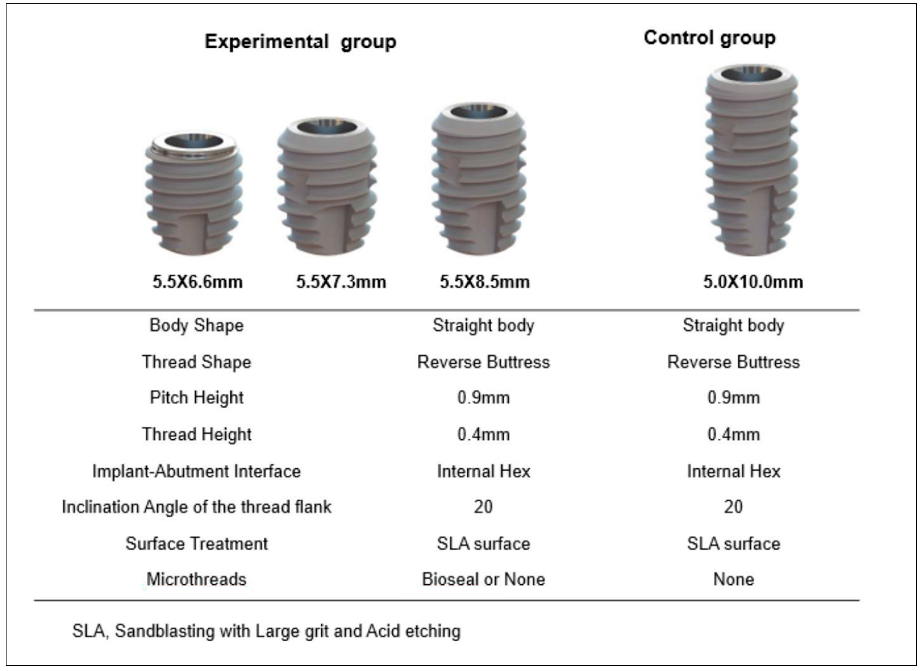
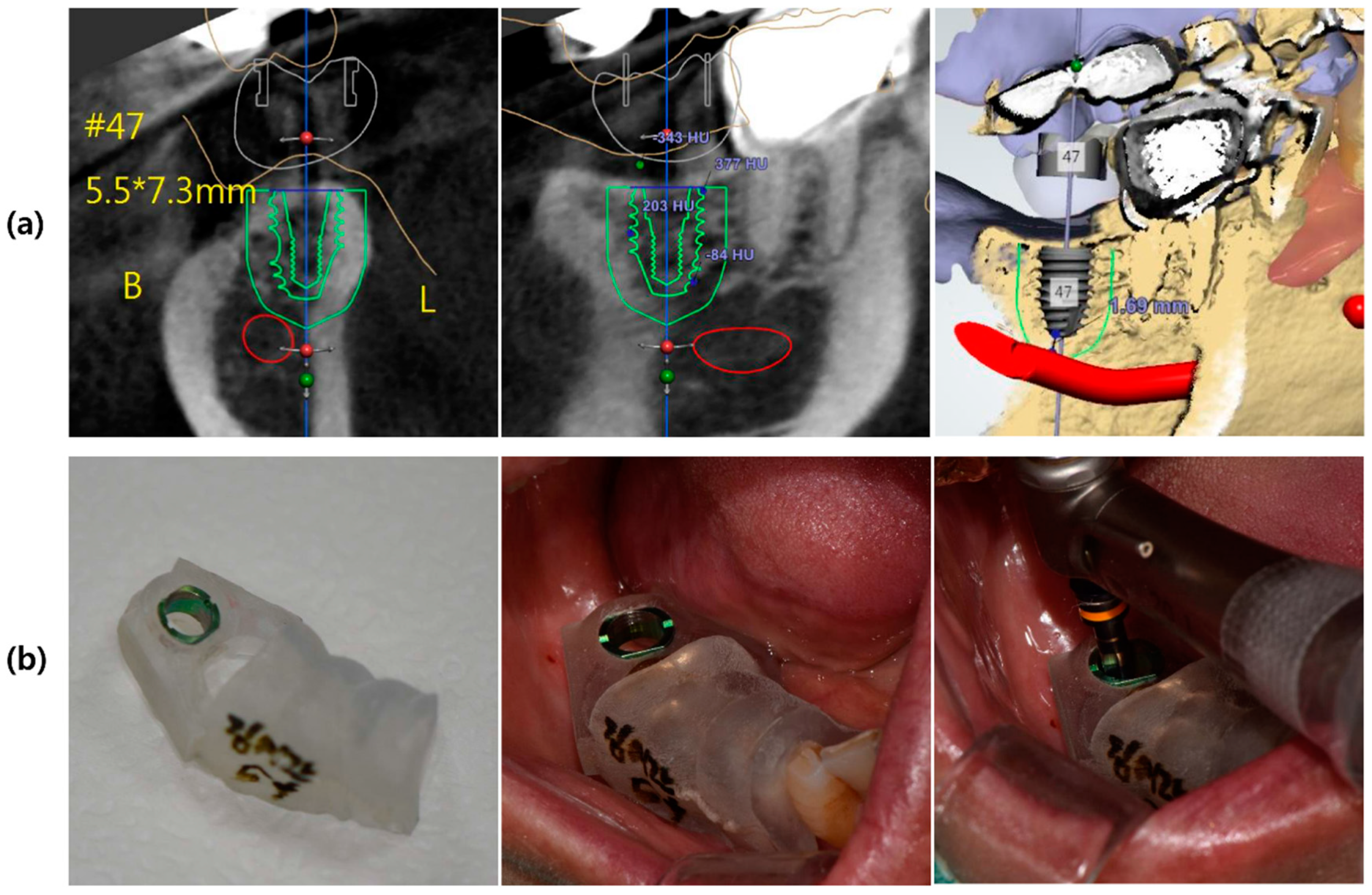
2.3.1. Virtual Planning, 3D Surgical Templates, Ti-Customized Abutment, and Temporary Prostheses

All patients underwent a CBCT scan (CS9300®; Carestream Health, Rochester, NY) and intraoral scan (Trios3®; 3Shape, Copenhagen, Denmark) prior to implant placement, for virtual surgical planning and the assessment of the bone dimensions around the implant site. The matching process of two scans and the surgical template were designed using the Implant Studio™ (3Shape, Copenhagen, Denmark). From this virtual planning, the surgical templates was produced by stereolithography (Objet 30®; Stratasys Ltd., MN, USA). At the aimed implant positions, titanium guiding sleeves were inserted. The height of the guiding sleeves was 4 mm; the distance from the lower margin of the sleeve and the coronal end of the implant was 5.0 mm (Figure 2). 3D designs of both customized titanium abutment and temporary prostheses were performed using the Dental Designer™ (3Shape, Copenhagen, Denmark) and fabricated by means of a CAD/CAM system. In order to use a customized titanium abutment as a scan body in digital impression, the bonding surface of all CAD-CAM customized implant abutments were airborne particle-abraded with 50-μm aluminum oxide (0.4 MPa, 10 mm in distance for 10 seconds).

2.3.2. Template-Guided Implant Placement and Evaluation of Implant Stability

Figure 3 illustrates the design of this clinical study. Patients were randomly assigned to one of the two implant types before surgery. The pre-operative examination included a panoramic radiograph, computerized tomography (CT), and intra-oral exam. After implant placement, each patient received antibiotics (Cefdinir, 100 mg three times daily) for 5 days and analgesics (Acetaminophen, 650 mg as needed) and 0.1% chlorhexidine mouthwash for use twice daily for 7 days. For each patient, single implants were placed in the mandibular molar region under local anesthesia according to the manufacturer’s protocol (NeoGudie® procedure). The implant bed was prepared in a minimally invasive procedure (flapless) using surgical template.
The peak insertion torque value was recorded as the maximum torque value (Ncm) reached at the end of the insertion of the implant. Primary stability was assessed by measuring implant stability quotient (ISQ) as an outcome variable. The resonance frequency analysis (RFA) was recorded using an Osstell Mentor® (Osstell AB, Göteborg, Sweden). The target values for the peak insertion torque and ISQ were 35–45 Ncm and greater than 65, respectively. Healing abutments were installed and periapical radiographs was taken.

2.3.3. Post-Operative Care

Adequate oral hygiene and a soft diet were recommended. The patients were instructed to use 0.1% chlorhexidine mouthwash and if necessary, analgesics for pain control. The ISQ measurements and periapical radiographs were taken at 1, 3, 4, 8, 12, 24, 36, and 48 weeks after surgery.

2.3.4. Prosthetic Procedure

At 1 week after implant placement, the fixtures that showed ISQ value of 65 or more were functionally loaded with pre-fabricated Ti customized abutments and provisional restorations. The pre-fabricated Ti customized abutments were secured to the implants at 20 Ncm and pre-fabricated PMMA provisional crowns were cemented with temporary cement. Occlusion was adjusted to prevent any eccentric occlusal contact and a periapical radiograph was taken.
After 8 weeks from implant installation, the final prosthetic procedure was commenced with a fully digital implant-prosthetic workflow. 6 participants were excluded because they could not fulfill the protocol standards, and 46 subjects were ready for the final impression stage. Intraoral digital impression was taken on the pre-abraded Ti customized abutment with the Trios 3® (3Shape, Copenhagen, Denmark) and monolithic zirconia definitive prosthesis was fabricated by means of a CAD/CAM system. On the day of delivery, after 12 weeks from the surgery, the definitive screw and cement retained prostheses (SCRP) were delivered on the fixtures that showed ISQ value of 65 or more. The occlusion was adjusted and the lateral contacts were removed.
On the day of delivery, the ISQ measurement with the Osstell device was repeated for each implant. A definitive fixed screw and cement retained prosthesis (SCRP) was delivered at 12 weeks after surgery (visit 7). The occlusion and lateral contacts were adjusted for the even distribution of the occlusal force over the fixed prosthesis. In addition, in all cases postoperative CBCT scans, directly performed after final prosthetic installation, with the same parameters were applied for comparative analysis.

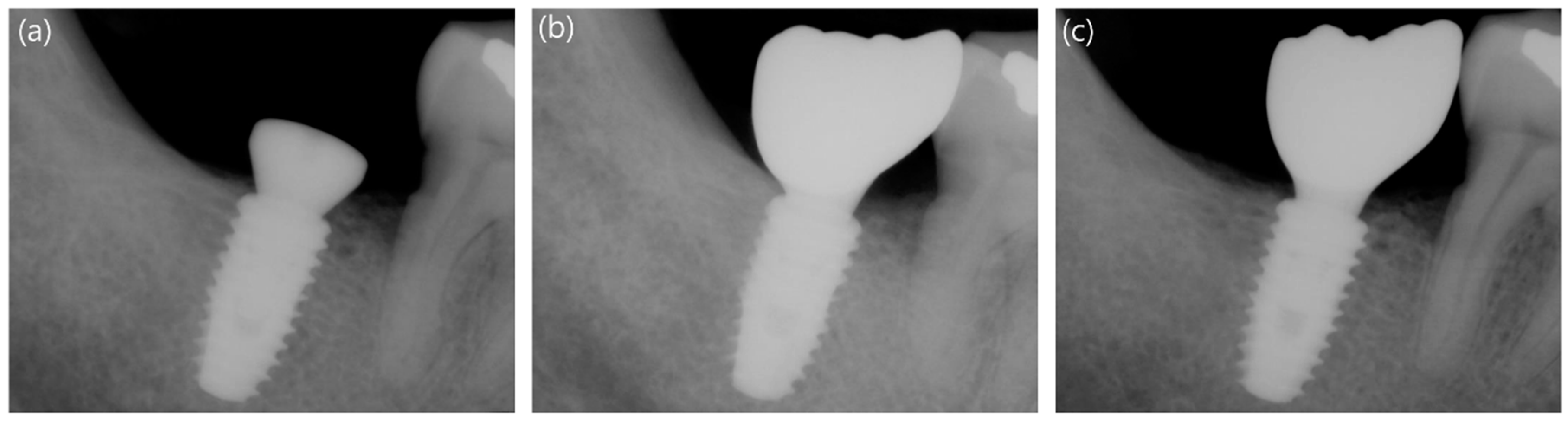
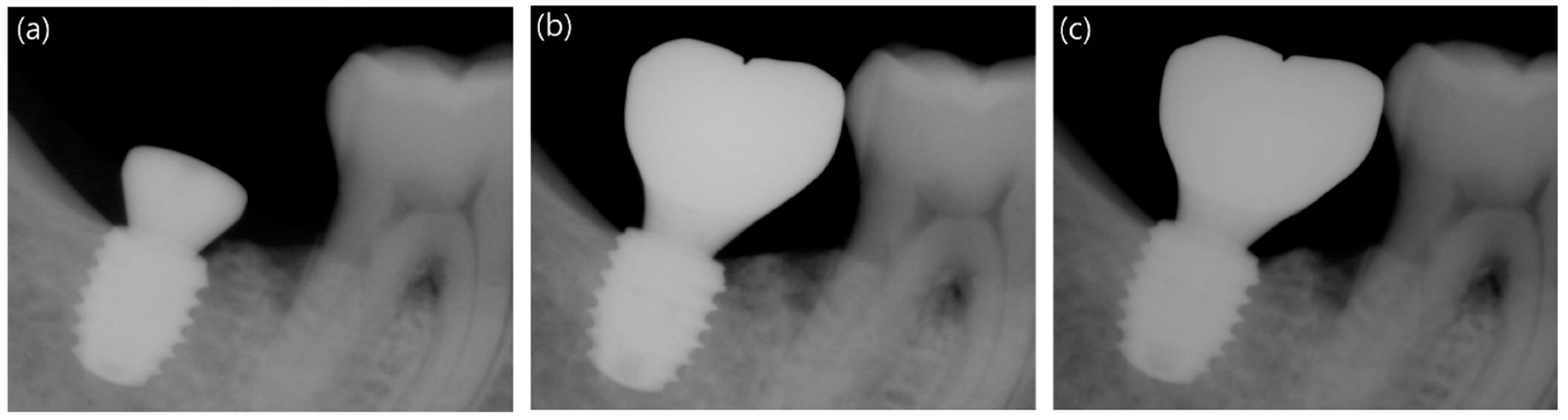
2.3.5. Measurement of Marginal Bone Loss

Peri-Implant marginal bone loss (PIMBL) was evaluated using standard periapical radiographs taken immediately after surgery and at 12 and 48 weeks after the implant installation (Figure 4 and Figure 5). In order to obtain the marginal bone level, the enlargement ratio of each image was calculated from the manufacturer-specified thread pitch of 0.9 mm that is known for each implant system used in this study. The distance from the fixture platform top (reference point) to the level of the alveolar bone crest was measured in the mesial and distal surfaces of the implant and converted to the actual value using the enlargement ratio. This value was then compared with the measurement taken at surgery (baseline).

2.3.6. Follow-Up Procedures and Implant Success

The patients were scheduled for recall visits at 9 and 12 months after implant surgery. The clinical and radiographic examinations were performed during the follow-up period. The occlusion was checked and the ISQ values were measured at the final appointment. In addition, soft tissue parameters, such as plaque index, sulcus bleeding index, and widths of keratinized mucosa (KM) were assessed and periapical radiographs was taken. The following criteria described by Buser et al. [24] were applied to evaluate implant success: (1) absence of clinically detectable mobility; (2) absence of pain or other symptoms of discomfort or ongoing pathologic processes; (3) absence of recurrence of peri-implantitis with suppuration; (4) no evidence of continuous radiolucency around the implant.

2.4. Statistical Analysis

The statistical analysis comparing the two groups was performed based on the Intention to Treat (ITT) and the Per Protocol (PP) analyses. The χ2 test for categorical variables and the independent sample two tailed t-test or the Mann-Whitney test for continuous variables were used for comparative evaluation depending on the normality (Shapiro-Wilk) of the distribution. For continuous variables, the mean values and standard deviation were calculated, and significant differences were detected. Two-way repeated measures analyses of variance were performed after the verification of sphericity using the Huynh-Feldt method to evaluate differences in the patterns of ISQ change over time. The level of significance was set at p-value < 0.05.

3. Results

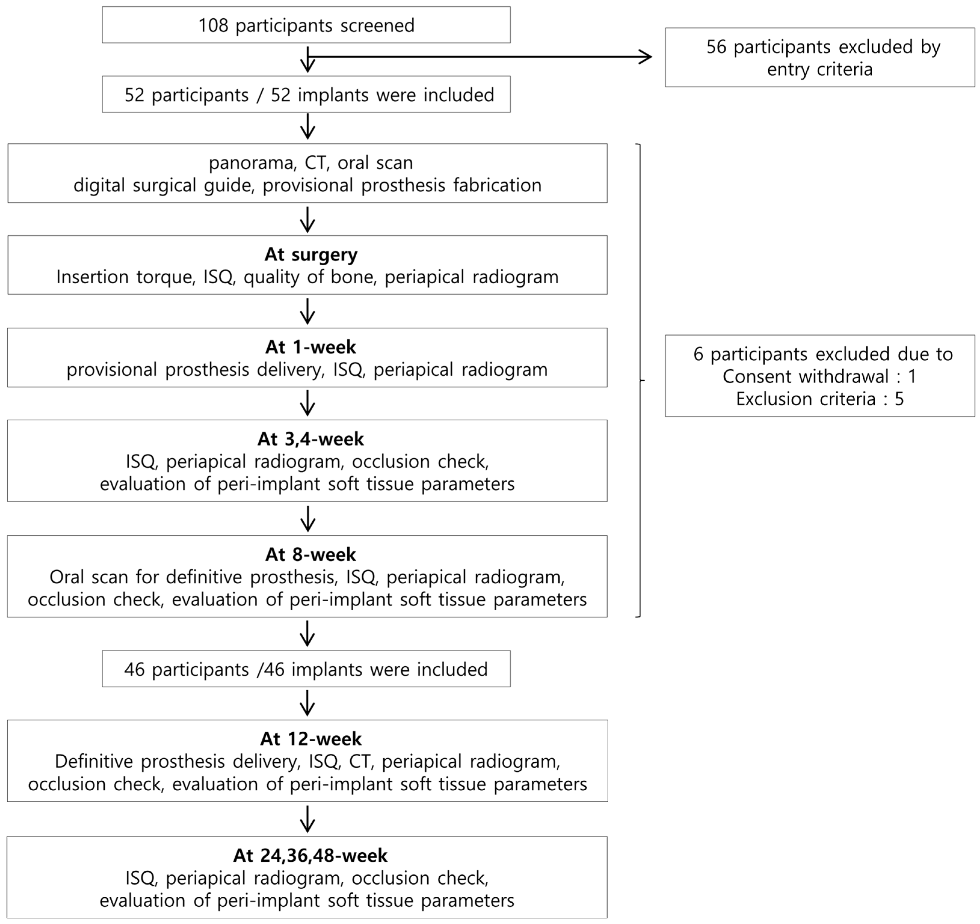
3.1. Participants and Implant Placed

Fifty-six of a total of 108 screened candidates were excluded by the entry criteria. During the 2 months after surgery, six participants dropped out because they could not fulfill the protocol standards. On the day of surgery, one subject withdrew the consent to continue participating in the study and five additional subjects were excluded due to low bone density (D4) and low initial stability values (ISQ < 65, insertion torque < 35) (Figure 3). As a result, the data from 46 implants in 46 participants were used for the final statistical analysis of the present study.

3.2. Demographic Characteristics of the Participants

The demographic and clinical characteristics of the study population for each implant system are presented in Table 1. The mean age of 19 patients (15 males and four females) in the control group was 55.42 ± 11.75, while the experimental group was composed of 27 patients (18 males and nine females) with a mean age of 52.06 ± 11.05 years. In both groups, the bone quality of the surgical sites was identified as D1, D2, or D3 based on Misch classification [25] during the drilling sequence. During initial drilling using a straight drill with a diameter of 2.2 mm, the drilling depth was divided into three parts to indicate bone quality in D113 format. D113 indicates that the bone is D1, D1, and D3 depending on the depth. The statistical analysis showed that there were no significant differences in age, sex, implant type, and bone quality between the two groups (p > 0.05).

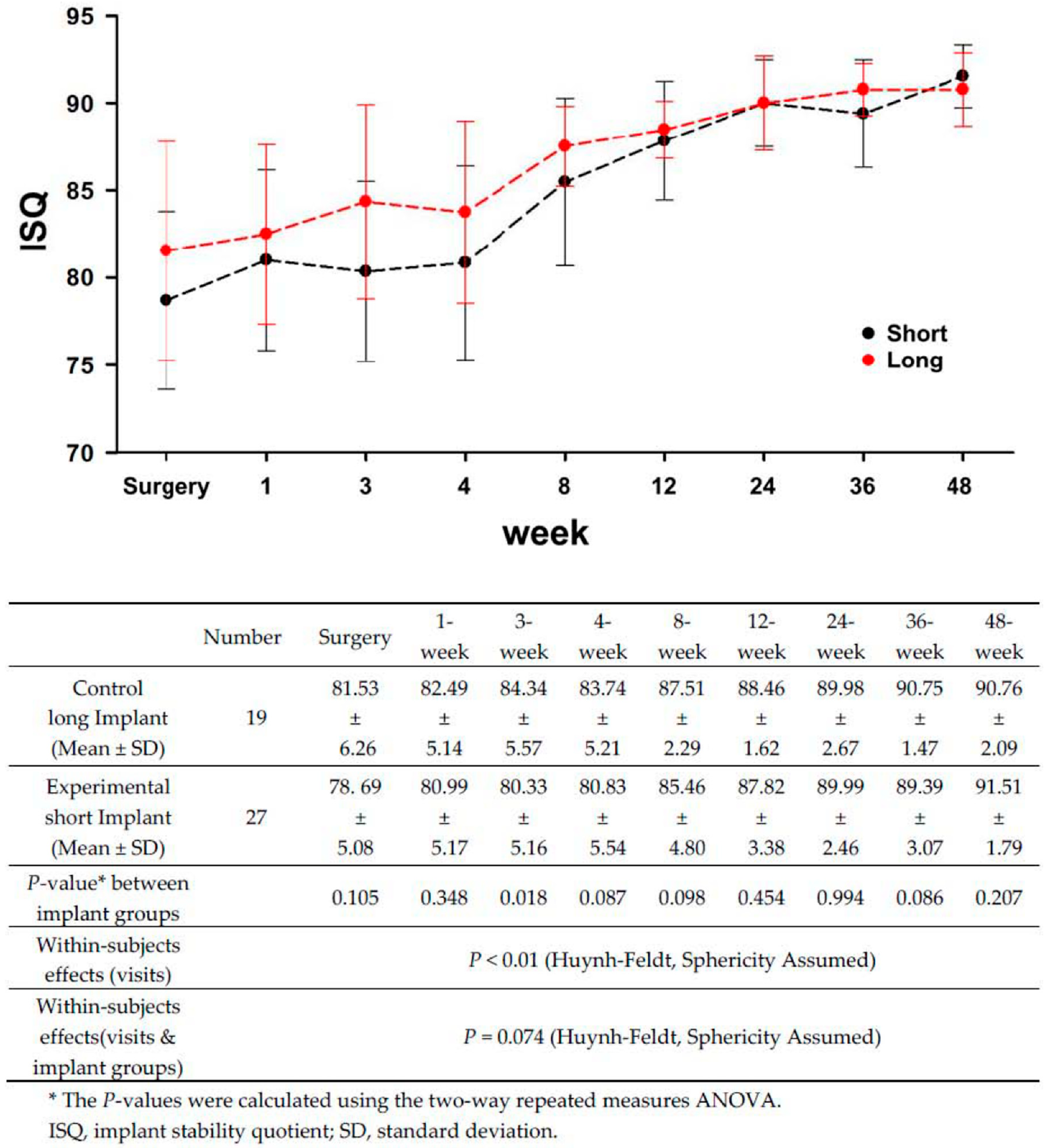
3.3. Comparison of Implant Stability between the Long and Short Implants

Primary stability was evaluated using the peak insertion torque and ISQ at surgery (Table 2). The control group had slightly greater average insertion torque and ISQ values at implant insertion than the experimental group, but no statistically significant differences were observed between the long and short implants (p-value > 0.05).
The ISQ values continued to be assessed during follow-up appointments for a post-operative period of 12 months (48 weeks), as shown in Figure 6. Up to 8 weeks after surgery, the ISQ values of the control group were steadily greater than those of the experimental group, but there were no statistically significant differences between the two groups (p-value > 0.05) except 3 week measurement (p-value = 0.018). The ISQ value did not decrease even after one week after the operation with the provisional restoration, and gradually increased with time. Likewise, within each implant group, the ISQ values showed a little change until 8 weeks but from the twelfth week to the forty-eighth week, the ISQ value remained constant, and both the control (long) and experimental (short) groups showed almost the same values (p-value > 0.05). There was no statistically significant difference, but ISQ values at 24 and 48 weeks were slightly higher in the experimental group. These results suggest that the stability dip, which usually appears within 4 weeks after surgery, had not been occurred in both implant groups.

3.4. Comparison of Marginal Bone Loss between the Long and Short Implants

Marginal bone loss after the implant insertion was evaluated for 46 implants using periapical radiographs taken at 12 weeks and 48 weeks after surgery (Table 3 and Figure 4 and Figure 5). The average marginal bone loss from the fixture platform top for the control and experimental groups was −0.07 ± 0.78 mm and 0.03 ± 0.63 mm after 12 weeks and 0.06 ± 0.82 mm and 0.05 ± 0.77 mm after 48 weeks, respectively. After a 12-week healing period, the distal surface exhibited slightly greater bone loss than the mesial side, but by the end of the trial, no differences in marginal bone loss between the two implant groups gained statistical significance (p-value > 0.05).

3.5. Evaluation of the Peri-Implant Soft Tissue Parameters and Success Rates of the Long and Short Implants

All of the mean values of the soft tissue parameters were clinically healthy within normal limits throughout the clinical trial (Table 4). There were no statistically significant differences in the soft-tissue parameters between the two implant groups (p-value > 0.05). The success criteria described by Buser et al. [24] were applied to evaluate implant success. The implants that did not fulfill the success criteria were considered failures. At the end of the 48-week follow-up period, all 46 implants fulfilled the strict success criteria. Consequently, the overall success rates were 100%.

4. Discussion

This clinical study has been performed with a complete digital workflow which includes computer aided implant surgery, immediate provisionalization, and definitive restoration. Previous studies demonstrated that the use of a surgical template significantly increases the accuracy and predictability of implant bed preparation compared to non-guided drilling [26]. The completely digital workflow, which applied in our study reduced treatment time, cost, and manual labor, eliminating conventional impressions and stone casts. Moreover, computer-aided surgery offers patients the benefits of successful implant placement without flap elevation as well as reduced postoperative pain and discomfort compared to conventional implant surgery [5].

4.1. Short Implants and Implant Stability

There is no consensus in the literature concerning its definition of a short implant but generally, the implants ranging from 6 to 10 mm have been considered as short implants. Authors have defined short implants as ≤7 mm [6,27], ≤8 mm [28], ≤10 mm [29,30], or ≤11 mm [31] long. Our study placed 6.6, 7.3, or 8.5 mm implants to the experiment group to compare with standard 10mm implant group as a control.
It is generally claimed that short implants have been associated with lower survival rates than standard length implants [32,33]. However, more recent studies revealed no apparent difference in performances between short and long implants, and suggested that the use of short implants may be a viable and effective alternative treatment option [17,34]. These results seem to be due to the technical innovations in both the surface characteristics and geometry (macro- and micro-) design of the implant, which helped to compensate for the unfavorable crown-to-implant ratio and lower surface area available for bone to implant contact [35].
A study performed by Anitua et al. [36] concluded that the immediate loading of short implants is not a risk factor for treatment success and this could be related to the good bone quality and the achievement of adequate primary stability. Lai et al. [37] have demonstrated that high survival rates for both the implants and the prostheses could be achieved after 5–10 years for short SLA implants (intra-bony length ≤8 mm) supporting single crowns, without severe marginal bone loss and complications. They recommended, however, that short implants in type IV bone sites should be applied with caution. Renouard and Nisand [28] also stressed the importance of surgical preparation, which conforms to bone quality and careful patient selection in terms of biomechanical conditions for short implant. Along with that, in a review performed by Schrott et al. [38], primary stability seems to be considered of paramount importance for immediate loading. For the same reasons, a high degree of primary stability is more strictly required for single implant [39,40]. Absolute optimal insertion torque or ISQ value was not identified in previous publications. Most publications proposed 30–35 Ncm to be the minimum insertion torque [41,42] and 60–65 as minimum ISQ value for successful early or immediate loading [19,43].
This study designed on clinical aspects following lower single molar replacement using short implants with immediate functional loading concept. In our experiment, type IV bone quality were excluded under strict inclusion criteria and the target values at surgery were achieved for the peak insertion torque and ISQ were 35–50 Ncm and greater than 65, respectively. The long and short implant groups showed the mean insertion torque of 40.53 ± 5.35 Ncm and 38.89 ± 4.85 Ncm and the mean ISQ value of 81.53 ± 6.26 and 78.69 ± 5.08, respectively, at surgery with no significant difference between two groups in neither insertion torque nor ISQ value (p-value > 0.05).

4.2. Implant Length and Stability Dip

Implant stability consists of primary and secondary stability [44]. Primary stability is obtained by mechanical engagement in the bone upon implant insertion, whereas secondary stability is related to the biological response to bone healing [39]. Primary stability is an important factor for implant success, especially when immediate loading is planned, because transmission of micromotion to the implant body can cause peri-implantal bone loss or osseointegration failure [44,45].
During the early healing period, primary stability constitutes most proportion of total stability, but thereafter biologic stability becomes dominant with new bone apposition [44]. Total stability is reported to decrease at the initial healing stage and rebound as healing progresses, showing a transient dip in total stability curve [46,47]. This study monitored the ISQ values longitudinally for a year to evaluate the course of stability and healing pattern. Short implant group showed excellent ISQ value high enough for required stability, without statistically significant differences to long implant group except during 3 weeks post-surgery. Additionally, both groups showed values far above the minimum ISQ value of 65, and an increasing tendency throughout 1-year monitoring period without a distinct stability dip. This result runs counter to previous studies reporting a transient dip in total stability during initial stage of healing [46,47].

4.3. Marginal Bone Loss and Success Rates of Long and Short Implants

Both the control and the experimental groups showed minimal marginal bone loss and no statistically significant differences during the whole observation period. No correlation between the implant length and marginal bone loss was observed. Marginal bone loss is a key factor in long-term implant stability and survival rate [48] and authors need to pay more attention to bone loss in short implant because of its relatively higher risk as a result of less reserve in bone height for the implant to be engaged in. Several authors insisted that the first year marginal bone loss should be included in the reports on marginal bone loss, because bone resorption rate is highest during first year of loading [49,50]. Therefore, minimal marginal bone loss shown in this study during initial year after the surgery may be interpreted positively for after coming prognosis. Peri-implant soft tissue observed was also clinically healthy with negligible plaque and calculus deposits and minimal tendency to bleed (Table 4).
The overall 48-week success rate in the present study was 100%, the same as the survival rate, as reviewed from the time of prosthetic delivery to the final follow-up appointment. In this study, short implants showed successful outcomes comparable to long implants in success rates, stability, and changes in marginal bone level, despite unfavorable conditions such as short length and immediate loading. These results were comparable to those of previous studies on immediately loaded single short implants [36,51]. Cannizzaro et al. [51] evaluated 6.5 mm-long flaplessly placed single implants immediately or early loaded at 6 weeks and concluded that flaplessly placed 6.5 mm-long single implants can be immediately loaded and remain successful up to 4 years after loading.
Correspondingly, to the previous studies, the successful outcome of this study can mainly be explained by good bone quality and primary stability. Under strict patient selection criteria, we screened out patients with smoking habit, parafunctional habits and D4 bone density known as disadvantageous factors for implant success and tried to achieve primary stability during surgery in terms of 35–45 Ncm insertion torque with surgical procedure variations such as under-drilling. Under-preparation of implant site, not following the whole standard drilling steps, has been suggested as a means to improve primary stability [52].
The implants used in the present study, Neobiotech CMI IS-III active®, are designed with various macrodesign features such as deep reverse buttress shaped thread, conical implant-abutment seal, micro grooves on implant collar, and self-tapping tapered apex. One of the key factors for successful stability and osseointegration is even stress distribution within peri-implant bone [53,54]. Previous studies have reported that the aforementioned macroscopic components diminish undesirable stress and strain around the implants and improve mechanical retention during the early phase of the healing process after operation [55,56]. For these reasons, a sharp drop of primary stability could be avoided not showing stability dip during healing period and it is concurrent with the result of our previous study using same implant system [57].
In this study, short implants with wider diameter (5.5 mm) were used comparing control group (5.0mm) to make up for the bone-to-implant contact surface of short length implants. Wide-diameter implant has been used to improve the success rate in compromised situations such as poor bone quality and/or quantity and replacement of a failing standard implant [58,59]. Several studies reported that failure rate was higher with standard-diameter short implants compared with wide-diameter short implants [11,58]. Furthermore, Simunek and coworkers [44] concluded that primary stability was more influenced by diameter than length. Accordingly with those studies, wide width of short implant group is assumed to be one of the contributing factors for the successful performance of short implants in our study.

5. Conclusions

The present study was performed with immediate loading protocol and used the completely digital pathway, short and standard-length implants supporting single prosthesis in the posterior mandible, showed no significant differences in terms of success rate, ISQ values, marginal bone loss, and peri-implant soft tissue parameters during the 1-year follow up period. Within the limitations of this study, the short implant supporting single crown with immediate loading protocol seems to be a successful treatment modality in the limited bone height mandible as long as adequate primary stability can be achieved; insertion torque of 35–45 Ncm and ISQ of more than 65. To consolidate this alternative solution for reduced bone, however, additional randomized controlled trials with larger sample sizes and longer follow-up periods are required.

Author Contributions

Conceptualization, supervision, Y.-J.L. and K.-T.K.; Writing—Original Draft preparation, data curation, Y.-W.B. and J.L.; visualization, Y.-J.L. and Y.-W.B.; Writing—Review and Editing, M.-J.K. and H.-B.K.

Acknowledgments

This research was supported by a grant of the Korea Health Technology R&D Project through the Korea Health Industry Development Institute (KHIDI), funded by the Ministry of Health and Welfare, Republic of Korea (grant number: HI16C1173). The APC was funded by “Dental Research Institute, School of Dentistry, Seoul National University, Seoul, Republic of Korea.

Conflicts of Interest

The authors declare no conflict of interest.

References

  1. Joda, T.; Brägger, U. Complete digital workflow for the production of implant-supported single-unit monolithic crowns. Clin. Oral Implants Res. 2014, 25, 1304–1306. [Google Scholar] [CrossRef] [PubMed]
  2. Joda, T.; Buser, D. Digital implant dentistry—A workflow in five steps. CAD/CAM 2013, 4, 16–20. [Google Scholar]
  3. Arunyanak, S.P.; Harris, B.T.; Grant, G.T.; Morton, D.; Lin, W.-S. Digital approach to planning computer-guided surgery and immediate provisionalization in a partially edentulous patient. J. Prosthet. Dent. 2016, 116, 8–14. [Google Scholar]
  4. Brodala, N. Flapless surgery and its effect on dental implant outcomes. Int. J. Oral Maxillofac. Implants 2009, 24, 118–125. [Google Scholar] [PubMed]
  5. Sicilia, A.; Botticelli, D. Computer-guided implant therapy and soft- and hard-tissue aspects. The Third EAO Consensus Conference 2012. Clin. Oral Implants Res. 2012, 23 (Suppl. 6), 157–161. [Google Scholar] [CrossRef]
  6. Neldam, C.A.; Pinholt, E.M. State of the art of short dental implants: A systematic review of the literature. Clin. Implant Dent. Relat. Res. 2012, 14, 622–632. [Google Scholar] [CrossRef]
  7. Tonetti, M.S.; Hämmerle, C.H. Advances in bone augmentation to enable dental implant placement: Consensus Report of the Sixth European Workshop on Periodontology. J. Clin. Periodontol. 2008, 35 (Suppl. 8), 168–172. [Google Scholar] [Green Version]
  8. Urban, I.A.; Jovanovic, S.A.; Lozada, J.L. Vertical ridge augmentation using guided bone regeneration (GBR) in three clinical scenarios prior to implant placement: A retrospective study of 35 patients 12 to 72 months after loading. Int. J. Oral Maxillofac. Implants 2009, 24, 502–510. [Google Scholar]
  9. Felice, P.; Soardi, E.; Pellegrino, G.; Pistilli, R.; Marchetti, C.; Gessaroli, M.; Esposito, M. Treatment of the atrophic edentulous maxilla: Short implants versus bone augmentation for placing longer implants. Five-month post-loading results of a pilot randomised controlled trial. Eur. J. Oral Implantol. 2011, 4, 191–202. [Google Scholar]
  10. Scarano, A.; Piattelli, A.; Murmura, G.; Iezzi, G.; Assenza, B.; Mancino, C. Delayed expansion of the atrophic mandible by ultrasonic surgery: A clinical and histologic case series. Int. J. Oral Maxillofac. Implants 2015, 30, 144–149. [Google Scholar]
  11. Bahat, O. Branemark system implants in the posterior maxilla: Clinical study of 660 implants followed for 5 to 12 years. Int. J. Oral Maxillofac. Implants 2000, 15, 646–653. [Google Scholar] [PubMed]
  12. Naert, I.; Koutsikakis, G.; Duyck, J.; Quirynen, M.; Jacobs, R.; Van Steenberghe, D. Biologic outcome of implant-supported restorations in the treatment of partial edentulism: Part 1: A longitudinal clinical evaluation. Clin. Oral Implants Res. 2002, 13, 381–389. [Google Scholar]
  13. Herrmann, I.; Lekholm, U.; Holm, S.; Kultje, C. Evaluation of patient and implant characteristics as potential prognostic factors for oral implant failures. Int. J. Oral Maxillofac. Implants 2005, 20, 220–230. [Google Scholar]
  14. Mangano, F.G.; Shibli, J.A.; Sammons, R.L.; Iaculli, F.; Piattelli, A.; Mangano, C. Short (8-mm) locking-taper implants supporting single crowns in posterior region: A prospective clinical study with 1-to 10-years of follow-up. Clin. Oral Implants Res. 2014, 25, 933–940. [Google Scholar] [PubMed]
  15. Rossi, F.; Botticelli, D.; Cesaretti, G.; De Santis, E.; Storelli, S.; Lang, N.P. Use of short implants (6 mm) in a single-tooth replacement: A 5-year follow-up prospective randomized controlled multicenter clinical study. Clin. Oral Implants Res. 2016, 27, 458–464. [Google Scholar] [CrossRef]
  16. Esposito; Cannizzaro, G.; Soardi, E.; Pellegrino, G.; Pistilli, R.; Felice, P. A 3-year post-loading report of a randomised controlled trial on the rehabilitation of posterior atrophic mandibles: Short implants or longer implants in vertically augmented bone? Eur. J. Oral Implantol. 2011, 4, 301–311. [Google Scholar]
  17. Telleman, G.; Raghoebar, G.M.; Vissink, A.; Den Hartog, L.; Huddleston Slater, J.J.; Meijer, H.J. A systematic review of the prognosis of short (<10 mm) dental implants placed in the partially edentulous patient. J. Clin. Periodontal. 2011, 38, 667–676. [Google Scholar] [CrossRef] [Green Version]
  18. Cochran, D.L.; Morton, D.; Weber, H.-P. Consensus statements and recommended clinical procedures regarding loading protocols for endosseous dental implants. Int. J. Oral Maxillofac. Implants 2004, 19, 109–113. [Google Scholar]
  19. Esposito, M.; Grusovin, M.G.; Maghaireh, H.; Worthington, H.V. Interventions for replacing missing teeth: Different times for loading dental implants. Cochrane Database Syst. Rev. 2013, CD003878. [Google Scholar] [CrossRef]
  20. Sanz-Sanchez, I.; Sanz-Martin, I.; Figuero, E.; Sanz, M. Clinical efficacy of immediate implant loading protocols compared to conventional loading depending on the type of the restoration: A systematic review. Clin. Oral Implants Res. 2015, 26, 964–982. [Google Scholar] [CrossRef]
  21. Benic, G.I.; Mir-Mari, J.; Hämmerle, F.; Christoph, H. Loading protocols for single-implant crowns: A systematic review and meta-analysis. Int. J. Oral Maxillofac. Implants 2014, 29, 222–238. [Google Scholar]
  22. World Medical Association. World Medical Association Declaration of Helsinki: Ethical principles for medical research involving human subjects. JAMA 2013, 310, 2191. [Google Scholar]
  23. Schulz, K.F.; Altman, D.G.; Moher, D. CONSORT 2010 statement: Updated guidelines for reporting parallel group randomised trials. BMC Med. 2010, 8, 18. [Google Scholar]
  24. Buser, D.; Mericske-stern, R.; Pierre Bernard, J.P.; Behneke, A.; Behneke, N.; Hirt, H.P.; Belser, U.C.; Lang, N.P. Long-term evaluation of non-submerged ITI implants. Part 1: 8-year life table analysis of a prospective multi-center study with 2359 implants. Clin. Oral Implants Res. 1997, 8, 161–172. [Google Scholar]
  25. Misch, C.E. Density of bone: Effect on treatment planning, surgical approach, and healing. In Contemporary Implant Dentistry; Mosby-Year Book, Inc.: St. Louis, MO, USA, 1993; pp. 469–485. [Google Scholar]
  26. Scherer, U.; Stoetzer, M.; Ruecker, M.; Gellrich, N.-C.; von See, C. Template-guided vs. non-guided drilling in site preparation of dental implants. Clin. Oral Investig. 2015, 19, 1339–1346. [Google Scholar]
  27. Piero, P.; Lorenzo, C.; Daniele, R.; Rita, G.; Luca, P.; Giorgio, P. Survival of short dental implants≤ 7 mm: A review. Int. J. Contemp. Dent. Med. Rev. 2015, 2015, 011015. [Google Scholar]
  28. Renouard, F.; Nisand, D. Impact of implant length and diameter on survival rates. Clin. Oral Implants Res. 2006, 17 (Suppl. 2), 35–51. [Google Scholar] [CrossRef] [PubMed] [Green Version]
  29. Feldman, S.; Boitel, N.; Weng, D.; Kohles, S.S.; Stach, R.M. Five-Year Survival Distributions of Short-Length (10 mm or less) Machined-Surfaced and Osseotite® Implants. Clin. Implant Dent. Relat. Res. 2004, 6, 16–23. [Google Scholar] [PubMed]
  30. Sun, H.L.; Wu, Y.R.; Huang, C.; Shi, B. Failure rates of short (≤10 mm) dental implants and factors influencing their failure: A systematic review. Int. J. Oral Maxillofac. Implants 2011, 26, 816–825. [Google Scholar]
  31. Das Neves, F.D.; Fones, D.; Bernardes, S.R.; do Prado, C.J.; Neto, A.J.F. Short implants-an analysis of longitudinal studies. Int. J. Oral Maxillofac. Implants 2006, 21, 86–93. [Google Scholar]
  32. Weng, D.; Jacobson, Z.; Tarnow, D.; Hürzeler, M.B.; Faehn, O.; Sanavi, F.; Barkvoll, P.; Stach, R.M. A prospective multicenter clinical trial of 3i machined-surface implants: Results after 6 years of follow-up. Int. J. Oral Maxillofac. Implants 2003, 18, 417–423. [Google Scholar] [PubMed]
  33. Winkler, S.; Morris, H.F.; Ochi, S. Implant survival to 36 months as related to length and diameter. Ann. Periodontol. 2000, 5, 22–31. [Google Scholar] [PubMed]
  34. Camps-Font, O.; Burgueno-Barris, G.; Figueiredo, R.; Jung, R.E.; Gay-Escoda, C.; Valmaseda-Castellon, E. Interventions for Dental Implant Placement in Atrophic Edentulous Mandibles: Vertical Bone Augmentation and Alternative Treatments. A Meta-Analysis of Randomized Clinical Trials. J. Periodontol. 2016, 87, 1444–1457. [Google Scholar] [CrossRef]
  35. Annibali, S.; Cristalli, M.P.; Dell’Aquila, D.; Bignozzi, I.; La Monaca, G.; Pilloni, A. Short dental implants: A systematic review. J. Dent. Res. 2012, 91, 25–32. [Google Scholar] [CrossRef]
  36. Anitua, E.; Flores, J.; Flores, C.; Alkhraisat, M.H. Long-term Outcomes of Immediate Loading of Short Implants: A Controlled Retrospective Cohort Study. Int. J. Oral Maxillofac. Implants 2016, 31, 1360–1366. [Google Scholar] [CrossRef] [Green Version]
  37. Lai, H.C.; Si, M.S.; Zhuang, L.F.; Shen, H.; Liu, Y.l.; Wismeijer, D. Long-term outcomes of short dental implants supporting single crowns in posterior region: A clinical retrospective study of 5–10 years. Clin. Oral Implants Res. 2013, 24, 230–237. [Google Scholar] [PubMed]
  38. Schrott, A.; Riggi-Heiniger, M.; Maruo, K.; Gallucci, G.O. Implant loading protocols for partially edentulous patients with extended edentulous sites—A systematic review and meta-analysis. Int. J. Oral Maxillofac. Implants 2014, 29, 239–255. [Google Scholar] [CrossRef]
  39. Atsumi, M.; Park, S.H.; Wang, H.L. Methods used to assess implant stability: Current status. Int. J. Oral Maxillofac. Implants 2007, 22, 743–754. [Google Scholar]
  40. Romanos, G.E. Bone quality and the immediate loading of implants-critical aspects based on literature, research, and clinical experience. Implant Dent. 2009, 18, 203–209. [Google Scholar] [CrossRef] [PubMed]
  41. Grandi, T.; Garuti, G.; Guazzi, P.; Tarabini, L.; Forabosco, A. Survival and success rates of immediately and early loaded implants: 12-month results from a multicentric randomized clinical study. J. Oral Implantol. 2012, 38, 239–249. [Google Scholar] [CrossRef] [PubMed]
  42. Margossian, P.; Mariani, P.; Stephan, G.; Margerit, J.; Jorgensen, C. Immediate loading of mandibular dental implants in partially edentulous patients: A prospective randomized comparative study. Int. J. Periodontics Restor. Dent. 2012, 32, e51–e58. [Google Scholar]
  43. Sennerby, L.; Meredith, N. Implant stability measurements using resonance frequency analysis: Biological and biomechanical aspects and clinical implications. Periodontology 2008, 47, 51–66. [Google Scholar] [CrossRef]
  44. Simunek, A.; Strnad, J.; Kopecka, D.; Brazda, T.; Pilathadka, S.; Chauhan, R.; Slezak, R.; Capek, L. Changes in stability after healing of immediately loaded dental implants. Int. J. Oral Maxillofac. Implants 2010, 25, 1085–1092. [Google Scholar]
  45. Javed, F.; Romanos, G.E. The role of primary stability for successful immediate loading of dental implants. A literature review. J. Dent. 2010, 38, 612–620. [Google Scholar] [CrossRef] [PubMed]
  46. Bischof, M.; Nedir, R.; Szmukler-Moncler, S.; Bernard, J.P.; Samson, J. Implant stability measurement of delayed and immediately loaded implants during healing. Clin. Oral Implants Res. 2004, 15, 529–539. [Google Scholar] [CrossRef] [PubMed]
  47. Huwiler, M.A.; Pjetursson, B.E.; Bosshardt, D.D.; Salvi, G.E.; Lang, N.P. Resonance frequency analysis in relation to jawbone characteristics and during early healing of implant installation. Clin. Oral Implants Res. 2007, 18, 275–280. [Google Scholar] [CrossRef]
  48. Pommer, B.; Heuberer, S.; Nahler, L.; Watzek, G. How much bone loss can be prevented by alveolar ridge preservation? Systematic literature review and meta-analysis. Implant News 2012, 9, 795–799. [Google Scholar]
  49. Simons, W.F.; De Smit, M.; Duyck, J.; Coucke, W.; Quirynen, M. The proportion of cancellous bone as predictive factor for early marginal bone loss around implants in the posterior part of the mandible. Clin. Oral Implants Res. 2015, 26, 1051–1059. [Google Scholar] [CrossRef]
  50. Weber, H.P.; Crohin, C.C.; Fiorellini, J.P. A 5-year prospective clinical and radiographic study of non-submerged dental implants. Clin. Oral Implants Res. 2000, 11, 144–153. [Google Scholar]
  51. Cannizzaro, G.; Felice, P.; Leone, M.; Ferri, V.; Viola, P.; Esposito, M. Immediate versus early loading of 6.5 mm-long flapless-placed single implants: A 4-year after loading report of a split-mouth randomised controlled trial. Eur. J. Oral Implantol. 2012, 5, 111–121. [Google Scholar]
  52. Norton, M.R. The influence of insertion torque on the survival of immediately placed and restored single-tooth implants. Int. J. Oral Maxillofac. Implants 2011, 26, 1333–1343. [Google Scholar] [PubMed]
  53. Geng, J.P.; Tan, K.B.; Liu, G.R. Application of finite element analysis in implant dentistry: A review of the literature. J. Prosthet. Dent. 2001, 85, 585–598. [Google Scholar] [CrossRef] [PubMed]
  54. Kim, Y.S.; Lim, Y.J. Primary stability and self-tapping blades: Biomechanical assessment of dental implants in medium-density bone. Clin. Oral Implants Res. 2011, 22, 1179–1184. [Google Scholar] [CrossRef]
  55. Kumar, A.B.T.; Khalia, N.; Gayathri, G.V.; Kumar, B.H.D.; Mehta, D.S. Peri-implant crestal bone preservation: Where do we stand? Int. J. Oral Implantol. Clin. Res. 2013, 4, 72–77. [Google Scholar]
  56. Ryu, H.S.; Namgung, C.; Lee, J.H.; Lim, Y.J. The influence of thread geometry on implant osseointegration under immediate loading: A literature review. J. Adv. Prosthodont. 2014, 6, 547–554. [Google Scholar] [CrossRef]
  57. Ryu, H.S.; Namgung, C.; Heo, Y.K.; Lee, J.H.; Lim, Y.J. Early loading of splinted implants supporting a two-unit fixed partial denture in the posterior maxilla: 13-month results from a randomized controlled clinical trial of two different implant systems. Clin. Oral Implants Res. 2016, 27, 1017–1025. [Google Scholar] [CrossRef]
  58. Langer, B.; Langer, L.; Herrmann, I.; Jorneus, L. The wide fixture: A solution for special bone situations and a rescue for the compromised implant. Part 1. Int. J. Oral Maxillofac. Implants 1993, 8, 400–408. [Google Scholar]
  59. Renouard, F.; Arnoux, J.P.; Sarment, D.P. Five-mm-diameter implants without a smooth surface collar: Report on 98 consecutive placements. Int. J. Oral Maxillofac. Implants 1999, 14, 101–107. [Google Scholar]
Figure 1. Characteristics of the implants systems used in this study: CMI IS-III active® (Neobiotech, Seoul, Korea), short and long implants.
Applsci 09 01282 g001
Figure 2. Computer aided surgery (a) 3D digital implant planning with software, (b) surgical template.
Applsci 09 01282 g002
Figure 3. Flow diagram of the controlled clinical trial protocol used in this study.
Applsci 09 01282 g003
Figure 4. Standard periapical radiographs of implants placed in a patient in the control group (CMI IS-III active® long implant, Neobiotech Co., Seoul, Korea): (a) at surgery, (b) at 12 weeks, and (c) at 48 weeks.
Applsci 09 01282 g004
Figure 5. Standard periapical radiographs of implants placed in a patient in the experimental group (CMI IS-III active® short implant, Neobiotech Co., Seoul, Korea): (a) at surgery, (b) at 12 weeks, and (c) at 48 weeks.
Applsci 09 01282 g005
Figure 6. Comparison of stability in terms of the pattern of change in implant stability quotient (ISQ) during the 48-week observation period after implant surgery.
Applsci 09 01282 g006
Table 1. Demographic data of study participants.
VariablesControl (Neobiotech CMI IS-III Active® Long Implant)Experimental (Neobiotech CMI IS-IIIActive® Short Implant)p-Value
Participant based (n = 46)Participant number19270.514
Age (mean ± SD)55.42 ± 11.7552.06 ± 11.050.305
20–6013180.740
Over 6069
Sex
Male /Female15/419/80.514
Implant based (n = 46)Implant number1927
Lower 1st molar / 2nd molar9/104/23
Implant Type 1.000
∅5.00 × 10 mm19/
∅5.50 × 8.5 mm/10
∅5.50 × 7.3 mm/9
∅5.00 × 6.6 mm/8
Bone quality 0.378
D11204
D12233
D21101
D22267
D22311
D23201
D23337
D33363
  • ‘Control’ indicates the Neobiotech CMI IS-III active® long implant and ‘Experimental’ the Neobiotech CMI IS-III active® short implant.
  • Data, except for age are presented as the number of implants or participants. The units of age are year.
  • The p-values were calculated using the χ2 test (Pearson Chi-Square) for all variables except age. The p-value for age was calculated using the Mann–Whitney test.
  • Bone quality was assessed based on the classification system of Misch (1993) during the drilling sequence. While drilling, divide the depth of the bone into three parts and evaluate the boat quality. D113 indicates that the bone is D1, D1, and D3 depending on the depth.
  • SD, standard deviation.
Table 2. Comparison of primary stability between the long and short implants.
Control Neobiotech CMI IS-III Active® Long ImplantExperimental Neobiotech CMI IS-III Active® Short Implant
Participant number1927p-value *
Insertion Torque (Ncm)
(Mean ± SD)
40.53 ± 5.3538.89 ± 4.850.298
ISQ at surgery
(Mean ± SD)
81.53 ± 6.2678.69 ± 5.080.120
  • * The p-values for insertion torque and ISQ were calculated by the t-test.
  • ISQ, implant stability quotient; SD, standard deviation.
Table 3. Comparison of marginal bone loss between the long and short implants.
Control Neobiotech CMI IS-III Active® Long ImplantExperimental Neobiotech CMI IS-III Active® Short Implant
Participant number1927
DurationAreaMean ± SD (mm)Mean ± SD (mm)p-value *
12-week follow upMesial−0.22 ± 0.98-0.15 ± 0.790.893
Distal0.08 ± 0.810.20 ± 0.780.728
Avg.−0.07 ± 0.780.03 ± 0.630.885
48-week follow upMesial−0.15 ± 0.94-0.13 ± 0.820.719
Distal0.27 ± 0.800.23 ± 0.920.573
Avg.0.06 ± 0.820.05 ± 0.770.655
  • * The p-values were calculated using the Mann–Whitney test.
  • Normality test was failed (Shapiro-Wilk, p < 0.05).
  • Area, the radiographic measurement area for calculation of marginal bone loss; Avg., the average value of mesial and distal bone loss; SD, standard deviation.
Table 4. Comparison of peri-implant soft tissue parameters between the long and short implants after 1-year follow-up
Control Neobiotech CMI IS-III Active® Long ImplantExperimental Neobiotech CMI IS-III Active® Short Implant
Participant number1927
ParametersMean ± SD (mm)Mean ± SD (mm)p-value *
Plaque index0.22 ± 0.200.32 ± 0.220.168
Calculus index0.00 ± 0.020.02 ± 0.050.465
Sulcus bleeding index0.03 ± 0.070.09 ± 0.110.051
Pocket Depth3.38 ± 0.633.31 ± 0.470.760
Width of keratinized mucosa (mm)2.17 ± 0.542.18 ± 0.420.928
  • * The p-values were calculated using the Mann–Whitney test.
  • Plaque index: score 0, no detection of plaque; score 1, plaque only recognized by running a probe across the smooth marginal surface of the implant; score 2, plaque can be seen by the naked eye; score 3, abundance of soft matter.
  • Calculus index: score 0, no detection of calculus; score 1, supragingival calculus covering ≤ 1/3 exposed tooth surface; score 2, supragingival calculus covering > 1/3 but < 2/3 tooth surface, flecks of subgingival calculus in cervical margin; score 3, supragingival calculus covering > 2/3 surface, continuous band of subgingival calculus.
  • Sulcus bleeding index: score 0, no bleeding when a periodontal probe is passed along the gingival margin adjacent to the implant; score 1, isolated bleeding spot visible; score 2, blood forms a confluent red line on margin; score 3, heavy or profuse bleeding.
  • SD, standard deviation.

