
The Effect of Cold Plasma on Selected Parameters of Bovine Colostrum
by
 , ,
, ,
Elżbieta Bogusławska-Wąs
1 ,
,
Alicja Dłubała
1,
Wojciech Sawicki
1,
Małgorzata Ożgo
2,* and
Adam Lepczyński
2 1
Department of Applied Microbiology and Human Nutrition Physiology, Faculty of Food Sciences and Fisheries, West Pomeranian University of Technology Szczecin, Papieża Pawła VI 3, 71-459 Szczecin, Poland
2
Department of Physiology, Cytobiology and Proteomics, Faculty of Biotechnology and Animal Husbandry, West Pomeranian University of Technology Szczecin, Klemensa Janickiego 29, 71-270 Szczecin, Poland
*
Author to whom correspondence should be addressed.
Appl. Sci. 2023, 13(9), 5490; https://doi.org/10.3390/app13095490
Submission received: 6 April 2023
/
Revised: 25 April 2023
/
Accepted: 26 April 2023
/
Published: 28 April 2023
(This article belongs to the Special Issue Effects of Processing on Food Composition, Nutritional Value and Sensory Quality)
Abstract
:The main problem in processing bovine colostrum is preserving as many beneficial compounds as possible, most of which have low thermal stability. The present study evaluates the possibility of using cold plasma (CP) as a decontamination technology and its effect on selected biologically active fractions of freeze-dried bovine colostrum. The plasma process was carried out in air, nitrogen, and oxygen environments. The results revealed that the sterilization process using CP caused slight changes in the colour of the samples expressed by the attributes ΔC, ΔL, Δh and ΔE. The decontamination effect depended on the gas used and the type of microorganism. The highest decontamination effects were gained under oxygen conditions, where reductions were obtained for total psychrophilic bacteria (THPC) by log 1.24, mesophilic bacteria (THMC) by log 1.02, Enterobacteriaceae by log 1.16, E. coli by log 0.96, yeast (TYMC) by log 0.92. A significantly lower decontaminating effect was obtained for Gram-positive bacteria and sporophytic forms. Additionally, the application of CP, regardless of the gas used, affected the modification of protein structure and reduction of immunoglobulin concentration. as proven by proteomics analyses (1-DE, 2-DE, MALDI–TOF MS). The same applied to β-lactoglobulin in air and oxygen and BSA in nitrogen and air.
1. Introduction
Colostrum is the first milk for newborn mammals. It is a valuable source of nutrients and contains numerous bioactive compounds crucial for mammal growth and development, such as immunoglobulins, lactoferrin, lactoperoxidase, lysozyme, lactalbumin, hormones, enzymes, and whey proteins [1]. Immunostimulators: lactoferrin, colostrin, cytokines, and leukocytes are particularly important in colostrum as they modulate the newborn immune system [2,3]. The composition of colostrum is highly variable in terms of protein, fat, cytokines, growth factors, carbohydrates, vitamins, and minerals content. It depends on many factors, such as the breed and age of the animal, prepartum nutrition, and the course of birth.
The functional features of colostrum proteins are determined by the properties and structure of their molecules. Their size, shape, spatial arrangement, ability to create conglomerates, susceptibility to denaturation, and amino acid sequences are among the most important features. Colostrum contains more than 30 different proteins, which can be divided into two groups: casein (75–80%) and whey proteins (up to 20%) [4]. Nineteen amino acids have been determined in colostrum proteins, including all essential amino acids [5].
The most critical group of colostrum proteins are whey globular proteins. In bovine colostrum, there are α-lactalbumin (15–20%) and β-lactoglobulin (55–65%) fractions, as well as immunoglobulins (9%), bovine serum albumin (BSA) (5.5%), lactoferrin, lactoperoxidase, phospholipoproteins, bioactive factors and enzymes [6]. In addition to their nutritional value, they also exhibit functional activities, including, most notably, immunomodulatory and antioxidant activity. They also regulate fatty acid synthesis in the liver, mitigating oxidative damage to DNA and oxidation of LDL [7].
Bovine colostrum has recently been used as a functional food, i.e., food of natural origin that benefits health. Unfortunately, most of the biologically active substances found in colostrum are thermolabile. Pasteurization degrades most desirable substances, including lactoferrin, depriving colostrum of its antigenic properties and ability to bind iron. In addition, spray drying of colostrum degrades lysozyme [8], and this technology does not guarantee the preservation of the health-promoting components of colostrum due to high temperatures. Therefore, freeze-drying is significantly more commonly used to stabilize colostrum.
The abundance of nutrients and biologically active substances in colostrum, especially proteins with defensive properties for newborns and therapeutic and prophylactic properties for older animals and humans, has prompted the development of new non-thermal and highly energy-efficient techniques that can be used to effectively reduce microbial contaminants in food [9,10,11] while maintaining the stability of biological components. Cold plasma is a technology with a high potential for application in the food processing sector.
Despite the fact it is essential from the quality and health and safety risk assessment perspective, there is a lack of information on the microbiological content of colostrum. According to Fecteau et al. [12], the microorganisms isolated from colostrum can be divided into four groups: (1) pathogens from the mammary gland, which include: S. uberis, S. dysgalactiae and S.aureus, (2) physiological flora of bovine skin and mucous membranes, e.g., A. pyogenes, Corynebacterium spp., Pasteurella spp., Streptococcus spp., Staphylococcus spp., and yeast. (3) faecal contaminants, e.g., Enterococcus spp., E. coli and other coliform bacteria (Klebsiella spp., Enterobacter spp.) and (4) environmental contaminants, e.g., Micrococcus spp., Bacillus spp., Acinetobacter spp., Pseudomonas spp., Flavobacterium spp. Therefore, how colostrum is sourced and handled after milking should be performed in a way that reduces contamination and inhibits and eliminates further growth of microorganisms without lowering its biological properties.
Several green technologies have been used in the food industry to decontaminate production lines and products from microorganisms that could degrade product quality, causing recalls and the emergence of food-borne illnesses. Over the past decade, the use of plasma has gained widespread application in the food industry as a relatively new and promising non-thermal decontamination technology [13]. Cold plasma (CP) is the application of plasma as ionized or partially ionized gases to inactivate bacterial cells. The plasma is created either by heating the gas in a closed chamber under deep vacuum conditions or by using radiofrequency or microwave energy to excite the gas molecules to produce free radicals and electrons, which are the main components of the plasma [14]. These components have a destructive effect on microorganisms, including bacterial spores, yeast, moulds, and viruses.
The antimicrobial effect of plasma is multimodal and includes both physical and chemical effects. However, the predominant mode of action is unknown. In addition, the impact of plasma on a microbial cell varies depending on the type of microorganisms (bacteria, fungi), working gas, kind of plasma, process parameters, substrate, condition of the substrate, and surface roughness [14]. For example, argon with oxygen and nitrogen emits four times more UV photons than pure argon [15]. Hence the mode of action could be significantly different in both cases. The advantage of cold plasma sterilization is that its components involved in destroying microorganisms are perishable. Therefore, there is no danger of them being present in the finished food.
Given recent developments in plasma technology, many distinct plasma systems have been designed. Among them can be distinguished low-temperature atmospheric-pressure plasma (APP) [16] or microwave-driven discharge (MWD). The dielectric barrier discharge (DBD) plasma system, which is widely considered the most commonly used APP system [17], generates plasma between two electrodes that are covered with dielectric layers [18].
Therefore, this study aims to evaluate the suitability of low-temperature plasma for inactivating microorganisms present in freeze-dried bovine colostrum while preserving the biological value of the raw material.
2. Materials and Methods
2.1. Materials
A total of 25 freeze-dried samples of bovine colostrum provided by a single manufacturer of bovine colostrum supplements were tested. The samples represented six batches. Each batch was sourced from a different farm according to the manufacturer’s declaration.
2.2. Microbiological Analysis
All microbiological analyses were performed in accordance with accepted standards ISO 93/A-86034/02 [19]. To determine the quantitative and qualitative diversity of microbiota of freeze-dried bovine colostrum samples (before and after plasma decontamination), 10 g weights were prepared and homogenized in 90 mL of buffered peptone water (Oxoid, Basingstoke, UK). Decimal dilutions were performed according to EN ISO ISO 6887-5: 2020-10 [20]. The following ISO standards were used to determine specific groups of microorganisms: total bacterial count (THMC and THPC) EN ISO 4833-2:2013-12 [21], total yeast and mould count (TYMC and TMMC) ISO 21527-1: 2009 [22] using DRBC (Oxoid, UK), the total count of Enterobacteriaceae (TEb) and E. coli (Ec) ISO 21528-2: 2017-08 [23] using ChromaCult coli® (Millipore, Burlington, MA, USA), total count of Staphylococcus spp. (TSt) ISO 6888-2:2001+A1: 200 [24], using Baird-Parker Agar (Oxoid, UK) aerobic spore-forming bacteria (TCBac) ISO 7932: 2005/A1:2020-09 [25] using MYP Agar (Oxoid, UK), the total count of Enterococcus spp. (TEcc) and total count of Streptococcus spp. (TStr) ISO 15788:2009 [26] using BEA (Oxoid, UK) as a confirmation medium, lactic acid bacteria (LAB) ISO 15214:2002 [27] using MRS Agar (Scharlau, Sentmenat, Spain).
2.3. Measurement of pH
The pH of colostrum samples was measured using a pH meter (AD 12, Adwa, Romania) in the mixture obtained by adding 10 mL of redistilled water to 10 g of sample and homogenizing for 30 s.
2.4. Colour Analysis
The colour of test samples was assessed with an objective method using colourimeter WR 18 (FRU®, Shenzhen Wave Optoelectronics Technology Co., Ltd., Shenzhen, China), based on white standard tile (L* = +92.4; a* = −0.04; b* = +1.9) and CIE L* a* b*, illuminant D65, observer 10°, illumination mode d/8 and calibre 8 mm. The hue (h) and (C) chroma of colour were calculated according to Dmytrów et al. [28]. The colour difference (ΔE) was calculated according to Pankaj et al. [29]. The values obtained were classified as very distinct (ΔE > 3), distinct (1.5 < ΔE < 3) and small difference (1.5 < ΔE) following Pankaj et al. [29].
2.5. Sensory Evaluation
Thirty participants took part in the sensory evaluation. Taste, odour, and texture were evaluated in accordance with ISO 22935-2:2013-07 [30] and ISO 22935-3:2013-07 [31]. The sensory characteristics of dried dairy products were included in the evaluation of each variant, as indicated by Drake et al. [32] and Abdalla et al. [33]. Samples of 10 g were placed in closed Petri dishes and served in random order. Distilled water (~20 °C) was used as a taste neutralizer between each sample. The results for each descriptor were expressed as an arithmetic mean and reported in conventional units on a scale of 1–5.
2.6. Cold Plasma Treatment
A cold vacuum, low-pressure plasma generator-Tetra 100 RF-PC-D (Diener Electronics-Plasma Surface Technology, Germany), was sterilized. The variable factors in the plasma process were the gases used: air, oxygen, and nitrogen. Before decontamination, the freeze-dried colostrum samples (10 g each) were packed in Tyvek® designed for safe cold plasma decontamination (DuPont, Poland, Warsaw) and sealed. All 25 samples were treated with cold plasma one at a time. Packages were sterilized from each batch in five independent replicates. The results obtained were averaged. Gas flow rate, chamber temperature and time to achieve vacuum were automated and resulted from keeping plasma conditions constant. Sterilization conditions are presented in Table 1.
2.7. Protein Extract Preparation
Samples of freeze-dried bovine colostrum were defatted by double centrifugation at 4 °C, 4500× g (Centrifuge MPW 351R. MPW Med. Instruments, Poland, Warsaw) first for 20 min, then again for 10 min. The lipid layer was removed each time. The next step was to precipitate the casein with 50% acetic acid. After precipitation, the samples were centrifuged at 4 °C, 3380× g for 25 min, and the obtained supernatant was transferred to Eppendorf tubes. The protein concentration in the samples was measured using the Protein Assay (Bio-Rad, Hercules, CA, USA) before proteomic analyses. Samples were stored at a temperature of –35 °C before further analysis.
2.8. Protein Profiling Using SDS-PAGE (1-DE)
The colostrum proteins were analyzed using 4% (w/v) stacking gel and 12% separating gels. To prepare the 12% separation gel solution, the following was added: 10 mL 30% acrylamide/Bis Solution 37.5: 1 (Bio-Rad, Hercules, CA, USA), 6.25 mL 1.5 M Tris HCl (pH 8.8) (Bio-Rad, Hercules, CA, USA), 250 μL SDS 10% (w/v) (Sigma-Aldrich, Co., St. Louis, MO, USA), 8.375 mL deionized water, 125 μL 10% APS (ammonium persulfate) (Sigma -Aldrich, Co., St. Louis, MO, USA), 127.5 μL N, N, N′, N′-Tetramethylenediamine (Sigma-Aldrich, Co., St. Louis, MO, USA). To prepare a 4% concentration gel solution, 1.65 mL of 30% acrylamide/Bis Solution 37.5:1 (Bio-Rad, Hercules, CA, USA), 3.15 mL of 0.5 M Tris HCl (pH 6.8) (Bio-Rad), 125 μL of 10% SDS solution (w/v) (Sigma-Aldrich, Co., St. Louis, MO, USA), 7.5 mL of deionized water, 62.5 μL of 10% APS (Sigma-Aldrich, Co., St. Louis, MO, USA), 12.5 μL of TEMED (S Sigma-Aldrich, Co., St. Louis, MO, USA) were added. The gels were stained with Coomassie brilliant blue R250 to visualize the protein bands. Finally, gels were photographed using GelDoc Go Gel Imaging System (Bio-Rad, Hercules, CA, USA). Computerized gel analysis was performed using KTE Gel Scan (Kucharczyk Electrophoretic Techniques, Poland, Warsaw). Molecular weight determination was performed by comparison of peptides against the protein molecular weight marker Precision Plus Protein Standards 10–250 kD (Bio-Rad, Hercules, CA, USA).
2.9. Two-Dimensional Electrophoresis (2-DE)
Bovine colostrum whey protein samples were separated using 2-DE electrophoresis in accordance with the protocol previously described by Lepczyński et al. [34]. Briefly, after the protein quantification using the Bradford method (Bio-Rad Protein Assay, Bio-Rad), the protein samples were combined with the sample buffer (7 M urea, 2 M thiourea, 4% w/v CHAPS, 0.2% w/v 3–10 carrier ampholytes, 1% w/v 1,4-dithiothreitol) to obtain 600 µg of total protein in the volume of 250 µL. The first dimension of the protein separation (IEF–isoelectric focusing) was run in 11 cm length IPG strips with nonlinear pH gradient 3–10 (Bio-Rad, Hercules, CA, USA) using Protean i12® IEF Cell (Bio-Rad, Hercules, CA, USA). Before direct, IEF protein samples were loaded into the strips using a combined rehydration method (6 h, 20 °C–passive; 50 V, 12 h, 20 °C–active). The IEF was run for a total of 35 kVh. After the separation, the IPG strips were equilibrated for 15 min in basal buffer (6 M urea, 0.5 M Tris/HCl, pH 6.8, 2% w/v SDS, 30% w/v glycerol) with 1% DTT addition (15 min). Next, the strips were washed in the basal buffer with a 2.5% iodoacetamide addition. The second-dimension separation (SDS-PAGE) was processed in 12% polyacrylamide gels using Protean PlusTM Dodeca CellTM electrophoretic chamber (Bio-Rad, Hercules, CA, USA) under the following condition: 40 V and subsequently at 100 V for 2.5 h and 16 h, respectively (20 °C).
After the 2-DE separation in gels, protein detection was performed with CBB G-250 according to the procedure described by Lepczyński et al. [35]. First, the gels were fixed for 3 h in a buffer containing: 50% v/v ethanol and 5% v/v phosphoric acid in ddH2O. Then, the gels were stained in Bio-Rad Protein Assay (Bio-Rad, Bio-Rad, CA, USA) 20-fold diluted in ddH2O (3 h). After that, the excess of the unbound dye was washed out in the ddH2O (3 × 15 min). After washing, gel images were equalized using GS-800™ Calibrated Densitometer (Bio-Rad, Hercules, CA, USA). Finally, densitometric analysis of the gels representing the samples was performed using PDQuest 8.0.1 advanced (Bio-Rad, Hercules, CA, USA) software.
2.10. MALDI—TOF (Matrix Assisted Laser Desorption/Ionisation-Time of Flight) Mass Spectrometry
After manual picking of all visualized spots from the 2-D gels, their proteins were identification identified by the aid of (MALDI-TOF) mass spectrometric analysis (Microflex (Bruker Daltonics, Bremen, Germany) mass spectrometer, with MALDI ionization type and ToF analyser according to the procedure as previously described by Ożgo et al. [36].
2.11. Statistical Analysis
All analyses were repeated three times. Significant differences (p < 0.05) were determined by using analysis of variance (ANOVA) with Tukey’s tests were carried out using Statistica 13.0 (StatSoft, Kraków, Poland), and p-value < 0.05 are considered to be statistically significant.
3. Results and Discussion
As the first secretion of the mammary gland, colostrum is characterized by many nutrients and biologically active compounds. However, it is also a source of microorganisms that can adversely affect its quality and consumer safety. Therefore, it has been assumed that the number of microorganisms in raw colostrum samples should not exceed 5 log cfu/mL for the total bacterial count and 4 log cfu/mL for coliforms [37].
In a study by Godden et al. [37] in heat-treated samples, these values were as high as 4.3 log cfu/mL and 2.0 log cfu/mL. This may pose a significant risk of pathogen transmission. Furthermore, the presence of microorganisms also decreases immunoglobulin activity due to the binding of bacteria to free immunoglobulins in the small intestine or blocking the transport of IgG molecules by intestinal epithelial cells [38]. As a result, the absorption of colostrum components is impaired, and the transfer of passive immunity is reduced. Therefore, bacterial contamination, together with Ig concentration (>50 mg/mL), is the basic criteria for assessing colostrum quality, i.e., its nutritional value and microbiological safety [37].
Microbiological analysis of freeze-dried colostrum samples showed the absence of pathogenic bacteria: Salmonella spp., S. aureus (coagulase positive), L. monocytogenes and Cronobacter spp. However, normal inhabitants of bovine skin and mucosa (NI), environmental contaminants (EC) and faecal contaminants (FC) were present. Total bacteria count (TCB), including individual samples, was determined in the log at 3.91–5.54 cfu/g. Twenty percent of the samples were above the accepted levels of microbial contamination, as defined by Godden et al. [35]. According to Table 2, the highest differences in the tested colostrum samples were determined for microorganisms included in the NI group. In individual microbial groups (THMC including TSt, TStr or LAB and TYMC), statistically significant differences (p ≤ 0.05) were confirmed, depending on the batch.
The differences in the FC group counts could affect the THMC count. In this case, the statistical significance of the results was determined for Enterobacteriaceae (TEb), including E. coli. In contrast to TEb, which was determined in all colostrum samples, the detection of E. coli was incidental. The counts of faecal coliforms (Ecc) were noticeable but not statistically significant. The comparable numbers of identified microorganisms were present in all samples regardless of their batch in the EC group.
In the present study, the application of cold plasma air, nitrogen and oxygen as the working gases resulted in a noticeable reduction in the total microbial count (TBc). Unfortunately, the differences were not statistically significant. Depending on the gas used, a decrease of 0.68 log cfu/g (air), 0.83 log cfu/g (nitrogen) and 0.92 log cfu/g was achieved. It is confirmed that the level of microbial inactivation relies on the mixture of different reactive forms of ions, free electrons and molecules generated in the plasma and reactive nitrogen species (RNS), oxygen species (ROS) and hydrogen species (RHS) are the most important active plasma agents formed. Their number and type depend on the plasmonizer system and the technological parameters, including the ionizing gas [39].
In the present study, the greatest decontamination effects were obtained in oxygen as the working gas. In the case of TBC, a minimum reduction of 1 log was not achieved. These effects of plasma treatment were confirmed for THMC (1.02 log reduction) and THPC (1.24 log reduction). Similar results were observed for the reduction of Enterobacteriaceae-by 1.16 log and slightly lower for E. coli-by 0.96 log (Table 3). Due to their cell wall structure, Gram-negative rods are more sensitive to reactive nitrogen and oxygen species (RNOS) than Gram-positive bacteria [40,41,42]. Our study confirms these findings. Inactivation of Enterococcus spp., Staphylococcus spp., and Streptococcus spp. was no greater than 0.78 logs (Tst), and the results were comparable and not dependent on the working gas used. Only for LAB, among which representatives of Lactococcus sp. and Lactobacillus sp. were determined, a gas-dependent level of cfu reduction was established. In this case, the plasma process was most effective (reduction of 0.88 logs) in the experiment with oxygen (Table 3). Lactic acid bacteria (LAB) are classified as EC group, and their natural occurrence in colostrum is usually considered a source of strains with probiotic functions [43]. However, the nutrients of colostrum available to LAB can promote the proliferation of these bacteria, leading to a drop in colostrum pH caused by the active fermentation process. The consequence is a reduction of the nutritional values of colostrum [44].
As it has already been established, the reaction of microorganisms to cold plasma varies. The single or synergistic interaction of reactive plasma components activates a series of successive mechanisms of cell inactivation [40,42,45]. The adsorption of reactive plasma molecules to the cell surface results in the formation of chemical compounds that generate changes in the cell membrane. This affects the regulation of transport in the cell and intracellular changes in pH [44]. There are three primary mechanisms: oxidative damage to cell proteins and cell membrane lipids and damage to DNA for RNOS action [40,46,47]. In addition, reactive oxygen and nitrogen forms can be transported across the cell membrane into the cell and oxidate intracellular components [48]. Therefore, as in Gram-negative bacteria, a thinner cell membrane structure results in a less pronounced resistance against these plasma stress factors. This is confirmed by the results of our study and Fröhling et al. [41], who showed significantly lower sensitivity of L. innocua than E. coli. The faintest bacterial decontamination effect in the colostrum samples tested was obtained in sporulating bacteria and moulds. Regardless of the gas used, bacterial counts were lower by 0.50–0.67 log and filamentous fungi by 0.45–0.49 log (Table 3).
Bacterial and mould spores are characterized by high resistance to destructive environmental factors, including those generated by CP. The multilayered structure of the envelopes and the presence of dipicolinic acid (DPA) and SASPs (small amide-soluble proteins) reduce their sensitivity to CP-generated factors [49]. Understanding the mechanisms of cold plasma action requires further research. According to Deng et al. [50], the destruction of spores, like vegetative forms, results from a violation of the integrity of the spore and leakage of cytoplasm. Reineke et al. [15] show that UV radiation plays an important function in spores’ inactivation. UV photons emitted during the plasma process cause dimerization of thymine and cytosine bases, which results in reduced bacterial reproduction. However, due to the UV-absorbing abilities of the outer structures of the spores, the DNA of bacterial spores is not degraded but only partially fragmented [51]. According to Wang et al. [52], proteins associated with bacterial germination, which are inactivated under the influence of plasma-generated components, are of greater importance in the process of inactivating spores.
One of the factors regulating changes in the colostrum is pH value. Any drastic change can affect undesirable changes in its taste, texture, odour, or shelf life. Despite differences in the colour parameters of the control samples used in our study, their pH was not affected and was determined within the range (6.61–6.71). The plasma processes, regardless of the gas used, did not affect pH changes (Table 4). Such an insignificant effect of plasma treatment on pH was also observed in other studies [53,54,55]. Reactive plasma radicals react only with surface water in solid and dry products, forming acidic ions or acids in limited amounts (depending on the working gas used). The observed changes in pH and acidity depend on the matrix type and usually apply to liquid environments [56,57].
Colostrum colour is considered one of the parameters indicating its quality. A paler colour is associated with a lower quality in terms of overall composition compared to more yellowish and darker colours [58]. Significant differences (at p ≤ 0.05) between the tested colostrum samples were already established in preliminary tests (Table 4). Depending on their origin (batch), they were characterized by a different colour profile. This was indicated by the ranges of marked colour attributes, i.e., brightness (L: 82.357–83.403), saturations (C: 17.180–19.474) and hue (h: 82.84–84.534). The plasma process carried out solely with nitrogen contributed to the statistically significant increase in the C parameter. In addition, lower values of L* (81.49) were recorded, with a simultaneous slight increase in b* (19.64) and reduction in a* (−2.04) compared to the untreated CP sample (Table 4). The cumulative effect of the colour parameters: brightness, redness/greenness (a*) and yellowness/blueness (b*) determines the total colour difference (ΔE) of the samples tested. It was found that using air and oxygen as working gases in the plasma process had no significant effect on the colour change of the colostrum samples (ΔE ≤ 1.5), obtaining ΔE values of 0.82 and 1.04, respectively.
In the experiments with nitrogen, the obtained value of ΔE: 1.90 indicated colour differences at a noticeable (1.5 < ΔE < 3) and statistically significant level (Table 4). Our results confirmed no significant changes in the colour of milk plasma treatment in air and nitrogen, as previously reported by Gurol et al. [59] and Yong et al. [60] and Chen et al. [6]. However, it should be noted that a different type of plasma was used in our experiment, and the exposure of the matrix to the plasma was much longer, which, according to Nikmaram et al. [61], may impact results.
Determined differences in the values of basic colour parameters in nitrogen may indicate non-enzymatic reactions leading to colour changes in the analysed samples. According to Hertwig et al. [42] this is likely due to CP-induced oxidation processes. The study by Segat et al. [62] found an effect of cold plasma on the oxidation of milk proteins resulting in slight colour changes, in this case, yellowing.
The sensory evaluation of plasma-treated products also included taste, aroma, and texture. The overall sensory results do not indicate that the plasma process affected the sensory characteristics of freeze-dried colostrum at a statistically significant level (Table 5). However, in the panelists’ evaluation, the highest overall sensory score was obtained for samples of plasma treatment in nitrogen. In contrast, the lowest rating was for samples of plasma treatment in oxygen, primarily due to an intense grassy and fishy odor and an unacceptable rancid, bitter aftertaste. Similar taste and odour sensations were also indicated for air-treated colostrum, but they obtained higher notes. The perceptible changes in the tested samples may be due to the reaction of reactive oxygen species formed during the plasma process, mainly with fatty acids. Free radicals, precursors of lipid hydroperoxides, cause their oxidation and breakdown primarily to aldehydes.
The accumulation of secondary oxidation products is the cause of the release of odors described by the panelists as fishy, rancid, and oxidized, which was also observed in their study by Yong et al. [60]. In addition, lipid peroxidation increased with higher voltage and longer treatment time [63]. According to the panelists, a caramel smell was perceptible in all plasma-treated samples, which was considered desirable.
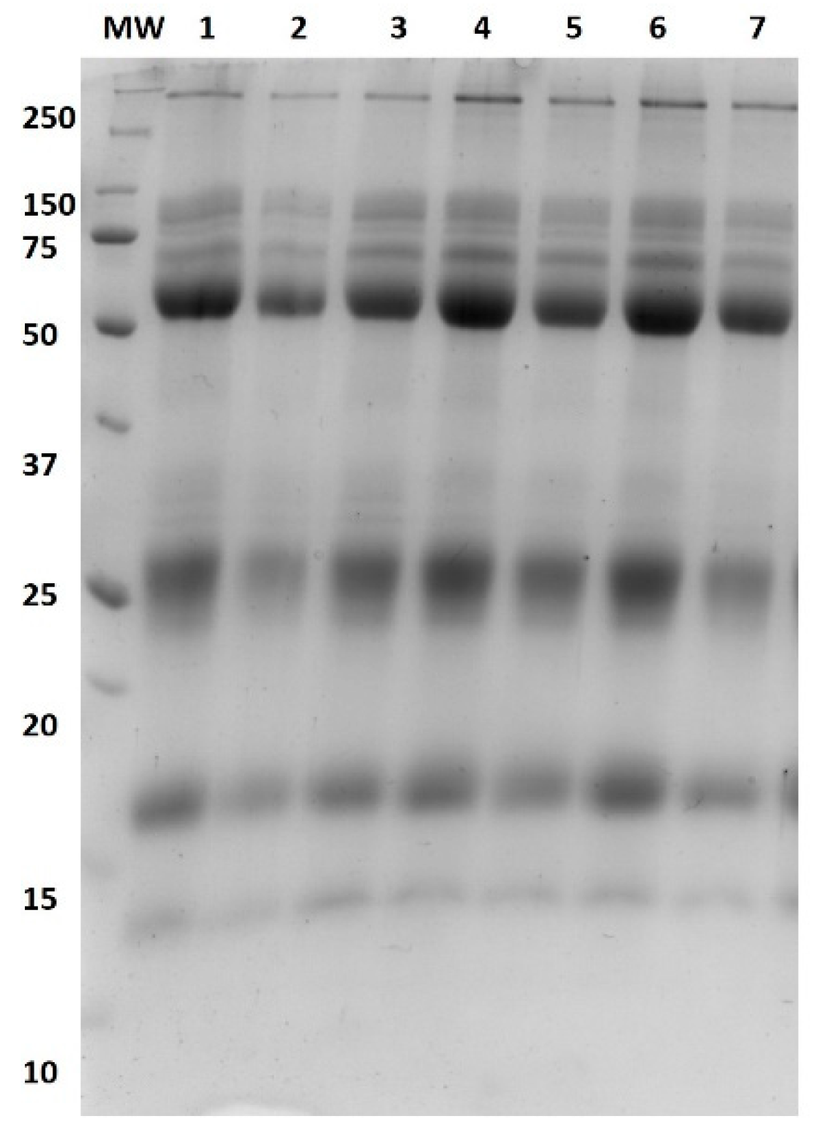
Based on the results, the bands from the separation 1-DE of bovine colostrum proteins were identified as lactoperoxidase (94 kDa), lactotransferrin (76 kDa), and serum albumin (62 kDa). Immunoglobulin G was identified as two different protein bands, namely IgG heavy chain and IgG light chain, with molecular weights of about 48 kDa and 24 kDa, respectively. Β-lactoglobulin and α-lactalbumin were identified as protein bands with molecular weights of 18 and 15 kD, respectively (Figure 1).
2-DE and MALDI TOF-based analysis created a protein profile for lyophilised bovine colostrum. We have generated a 2-D map of this medium protein in the range of isoelectric points (pI) from 3 to 10 and molecular masses (Mr) from 250 to less than 20 kDa. The protein profile comprised ca. 90 distinct protein spots, among which we successfully identified 54 representing 15 gene expression products. The representative 2-D map of lyophilized bovine colostrum whey protein is presented in Figure 2. Most of these proteins are characteristic of domestic cattle (Bos taurus). In addition, nearly 50% of gene products were identified as peptides representing immunoglobulins. Among the other identified gene products, most are prevalent as colostrum/milk proteins: serum albumin, transferrin, β-lactoglobulin, casein S1 and prepronociceptin. The list of identified proteins, including detailed information on the identification parameters of each protein spot, is given in Table 6.
Food processing technologies have a significant impact not only on food safety but also on the structure of proteins and their interactions [64]. For example, studies show that cold plasma induces changes in the structure and functionality of various proteins.
As a part of this study, a comparative densitometric analysis of proteome maps of lyophilized bovine colostrum whey protein after its treatment with cold plasma in different atmospheric environments in contrast to the untreated sample was also performed. The analysed protein profile representatives are shown in Figure 2. The untreated sample is shown on panel C, and the cold plasma-treated samples in the presence of nitrogen, oxygen and atmospheric air are presented on panels N2, O2 and A, respectively. In addition, the comparative analysis was performed for the identified protein spots; its results are shown in Table 6.
Densitometric analysis indicated changes occurring in various fractions regardless of the ionizing gas used compared to control samples. Immunoglobulins were noticeable in all samples subjected to plasma sterilization. However, a reduction in the intensity of these spots was observed for all test variants, indicating a reduction in the amount of this fraction.
Samples of freeze-dried bovine colostrum treated with plasma sterilization showed reduced β-lactoglobulin. Plasma sterilization using air, nitrogen and oxygen as ionizing gas had a negative effect on serum albumin fractions. Similar results for β-lactoglobulin and serum albumin bands were obtained by Ng et al. [65]. They attributed the disappearance of protein bands to forming new aggregates involving disulfide bonds and hydrophobic and electrostatic interactions. It has been suggested that cold plasma treatment causes aggregation of whey proteins [62]. The results indicated that the ionizing gases used in our study had a negative effect on the fraction of α-lactalbumin and serotransferrin.
In the 1-DE gels, bands of protein fractions were presently smeared and wider compared to the control sample. This may be caused by some cold plasma-induced structural modification resulting in molecular weight changes. These changes are most likely caused by reactive oxygen and nitrogen species and UV radiation, which can potentially modify protein structures [66]. This is supported by the study of Zhou et al. [67], in which modification of amino acids by cold plasma-induced reactions, e.g., oxidation, hydroxylation, dehydrogenation, sulfonation and dimerization, were found.
In addition, literature data [68,69,70] indicate protein aggregation as a consequence of plasma treatment, which can lead to the disappearance of protein bands in SDS-PAGE. Oxidation induces the unfolding of proteins, which increases their surface hydrophobicity, and causes protein aggregation and polymerization due to the formation of disulfide bonds, dityrosine bonds and other intermolecular bonds [71].
Segat et al. [62] treated whey proteins with dielectric barrier discharge (DBD) plasma and showed an increase in carbonyl groups, surface hydrophobicity, and aperiodic structures. This resulted in a decrease in free SH groups and improved foaming and emulsifying ability.
4. Conclusions
Cold plasma is a technology with an extensive range of applications. The variety of molecules activated during plasma generation, the near-zero electrical charge (net), and the low temperature-usually below 40 °C—are only some factors influenced by the type of gas supply and the configuration of the device. Many variables involved in the plasma process and the kind of matrix affect the decontamination results. The differences in parameters in the plasma technology limit the ability to compare the results, especially in the case of colostrum, which until now has not been widely used as the matrix in the plasma process.
As a food supplement, colostrum is available in powder form produced by freeze-drying or spray-drying. The technologies are not intended to eliminate microbial contaminants but mainly to increase their stability, especially during storage. Yet the nutritional values of colostrum and its basic parameters depend on the level of microbial contamination. The application of CP has the potential to inactivate microorganisms while keeping biologically active compounds of colostrum intact. However, our study’s results on the decontamination efficiency of plasma are similar to those described in other publications. It could be explained by the fact that product-plasma interactions were limited to the surface of the matrix due to the powder–like state of the product. Our results reflected the dependence of the limited penetration depth of plasma components on the effectiveness of microbial inactivation throughout the sample volume. In addition, reactive plasma molecules in contact with a moist environment produce more oxidizing radicals (ex. OH*, H*, H2O2, O3) by which the level of inactivation of microorganisms such as bacteria, yeast, and molds increases. Our studies indicate that using oxygen and nitrogen as working gases generates conditions conducive to the highest reduction of the determined microorganisms. Unfortunately, they negatively affected the sensory characteristics of the colostrum samples. The plasma also induced changes in protein fractions.
In general, cold plasma is an emerging and highly promising technology for dairy product processing, including colostrum, with minimal change in quality. However, further studies are required to prove if, by adjusting cold plasma treatment conditions, including the type of gas, voltage, treatment time and plasma source, non-thermal inactivation of microorganisms in commercial colostrum preparations can be achieved without affecting the sensory properties of the product.
Author Contributions
Conceptualization, E.B.-W.; methodology, E.B.-W., A.D., A.L., M.O. and W.S.; formal analysis, E.B.-W., A.D., M.O. and A.L.; writing—original draft preparation, review and editing E.B.-W., A.D., A.L. and W.S.; visualization E.B.-W. All authors have read and agreed to the published version of the manuscript.
Funding
This research was funded by Intelligent Development Operational Program POIR 02.03.02–30–0065-20-00.
Institutional Review Board Statement
Not applicable.
Informed Consent Statement
Not applicable.
Data Availability Statement
Any data or material supporting this study’s findings can be made available by the corresponding author upon request.
Acknowledgments
The authors are grateful to Dagmara Mędrala-Klein, for her kindness and English editing.
Conflicts of Interest
The authors declare no conflict of interest.
References
- Brian, A.M.; Patric, F.F.; Paul, S.; Alan, K. Composition and properties of bovine colostrum: A review. Dairy Sci. Technol. 2016, 96, 133–158. [Google Scholar]
- Pakkanen, R.; Aalto, J. Growth factors and antimicrobial factors of bovine colostrum. Int. Dairy J. 1997, 7, 258–297. [Google Scholar] [CrossRef]
- Shen, R.L.; Thymann, T.; Østergaard, M.V.; Støy, A.C.; Krych, Ł.; Nielsen, D.S.; Lauridsen, C.; Hartmann, B.; Holst, J.J.; Burrin, D.G.; et al. Early gradual feeding with bovine colostrum improves gut function and NEC resistance relative to infant formula in preterm pigs. Am. J. Physiol. Gastrointest. Liver. Physiol 2015, 309, 310–320. [Google Scholar] [CrossRef]
- Playford, R.J.; Weiser, M.J. Bovine Colostrum: Its Constituents and Uses. Nutrients 2021, 13, 265. [Google Scholar] [CrossRef]
- Płusa, T. Immunomodulatory proteins in colostrum. Pol. Merkur. Lekarski. 2009, 26, 8–10. [Google Scholar]
- Chen, D.; Peng, P.; Zhou, N.; Cheng, Y.; Min, M.; Ma, Y.; Mao, Q.; Chen, P.; Chen, C.; Ruan, R. Evaluation of Cronobacter sakazakii inactivation and physicochemical property changes of non-fat dry milk powder by cold atmospheric plasma. Food Chem. 2019, 290, 270–276. [Google Scholar] [CrossRef]
- Chiang, S.H.; Chang, C.Y. Antioxidant properties of caseins and whey proteins from colostrums. J. Food Drug. Anal. 2005, 13, 57–63. [Google Scholar] [CrossRef]
- Wieczorek—Dąbrowska, M.; Wójcik, P.; Malinowski, E. Importance of cow colostrum and factors determining its quality. Balice Przegląd Hod. 2013, 81, 9–10. [Google Scholar]
- Kim, J.S.; Lee, E.J.; Choi, E.H.; Kim, Y.J. Inactivation of Staphylococcus aureus on the beef jerky by radio-frequency atmospheric pressure plasma discharge treatment. Innov. Food Sci. Emerg. Technol. 2014, 22, 124–130. [Google Scholar] [CrossRef]
- Toepfl, S.; Mathys, A.; Heinz, V.; Knorr, D. Review: Potential of High Hydrostatic Pressure and Pulsed Electric Fields for Energy Efficient and Environmentally Friendly Food Processing. Food Rev. Int. 2006, 22, 405–423. [Google Scholar] [CrossRef]
- van Boekel, M.; Fogliano, V.; Pellegrini, N.; Stanton, C.; Scholz, G.; Lalljie, S.; Somoza, V.; Knorr, D.; Jasti, P.R.; Eisenbrand, G. A review on the beneficial aspects of food processing. Mol. Nutr. Food Res. 2010, 54, 1215–1247. [Google Scholar] [CrossRef]
- Fecteau, G.; Baillargeon, P.; Higgins, R.; Paré, J.; Fortin, M. Bacterial contamination of colostrum fed to newborn calves in Québec dairy herds. Can. Vet. J. 2002, 43, 523–527. [Google Scholar]
- Nwabor, O.F.; Onyeaka, H.; Miri, T.; Obileke, K.; Anumudu, C.; Hart, A. A Cold Plasma Technology for Ensuring the Microbiological Safety and Quality of Foods. Food Eng. Rev. 2022, 14, 535–554. [Google Scholar] [CrossRef]
- Yepez, X.V.; Misra, N.N.; Keener, K.M. Nonthermal Plasma Technology. In Food Safety Engineering. Food Engineering Series; Demirci, A., Feng, H., Krishnamurthy, K., Eds.; Springer: New York, NY, USA, 2020; pp. 607–628. [Google Scholar]
- Reineke, K.; Langer, K.; Hertwig, C.; Ehlbeck, J.; Schlüter, O. The impact of different process gas compositions on the inactivation effect of an atmospheric pressure plasma jet on Bacillus spores. Innov. Food Sci. Emerg. Technol. 2015, 30, 112–118. [Google Scholar] [CrossRef]
- Kim, H.-J.; Yong, H.I.; Park, S.; Choe, W.; Jo, C. Effects of dielectric barrier discharge plasma on pathogen inactivation and the physicochemical and sensory characteristics of pork loin. Curr. Appl. Phys. 2013, 13, 1420–1425. [Google Scholar] [CrossRef]
- Lee, H.J.; Jung, S.; Jung, H.; Park, S.; Choe, W.; Ham, J.S.; Jo, C. Evalution of dielectric barier discharge plasma system for inactivatating pathogens on cheese slices. J. Anim. Sci. Technol. 2012, 54, 191–198. [Google Scholar] [CrossRef]
- Moreau, M.; Orange, N.; Feuilloley, M.G.J. Non-thermal plasma technologies: New tools for bio-decontamination. Biotechnol. Adv. 2008, 26, 610–617. [Google Scholar] [CrossRef]
- PN—93/A-86034/02; Milk and Dairy Products. Microbiological Testing. General Principles of Testing. Polish Committee for Standardization: Warsaw, Poland, 2018.
- ISO 6887-5:2020-10; Microbiology of the Food Chain—Preparation of Samples, Stock Suspension and Decimal Dilutions for Microbiological Testing—Part 5: Specific Rules for the Preparation of Milk and Milk Products. Polish Committee for Standardization: Warsaw, Poland, 2020.
- ISO 4833-2:2013-12; Microbiology of the Food Chain—Horizontal Method for the Determination of Microbial Counts—Part 2: Determination of Counts by Surface Culture at 30 Degrees C. Polish Committee for Standardization: Warsaw, Poland, 2013.
- ISO 21527-1:2009; Microbiology of Food and Feed—Horizontal Method for the Determination of Yeast and Mold Counts—Part 1: Method for Counting Colonies in Products with Water Activity Higher than 0.95. Polish Committee for Standardization: Warsaw, Poland, 2009.
- ISO 21528-2:2017-08; Food Chain Microbiology—Horizontal Method for Detection and Enumeration of Enterobacteriaceae—Part 2: Colony Counting Method. Polish Committee for Standardization: Warsaw, Poland, 2017.
- ISO 6888-2:2001+A1:200; Microbiology of Food and Feed—Horizontal Method for the Determination of Coagulase-Positive Staphylococci (Staphylococcus Aureus and Other Species)—Part 2: Method Using Rabbit Plasma Agar Medium and Fibrynogen. Polish Committee for Standardization: Warsaw, Poland, 2004.
- ISO 7932:2005/A1:2020-09; Microbiology of Food and Feed—Horizontal Method for Determining the Number of Putative Bacillus Cereus—Method of Counting Colonies at 30 Degrees C. Polish Committee for Standardization: Warsaw, Poland, 2020.
- PN-EN 15788:2009; Detection and Enumeration of Enterococcus (E. faecium) spp. Polish Committee for Standardization: Warsaw, Poland, 2009.
- ISO 15214:2002; Microbiology of Food and Feed—Horizontal Method for the Determination of the Number of Mesophilic Lactic Fermentation Bacteria—Plate Method at 30 Degrees C. Polish Committee for Standardization: Warsaw, Poland, 2002.
- Dmytrów, I.; Szymczak, M.; Szkolnicka, K.; Kamiński, P. Development of Functional Acid Curd Cheese (Tvarog) with Antioxidant Activity Containing Astaxanthin from Shrimp Shells Preliminary Experiment. Foods 2021, 10, 895. [Google Scholar] [CrossRef]
- Pankaj, S.K.; Bueno-Ferrer, C.; Misra, N.N.; O’Neill, L.; Tiwari, B.K.; Bourke, P.; Cullen, P.J. Physicochemical characterization of plasma-treated sodium caseinate film. Food Res. Int. 2014, 66, 438–444. [Google Scholar] [CrossRef]
- ISO 22935-2:2013-07; Milk and Dairy Products—Sensory Analysis—Part 2: Recommended Methods for Sensory Evaluation. Polish Committee for Standardization: Warsaw, Poland, 2013.
- ISO 22935-3:2013-07; Milk and Dairy Products—Sensory Analysis—Part 3: Guidelines for Evaluating the Conformity of the Properties of Sensory Attributes with Product Specifications Using the Scoring Method. Polish Committee for Standardization: Warsaw, Poland, 2013.
- Drake, M.A.; Karagul-Yuceer, Y.; Cadwallader, K.R.; Civtlle, G.V.; Tong, P.S. Determination of the sensory attributes of dried milk powders and dairy ingredients. J. Sens. Stud. 2003, 18, 199–2016. [Google Scholar] [CrossRef]
- Abdalla, A.K.; Smith, J.K.; Lucey, K. Physical Properties of Nonfat Dry Milk and Skim Milk Powder. Int. J. Dairy Sci. 2017, 12, 149–154. [Google Scholar] [CrossRef]
- Lepczyński, A.; Ożgo, M.; Dratwa-Chałupnik, A.; Robak, P.; Pyć, A.; Zaborski, D.; Herosimczyk, A. An update on medium- and low-abundant blood plasma proteome of horse. Animal 2018, 12, 76–87. [Google Scholar] [CrossRef]
- Lepczyński, A.; Ożgo, M.; Michałek, K.; Dratwa-Chałupnik, A.; Grabowska, M.; Herosimczyk, A.; Liput, K.P.; Poławska, E.; Kram, A.; Pierzchała, M. Effects of three-month feeding high fat diets with different fatty acid composition on myocardial proteome in mice. Nutrients 2021, 13, 330. [Google Scholar] [CrossRef]
- Ozgo, M.; Lepczynski, A.; Herosimczyk, A. Two-dimensional gel-based serum protein profile of growing piglets. Turk. J. Biol. 2015, 39, 320–327. [Google Scholar] [CrossRef]
- Godden, S.M.; Lombard, J.E.; Woolums, A.R. Colostrum Management for Dairy Calves. Vet. Clin. North. Am. Food. Anim. Pract. 2019, 35, 535–556. [Google Scholar] [CrossRef]
- Lorenz, I.; Mee, J.F.; Earley, B.; More, S.J. Calf health from birth to weaning. General aspects of disease prevention. Ir. Vet. J. 2011, 16, 3–8. [Google Scholar] [CrossRef]
- Bourke, P.; Ziuzina, D.; Han, L.; Cullen, P.J.; Gilmore, B.F. Microbiological interactions with cold plasma. J. Appl. Microbiol. 2017, 123, 308–324. [Google Scholar] [CrossRef]
- Stoffels, E.; Sakiyama, Y.; Graves, D.B. Cold atmospheric plasma: Charged species and their interactions with cells and tissues. IEEE Trans. Plasma Sci. 2008, 36, 1441–1457. [Google Scholar] [CrossRef]
- Fröhling, A.; Baier, M.; Ehlbeck, J.; Knorr, D.; Schlüter, O. Atmospheric pressure plasma treatment of Listeria innocua and Escherichia coli at polysaccharide surfaces: Inactivation kinetics and flow cytometric characterization. Innov. Food Sci. Emerg. Technol. 2012, 13, 142–150. [Google Scholar] [CrossRef]
- Hertwig, C.; Reineke, K.; Ehlbeck, J.; Erdoğdu, B.; Rauh, C.; Schlüter, O. Impact of remote plasma treatment on natural microbial load and quality parameters of selected herbs and spices. J. Food Eng. 2015, 167, 12–17. [Google Scholar] [CrossRef]
- Cummins, C.; Lorenz, I.; Kennedy, E. Short communication: The effect of storage conditions over time on bovine colostral immunoglobulin G concentration, bacteria, and pH. J. Dairy Sci. 2016, 99, 4857–4863. [Google Scholar] [CrossRef] [PubMed]
- Fasse, S.; Jarmo, A.; Björn, F.; Gun, W. Bovine Colostrum for Human Consumption—Improving Microbial Quality and Maintaining Bioactive Characteristics through Processing. Dairy 2021, 2, 556–575. [Google Scholar] [CrossRef]
- Moisan, M.; Barbeau, J.; Moreau, S.; Pelletier, J.; Tabrizian, M.; Yahia, L.H. Low-temperature sterilization using gas plasmas: A review of the experiments and an analysis of the inactivation mechanisms. Int. J. Pharm. 2001, 226, 1–21. [Google Scholar] [CrossRef]
- Schlüter, O.; Fröhling, A. Non-Thermal Processing Cold Plasma for Bioefficient Food Processing, Reference Module in Food Science Encyclopedia of Food Microbiology, 2nd ed.; Elsevier: New York, NY, USA, 2014; pp. 948–953. [Google Scholar]
- Li, J.; Sakai, N.; Watanabe, M.; Hotta, E.; Wachi, M. Study on plasma agent effect of a direct-current atmospheric pressure oxygen-plasma jet on inactivation of E. coli using bacterial mutants. IEEE Trans. Plasma Sci. 2013, 41, 935–941. [Google Scholar] [CrossRef]
- Joshi, S.G.; Cooper, M.; Yost, A.; Paff, M.; Ercan, U.K.; Fridman, G.; Friedman, G.; Fridman, A.; Brooks, A.D. Nonthermal dielectric-barrier discharge plasma-induced inactivation involves oxidative DNA damage and membrane lipid peroxidation in Escherichia coli. Antimicrob. Agents. Chemother. 2011, 55, 1053–1062. [Google Scholar] [CrossRef]
- Hertwig, C.; Meneses, N.; Mathys, A. Cold atmospheric pressure plasma and low energy electron beam as alternative nonthermal decontamination technologies for dry food surfaces: A review. Trends Food Sci. Technol. 2018, 77, 131–142. [Google Scholar] [CrossRef]
- Deng, X.; Shi, J.; Kong, M.G. Physical mechanisms of inactivation of Bacillus subtilis spores using cold atmospheric plasmas. IEEE Trans. Plasma Sci. 2006, 34, 1310–1316. [Google Scholar] [CrossRef]
- Tseng, S.; Abramzon, N.; Jackson, J.O.; Lin, W.J. Gas discharge plasmas are effective in inactivating Bacillus and Clostridium spores. Appl. Microbiol. Biotechnol. 2012, 93, 2563–2570. [Google Scholar] [CrossRef]
- Wang, S.; Doona, C.J.; Setlow, P.; Li, Y.Q. Use of Raman Spectroscopy and Phase-Contrast Microscopy to Characterize Cold Atmospheric Plasma Inactivation of Individual Bacterial Spores. Appl. Environ. Microbiol. 2016, 82, 5775–5784. [Google Scholar] [CrossRef]
- Segat, A.; Misra, N.N.; Cullen, P.J.; Innocente, N. Effect of atmospheric pressure cold plasma (ACP) on activity and structure of alkaline phosphatase. Food Bioprod. Process. 2016, 98, 181–188. [Google Scholar] [CrossRef]
- Manoharan, D.; Stephen, J.; Radhakrishnan, M. Study on low-pressure plasma system for continuous decontamination of milk and its quality evaluation. J. Food Process. Preserv. 2020, 45, 234–245. [Google Scholar] [CrossRef]
- Wan, Z.; Misra, N.N.; Li, G.; Keener, K.M. High voltage atmospheric cold plasma treatment of Listeria innocua and Escherichia coli K-12 on Queso Fresco (fresh cheese). LWT 2021, 146, 111406. [Google Scholar] [CrossRef]
- Oehmigen, K.; Hahnel, M.; Brandenburg, R.; Wilke, C.; Weltmann, K.D.; von Woedtke, T. The Role of Acidification for Antimicrobial Activity of Atmospheric Pressure Plasma in Liquids. Plasma Process. Polym. 2010, 7, 250–257. [Google Scholar] [CrossRef]
- Traylor, M.J.; Pavlovich, M.J.; Karim, S.; Hait, P.; Sakiyama, Y.; Clark, D.S.; Graves, D.B. Long-term antibacterial efficacy of air plasma-activated water. J. Phys. Appl. Phys. 2011, 44, 134–145. [Google Scholar] [CrossRef]
- Gross, J.J.; Kessler, E.C.; Bruckmaier, R.M. Colour measurement of colostrum for estimation of colostral IgG and colostrum composition in dairy cows. J. Dairy. Res. 2014, 81, 440–444. [Google Scholar] [CrossRef] [PubMed]
- Gurol, C.; Ekinci, F.Y.; Aslan, N.; Korachi, M. Low Temperature Plasma for decontamination of E. coli in milk. Int. J. Food. Microbiol. 2012, 157, 1–5. [Google Scholar] [CrossRef]
- Yong, H.I.; Kim, H.-J.; Park, S.; Kim, K.; Choe, W.; Yoo, S.J.; Jo, C. Pathogen inactivation and quality changes in sliced cheddar cheese treated using flexible thin-layer dielectric barrier discharge plasma. Food Res. Int. 2015, 69, 57–63. [Google Scholar] [CrossRef]
- Nikmaram, N.; Keener, K.M. The effects of cold plasma technology on physical, nutritional, and sensory properties of milk and milk products. LWT 2022, 154, 112729. [Google Scholar] [CrossRef]
- Segat, A.; Misra, N.N.; Cullen, P.J.; Innocente, N. Atmospheric pressure cold plasma (ACP) treatment of whey protein isolate model solution. Innov. Food Sci. Emerg. Technol. 2015, 29, 247–254. [Google Scholar] [CrossRef]
- Wu, X.; Luo, Y.; Zhao, F.; Murad, M.S.; Mu, G. Influence of dielectric barrier discharge cold plasma on physicochemical property of milk for sterilization. Plasma Process. Polym. 2021, 18, 245–256. [Google Scholar] [CrossRef]
- Singh, P.K.; Huppertz, T. Chapter 8—Effect of Nonthermal Processing on Milk Protein Interactions and Functionality. In Milk Proteins; Boland, M., Singh, H., Eds.; Academic Press: Toronto, ON, Canada, 2020; pp. 293–324. [Google Scholar]
- Ng, S.W.; Lu, P.; Rulikowska, A.; Boehm, D.; O’Neill, G.; Bourke, P. The effect of atmospheric cold plasma treatment on the antigenic properties of bovine milk casein and whey proteins. Food Chem. 2021, 342, 128283. [Google Scholar] [CrossRef] [PubMed]
- Ramazzina, I.; Tappi, S.; Rocculi, P.; Sacchetti, G.; Berardinelli, A.; Marseglia, A.; Rizzi, F. Effect of Cold Plasma Treatment on the Functional Properties of Fresh-Cut Apples. J. Agric. Food. Chem. 2016, 64, 8010–8018. [Google Scholar] [CrossRef] [PubMed]
- Zhou, R.; Zhuang, J.; Zong, Z.; Zhang, X.; Liu, D.; Bazaka, K.; Ostrikov, K. Interaction of Atmospheric-Pressure Air Microplasmas with Amino Acids as Fundamental Processes in Aqueous Solution. PLoS ONE 2016, 11, 134–148. [Google Scholar] [CrossRef]
- Held, S.; Tyl, C.E.; Annor, G.A. Effect of radio frequency cold plasma treatment on intermediate wheatgrass (Thinopyrum intermedium) flour and dough properties in comparison to hard and soft wheat (Triticum aestivum L.). J. Food. Qual. 2019, 34, 156–176. [Google Scholar] [CrossRef]
- Ji, H.; Dong, S.; Han, F.; Li, Y.; Chen, G.; Li, L.; Chen, Y. Effects of Dielectric Barrier Discharge (DBD) Cold Plasma Treatment on Physicochemical and Functional Properties of Peanut Protein. Food Bioprocess Technol. 2018, 11, 344–354. [Google Scholar] [CrossRef]
- Zhang, S.; Huang, W.; Feizollahi, E.; Roopesh, M.S.; Chen, L. Improvement of pea protein gelation at reduced temperature by atmospheric cold plasma and the gelling mechanism study. Innov. Food Sci. Emerg. Technol. 2021, 67, 102–112. [Google Scholar] [CrossRef]
- Zhang, W.; Xiao, S.; Ahn, D.U. Protein oxidation: Basic principles and implications for meat quality. Crit. Rev. Food. Sci. Nutr. 2013, 53, 1191–1201. [Google Scholar] [CrossRef]
Figure 1.
Results of 1D electrophoretic separation of samples and bovine colostrum; MW Molecular weight 10–250 kD, lane 1 N2 cold plasma, lane 2 O2 cold plasma, lane three air cold plasma, lanes 4–7 control condition.
Figure 1.
Results of 1D electrophoretic separation of samples and bovine colostrum; MW Molecular weight 10–250 kD, lane 1 N2 cold plasma, lane 2 O2 cold plasma, lane three air cold plasma, lanes 4–7 control condition.

Figure 2.
2-DE map of the differentially expressed protein spots found in the colostrum spot numbers corresponds to those in Table 6. Control condition (C) nitrogen cold plasma (N2), air (A) and oxygen (O2).
Figure 2.
2-DE map of the differentially expressed protein spots found in the colostrum spot numbers corresponds to those in Table 6. Control condition (C) nitrogen cold plasma (N2), air (A) and oxygen (O2).


Table 1.
Conditions for generating and maintaining cold plasma depend on the ionizing gas used.
| Sample | Gas Flow | Time to Obtain Vacuum | Temperature | Power | Process Time | Pressure in the Chamber |
|---|---|---|---|---|---|---|
| [cm3/min] | [min] | [°C] | [W] | [min] | [mbar] | |
| Air | 20–50 | 60 | 27–30 | 300 | 60 | 0.6 |
| Nitrogen | 20–48 | 60 | 28–32 | |||
| Oxygen | 20–50 | 70 | 26–30 |
Table 2.
Microbiotic diversity of freeze-dried bovine colostrum samples.
| Bacteria | Average | LL-UL | SEM | p-Value |
|---|---|---|---|---|
| Normal inhabitants of bovine skin and mucosa (NI) | log cfu/g | |||
| Mesophilic bacteria (THMC) | 4.70 * | 3.21–4.86 | 0.082 | 0.001 |
| Staphylococcus spp. (TSt) | 1.89 * | 0.5–3.29 | 0.148 | 0.031 |
| Streptococcus spp. (TStr) | 1.87 * | 1.00–2.30 | 0.135 | 0.049 |
| Yeast (TYMC) | 2.95 * | 1.61–3.25 | 0.019 | 0.046 |
| LAB | 2.94 * | 1.29–3.72 | 0.006 | 0.001 |
| Environmental contaminants (EC) | ||||
| Psychrotrophic bacteria (THPC) | 5.24 | 4.56–5.78 | 0.366 | 0.897 |
| Bacillus spp. (TCBac) | 2.96 | 2.48–3.02 | 0.313 | 0.604 |
| Moulds (TMMC) | 1.97 | 1.74–2.24 | 0.185 | 0.091 |
| Faecal contaminants (FC) | ||||
| Enterobacteriacea (TEb) | 2.43 * | 1.18–2.98 | 0.267 | 0.049 |
| E. coli (Ec) | 1.38 * | 0.00–2.00 | 0.348 | 0.048 |
| Enterococcus spp. (TEcc) | 2.37 | 1.69–2.59 | 0.134 | 0.167 |
*—statistically significant differences between samples, LL—lower limit, UL—upper limit.
Table 3.
The level of microorganisms’ reduction after plasma-induced freeze-dried colostrum samples.
Table 3.
The level of microorganisms’ reduction after plasma-induced freeze-dried colostrum samples.
| Normal Inhabitants of Bovine Skin and Mucosa | Environmental Contaminants | Faecal Contaminants | |||||||||
|---|---|---|---|---|---|---|---|---|---|---|---|
| Sample | THMC | TSt | TStr | TYMC | LAB | THPC | TCBac | TMMC | TEb | Ec | TEcc |
| log cfu/g | |||||||||||
| Air | 0.72 | 0.61 | 0.54 | 0.91 | 0.16 a | 0.73 | 0.76 | 0.45 | 0.68 a | 0.64 | 0.79 |
| Nitrogen | 1.19 | 0.78 | 0.65 | 0.62 | 0.56 | 1.09 | 0.53 | 0.49 | 1.14 | 0.88 | 0.75 |
| Oxygen | 1.02 | 0.73 | 0.75 | 0.92 | 0.88 b | 1.24 | 0.81 | 0.45 | 1.16 b | 0.96 | 0.74 |
| SEM | 0.127 | 0.149 | 0.158 | 0.174 | 0.084 | 0.124 | 0.369 | 0.681 | 0.079 | 0.079 | 0.019 |
| p-value | 0.101 | 0.122 | 0.326 | 0.094 | 0.032 | 0.701 | 0.112 | 0.843 | 0.043 | 0.133 | 0.052 |
a,b—statistically significant differences. THMC—total count of mesophilic bacteria, THPC—total count of psychrortophic bacteria, TYMC and TMMC—total count of yeast and mould, TEb—total count of Enterobacteriaceae, Ec—total count of E. coli, TSt—total count of Staphylococcus spp., TCBac—total count of aerobic spore-forming bacteria, TEcc—total count of Enterococcus spp., TStr—total count of Streptococcus spp., LAB—total count of lactic acid bacteria (LAB).
Table 4.
Changes in pH and colour parameters of freeze-dried bovine colostrum after plasma process.
| Samples | pH | a*(-) | b* | L* | h (-) | C | ΔE |
|---|---|---|---|---|---|---|---|
| Control | 6.656 | 1.94 | 18.29 | 82.84 a | 83.92 | 18.40 a | - |
| Air | 6.663 | 2.31 | 18.43 | 82.11 | 82.87 | 18.57 | 0.82 A |
| Nitrogen | 6.654 | 2.04 | 19.64 | 81.49 b | 84.18 | 19.74 b | 1.90 B,C |
| Oxygen | 6.667 | 1.87 | 19.27 | 82.49 | 84.46 | 19.36 | 1.04 D |
| SEM | 0.033 | 0.039 | 0.554 | 0.166 | 0.855 | 0.338 | 0.159 |
| p-value | 0.992 | 0.056 | 0.082 | 0.012 | 0.055 | 0.000 | 0.000 |
a,b statistically significant differences among the control and plasma samples, A,B,C,D statistically significant differences among plasma samples.
Table 5.
Effect of cold plasma on sensory qualities of freeze-dried bovine colostrum.
| Sample | Smell | Taste | Structure | Overall Sensory Quality |
|---|---|---|---|---|
| Control | 3.954 | 3.944 | 3.978 | 3.972 |
| Air | 3.833 | 3.899 | 3.944 | 3.745 |
| Nitrogen | 4.012 | 4.051 | 3.816 | 4.034 |
| Oxygen | 3.675 | 3.749 | 3.801 | 3.933 |
| SEM | 0.450 | 0.801 | 0.394 | 0.654 |
| p-value | 0.178 | 0.450 | 0.765 | 0.125 |
Table 6.
Proteins identified in the freeze-drying of bovine colostrum with their abundance in the control condition (C) and treated with cold plasma in the presence of nitrogen (N2), air (A), and oxygen (O2). Spot number indicates the number labelling the spots in Figure 2. For each identified protein spot: the protein name, gene name, and accession number are shown in Uniprot/NCBI database. Also, the identification parameters using MALDI-ToF MS are given: several peptides matched per gene product (Pep. Mach.), percentage of the sequence coverage (Seq. %), the Mascot score (score) and predicted pI and Mr.
| No. | Protein Name | Gene Name | Accession Number | C | N2 | N2/C | A | A/C | O2 | O2/C | Pep. Mach | Seq.% /Score | pI/Mr pH/kDa | Sp. |
|---|---|---|---|---|---|---|---|---|---|---|---|---|---|---|
| 1 | Immunoglobulin gamma | IGHG | AQT27060 | 251.6 | 25.8 | 0.1 | 14.9 | 0.06 | 51 | 0.2 | 28 | 65/82 | 6.21/51.09 | B |
| 2 | heavy chain | 144.1 | 9.6 | 0.07 | 7.3 | 0.05 | 31.7 | 0.22 | 18 | 42/87 | ||||
| 3 | 174.4 | 146 | 0.84 | 118.9 | 0.68 | 50.4 | 0.29 | 18 | 42/100 | |||||
| 4 | 173.6 | 82.6 | 0.48 | 31.2 | 0.18 | 64.7 | 0.37 | 16 | 42/82 | |||||
| 5 | 152.1 | 121.4 | 0.8 | 257.9 | 1.7 | 52.3 | 0.34 | 15 | 41/84 | |||||
| 6 | 57.9 | 125.4 | 2.17 | 203.3 | 3.51 | 89.8 | 1.55 | 14 | 41/84 | |||||
| 7 | 43.8 | 7.8 | 0.18 | 133.6 | 3.05 | 34.5 | 0.79 | 14 | 41/86 | |||||
| 8 | l-lactate dehydrogenase A chain | LDHA | P00339 | 58.6 | 22.8 | 0.39 | 38.2 | 0.65 | 23.4 | 0.4 | 13 | 58/64 | S | |
| 9 | Sclerostin domain-containing protein 1 | SOSTDC1 | Q9CQN4 | 28 | 16.9 | 0.61 | 41.3 | 1.48 | 16.5 | 0.59 | 7 | 33/63 | 9.87/23.73 | M |
| 10 | Serotransferrin | TRF | Q299443 | 15.9 | 4.4 | 0.28 | 11.4 | 0.72 | 0.3 | 0.02 | 39 | 63/78 | 6.75/79.87 | B |
| 11 | 24.7 | 24.3 | 0.98 | 50.5 | 2.04 | 19.8 | 0.8 | 40 | 62/63 | |||||
| 12 | 42 | 25.2 | 0.6 | 85.7 | 2.04 | 116.9 | 2.78 | 41 | 65/66 | |||||
| 13 | 31.2 | 40.7 | 1.3 | 62.5 | 2 | 57.2 | 1.83 | 41 | 66/99 | |||||
| 14 | 38 | 22.1 | 0.58 | 28.8 | 0.76 | 2.5 | 0.06 | 17 | 33/61 | |||||
| 15 | 23.7 | 24.2 | 1.02 | 12.3 | 0.52 | 15.4 | 0.65 | 45 | 68/81 | |||||
| 16 | 10.2 | 18.1 | 1.78 | 23.1 | 2.27 | 13.6 | 1.34 | 43 | 68/120 | |||||
| 17 | 16 | - | - | - | - | - | - | 36 | 62/82 | |||||
| 18 | Secreted immunoglobulin mu 2 heavy chain constant region, partial | IGMG | ANN46371 | 87.4 | 17.9 | 0.2 | 73.1 | 0.84 | 23.4 | 0.27 | 23 | 74/93 | 5.48/50.30 | B |
| 19 | Immunoglobulin mu heavy chain constant region, partial | IGMG | AAO37096 | 99.6 | 63.2 | 0.63 | 114.5 | 1.15 | 77 | 0.77 | 22 | 69/88 | 5.49/49.85 | B |
| 20 | Secreted immunoglobulin mu 2 heavy chain constant region, partial | IGMG | ANN46371 | 49.7 | 20.8 | 0.42 | 0.3 | 0.01 | 14.1 | 0.28 | 19 | 66/71 | 5.48/50.30 | B |
| 21 | Serum albumin precursor | ALB | NP851335 | 30.1 | 4.2 | 0.14 | 14.4 | 0.48 | 2.6 | 0.09 | 24 | 43/98 | 5.82/71.27 | B |
| 22 | Serum albumin | ALB | P02769 | 379.9 | 62 | 0.16 | 54.5 | 0.14 | 71.7 | 0.19 | 17 | 34/75 | 5.82/71.24 | B |
| 23 | 465.7 | 248.2 | 0.53 | 334.9 | 0.72 | 206.5 | 0.44 | 17 | 31/75 | |||||
| 24 | 351.2 | 264.6 | 0.75 | 738.1 | 2.1 | 328.3 | 0.93 | 21 | 41/87 | |||||
| 25 | 604.4 | 69.7 | 0.12 | 99.9 | 0.17 | 162.6 | 0.27 | 18 | 34/76 | |||||
| 26 | Immunoglobulin gamma | IGHG | AQT27060 | 596.1 | 363.9 | 0.61 | 475.3 | 0.8 | 111.5 | 0.19 | 16 | 48/22 | 6.21/51.09 | B |
| 27 | heavy chain | 866.9 | 105.1 | 0.12 | 390.6 | 0.45 | 1891 | 2.18 | 24 | 53/80 | ||||
| 28 | Secreted immunoglobulin gamma1 heavy chain constant region, partial | IGHG | ANN46377 | 2237 | 2301 | 1.03 | 2428 | 1.09 | 287.2 | 0.13 | 14 | 60/79 | 6.07/36.32 | B |
| 29 | Immunoglobulin gamma | IGHG | AQT27060 | 2275 | 1017 | 0.45 | 1618 | 0.71 | 2225 | 0.98 | 26 | 56/89 | 6.21/50.09 | B |
| 30 | heavy chain | 5708 | 4441 | 0.78 | 4624 | 0.81 | 3248 | 0.57 | 15 | 48/82 | 6.21/50.09 | B | ||
| 31 | 69.1 | 759.5 | 11 | 160.6 | 2.33 | 1212 | 17.6 | 23 | 53/91 | |||||
| 32 | 1777 | 657.4 | 0.37 | 5782 | 3.25 | 627 | 0.35 | 16 | 49/109 | |||||
| 33 | 94 | 759.5 | 8.08 | 160.6 | 1.71 | 1212 | 12.9 | 17 | 50/107 | |||||
| 34 | 698.5 | 990.2 | 1.42 | 1057 | 1.51 | 779.5 | 1.12 | 16 | 47/87 | |||||
| 35 | 1087 | 566.7 | 0.52 | 1282 | 1.18 | 1709 | 1.57 | 15 | 41/81 | |||||
| 36 | 952.9 | 2962 | 3.11 | 413.2 | 0.43 | 1967 | 2.06 | 15 | 48/80 | |||||
| 37 | 1122 | 100.3 | 0.09 | 219.6 | 0.2 | 494.7 | 0.44 | 16 | 53/90 | |||||
| 38 | t-RNA specific adenosine | ADAT2 | Q5E9J7 | 55.8 | 24.9 | 0.45 | 49.7 | 0.89 | 41.3 | 0.74 | 14 | 61/61 | 6.33/21.57 | B |
| 39 | deaminase 2 | 29.4 | 11.5 | 0.39 | 14.9 | 0.51 | 17.9 | 0.61 | 14 | 61/58 | ||||
| 40 | Immunoglobulin gamma | IGHG | AQT27060 | 43.2 | 18.5 | 0.43 | 35.8 | 0.83 | 12.3 | 0.28 | 26 | 58/83 | 6.21/50.09 | B |
| 41 | heavy chain | 51.4 | 21.8 | 0.42 | 30.3 | 0.59 | 10.8 | 0.21 | 15 | 43/91 | ||||
| 42 | IgG heavy chain precursor (B/MT.4A.17.H5.A5) | IGHG | CAA44699 | 25.7 | 18.7 | 0.73 | 21.5 | 0.83 | 5.6 | 0.22 | 22 | 58/91 | 6.10/51.39 | B |
| 43 | Alpha-S1-casein | CSN1S1 | P02662 | 283.9 | 58.1 | 0.2 | 45.7 | 0.16 | 34.6 | 0.12 | 6 | 35/59 | 4.98/24.57 | B |
| 44 | T-complex protein 1 subunit epsilon isoform d | TCP1 | NP_001293084 | 668.8 | 643.3 | 0.96 | 2000 | 2.99 | 863.8 | 1.29 | 29 | 72/81 | 5.86/49.95 | H |
| 45 | Unnamed protein product, partial | CAF90119 | CAF90119 | 1558 | 1783 | 1.14 | 1941 | 1.25 | 710 | 0.46 | 12 | 64/95 | 9.78/13.15 | T |
| 46 | PREDICTED: nck-associated protein 5 isoform X5 | XP_012901706 | 4206 | 1057 | 0.25 | 1757 | 0.42 | 1033 | 0.25 | 95 | 45/83 | 8.25/22.35 | MP | |
| 47 | hypothetical protein EI555_020003, partial | TKC51465 | TKC51465 | 1185 | 191.5 | 0.16 | 489.4 | 0.41 | 840.7 | 0.71 | 21 | 61/82 | 10.5/46.66 | MM |
| 48 | Kv channel-interacting protein 1 | KCIP1 | Q9NZI2 | 1325 | 50 | 0.04 | 253.6 | 0.19 | 433.9 | 0.33 | 26 | 81/66 | 5.10/26.97 | H |
| 49 | Coiled-coil domain- | CCDC183 | XP_030896 | 3331 | 1156 | 0.35 | 1247 | 0.37 | 3818 | 1.15 | 38 | 67/85 | 9.90/32.80 | L |
| 50 | containing protein 183 | 878 | 2019 | 749.5 | 0.37 | 1594 | 0.79 | 2021 | 1 | 37 | 68/86 | |||
| 51 | isoform X2 | 4315 | 2094 | 0.49 | 144.7 | 0.03 | 2743 | 0.64 | 41 | 71/89 | ||||
| 52 | 44.6 | 181.5 | 4.07 | 187.3 | 4.2 | 928.3 | 20.8 | 35 | 76/82 | |||||
| 53 | Beta-lactoglobulin | LGB | P02754 | 155.1 | 22.3 | 0.14 | 21.7 | 0.14 | 6.9 | 0.04 | 23 | 96/62 | 4.93/20.27 | B |
| 54 | Prepronociceptin | PNOC | O62647 | 31 | 4.2 | 0.14 | 10.9 | 0.35 | 0.2 | 0.01 | 5 | 46/56 | 9.67/20.64 | B |
Pep. Mach.–peptides matched, Seq %-sequence coverage %, score–Mascot score value, pI/Mr–isoelectric point/molecular range, Sp.–species, B-Bos taurus; S–sus scrofa, M–Mus musculus, H-Homo sapiens, T-Tetraodon nigroviridis, MP-Mustela putorius furo, MM-Monodon Monoceros, L-Leptonychotes weddellii.
Disclaimer/Publisher’s Note: The statements, opinions and data contained in all publications are solely those of the individual author(s) and contributor(s) and not of MDPI and/or the editor(s). MDPI and/or the editor(s) disclaim responsibility for any injury to people or property resulting from any ideas, methods, instructions or products referred to in the content. |
© 2023 by the authors. Licensee MDPI, Basel, Switzerland. This article is an open access article distributed under the terms and conditions of the Creative Commons Attribution (CC BY) license (https://creativecommons.org/licenses/by/4.0/).
Share and Cite
MDPI and ACS Style
Bogusławska-Wąs, E.; Dłubała, A.; Sawicki, W.; Ożgo, M.; Lepczyński, A. The Effect of Cold Plasma on Selected Parameters of Bovine Colostrum. Appl. Sci. 2023, 13, 5490. https://doi.org/10.3390/app13095490
AMA Style
Bogusławska-Wąs E, Dłubała A, Sawicki W, Ożgo M, Lepczyński A. The Effect of Cold Plasma on Selected Parameters of Bovine Colostrum. Applied Sciences. 2023; 13(9):5490. https://doi.org/10.3390/app13095490
Chicago/Turabian StyleBogusławska-Wąs, Elżbieta, Alicja Dłubała, Wojciech Sawicki, Małgorzata Ożgo, and Adam Lepczyński. 2023. "The Effect of Cold Plasma on Selected Parameters of Bovine Colostrum" Applied Sciences 13, no. 9: 5490. https://doi.org/10.3390/app13095490
Note that from the first issue of 2016, this journal uses article numbers instead of page numbers. See further details here.