Article

The Impact of Calcitriol on Orthodontic Tooth Movement: A Cumulative Systematic Review and Meta-Analysis

1
Department of Orthodontic, Faculty of Dentistry, University of Baghdad, Baghdad 01110, Iraq
2
Department of Conservative Dentistry with Endodontics, Medical University of Silesia, 40-055 Katowice, Poland
3
Department of Orthodontics, Faculty of Dentistry, Oral & Craniofacial Sciences, King’s College London, London SE1 9RT, UK
4
Department of Basic and Clinical Oral Sciences, Faculty of Dentistry, Umm Al-Qura University, Makkah 24381, Saudi Arabia
*
Author to whom correspondence should be addressed.
These authors contributed equally to this work.
Appl. Sci. 2021, 11(19), 8882; https://doi.org/10.3390/app11198882
Submission received: 24 August 2021 / Revised: 16 September 2021 / Accepted: 20 September 2021 / Published: 24 September 2021
(This article belongs to the Special Issue New Materials and Technologies in Orthodontics)

Abstract

:

Featured Application

The clinical use of vitamin D3 could allow for more predictable treatment outcome during orthodontic therapy, accelerating dentition alignment.

Abstract

A cumulative review with a systematic approach aimed to provide a comparison of studies’ investigating the possible impact of the active form of vitamin D3, calcitriol (CTL), on the tooth movement caused by orthodontic forces (OTM) by evaluating the quality of evidence, based on collating current data from animal model studies, in vivo cell culture studies, and human clinical trials. Methods: A strict systematic review protocol was applied following the application of the International Prospective Register of Systematic Reviews (PROSPERO). A structured search strategy, including main keywords, was defined during detailed search with the application of electronic database systems: Medline/Pubmed, EMBASE, Scopus, Web of Science, and PsycINFO. In addition, a search was carried out with the use of ClinicalTrials.gov search in order to include ongoing or recently completed trials. The Oxford Level of Evidence and the Grading of Recommendations Assessment, Development, and Evaluation (GRADE) approach was utilized to critically evaluate the risk of bias and relative quality of studies included. Meta-analysis with the use of RevMan5 software, random effect, and inverted variable method allowed the quantification of cumulative results. Results: Twenty-seven studies were identified which fulfilled inclusion criteria, including two clinical studies. The assessed level of evidence was variable and inconsistent, predominantly being moderate or low due to a significant difference in study design, sample size, and study protocols. Data synthesis rendered from meta-analysis involving various CTL doses demonstrated slight discrepancies in tooth movement between control and experimental groups (mean difference = 0.27; 95% CI: 0.01–0.53, std mean difference = 0.49; 95% CI: 0.09–0.89), as well as relatively moderate heterogenicity. Conclusions: Although it has been suggested that CTL could accelerate OTM in animal studies and clinical context, these scarce data were supported by a low level of evidence and the studies were carried out using inadequate sample size. Well-powered RCT studies would help to overcome the lack of robustness of the research.

1. Introduction

The length of orthodontic treatment is a significant concern for both orthodontists and patients, with an average duration of approximately 24–36 months; however, it can be even further extended [1,2]. Increased duration of orthodontic treatment is associated with patient’s compliance and usually more prevalent clinical side effects, such as external root resorption and enamel demineralization in the form of white spot lesions [3,4]. As a result, orthodontists have been interested in exploring various non-surgical and surgical approaches that could potentially reduce treatment time by accelerating tooth movement [5,6,7]. Non-surgical approaches are non-invasive, yet they provide low quality evidence to support their effectiveness [5,8]. Although some surgical approaches have been promisingly associated with increased rates of OTM, they are less appealing for patients due to their invasive nature and potential for postoperative discomfort [9].
The sequence of biological events that occur during OTM has been investigated recently [10], revealing the mechanism by which various endogenous molecules, including neurotransmitters, inflammatory mediators, and cytokines, coordinate local responses to applied orthodontic forces, as well as both initiate and maintain OTM [11,12]. Moreover, they provide possible targets for pharmacological therapies that may have the potential to accelerate or suppress tooth movement during and after orthodontic treatment [13].
It is well established that the active form of vitamin D3, calcitriol, has an anabolic effect on bone metabolism, and its receptors are present on osteoblasts, osteoclast precursors, and osteoclasts. It appears to enhance bone turnover in the same way as parathyroid hormone does, by stimulating prostaglandin production in osteoblasts [14]. In vitro studies have also shown that CTL regulates the production of collagen type I, alkaline phosphatase, osteocalcin and osteoblastic proliferation [15,16]. In addition, evidence from animal studies suggests that CTL can enhance alveolar bone remodeling and thereafter accelerate OTM [17,18], which is consistent with a clinical study reporting the systemic impact of CTL on enhancement of tooth movement [19] (Figure 1). In general, the stimulatory effect of CTL on osteoblasts may stabilize OTM.
Despite the well-established role of CTL in the metabolism and remodeling of mineralized hard tissue, the evidence for its role in stimulating and enhancing tooth movement in humans remains unclear [14]. This comprehensive review attempts to summarize the existing evidence related to the influence of CTL on tooth movement initiated during orthodontic treatment, with the use of quality assessment methods and quantitative data synthesis. To date, this review herewith is the first comprehensive analysis of both animal and human studies’ results evaluating the impact of CTL on tooth movement generated by orthodontic forces.

2. Materials and Methods

The systematic review protocol was approved and registered by the International Prospective Register of Systematic Reviews (PROSPERO) (CRD: 42020166878). All clinical studies were selected according to the PICO mode including participants (P) at any age who received orthodontic treatment in conjunction with calcitriol administration. Intervention (I): the effect of calcitriol during orthodontic treatment; control group (C) those who received orthodontic treatment without calcitriol administration; outcomes (O): accelerated tooth movement.

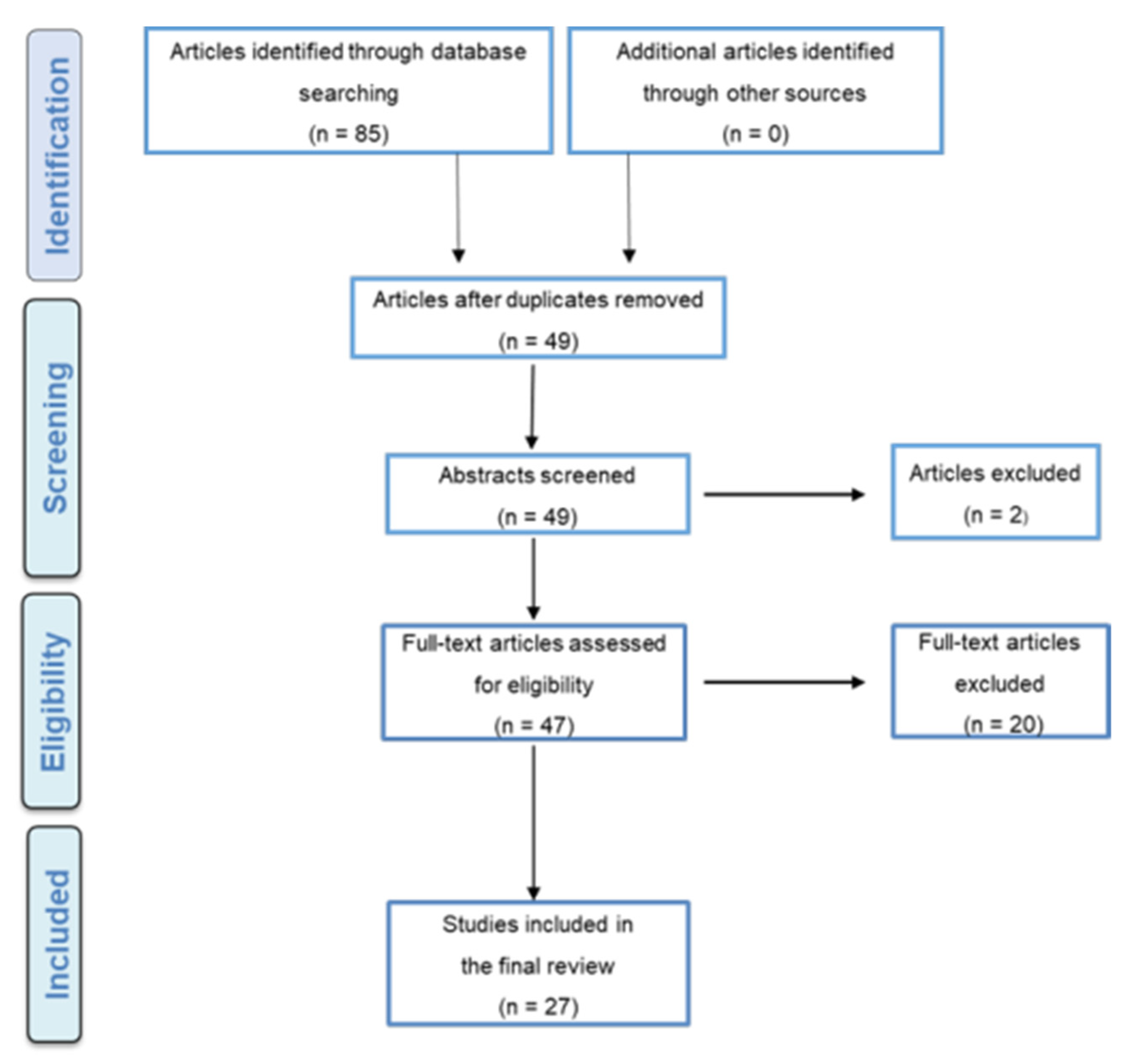
2.1. Search Strategy

Three databases were searched (Ovid MEDLINE(R) (1946 to 2020 Week 03); EMBASE (1974 to 2020 Week 03); and PsycINFO (1806 to 2018 Week 03). PROSPERO was searched for ongoing or recently completed systematic reviews. The International Clinical Trials Registry Platform Search Portal and ClinicalTrials.gov were also searched for ongoing or recently completed trials to optimize the extraction of data. Supporting Materials 1 presents the search strategy keywords for each database. The references of each included paper were used to search for additional papers.

2.2. Inclusion and Exclusion Criteria

The following type of studies were included in systematic review: randomized (RCTs) and non-randomized controlled trials, comparative cohort studies, cross-sectional studies, case–control studies, and peer-reviewed quantitative. Non-peer-reviewed published studies, case reports, case series, conference papers, commentaries, and editorial letters were excluded. Only studies published in the English language were considered for analysis.

2.3. Studies Selection

The reference management software EndNote (X7.7.1, Mac OS High Sierra, Clarivate Analytics, Philadelphia, PA, USA) was used to upload eligible studies. Duplicate publications were excluded by means of the EndNote filtering tool. Two independent researchers (AA and MA) screened abstracts for all titles that could be indicators for eligible studies, according to the inclusion criteria discussed above. In cases where the abstract was not available, the full article was obtained for further independent screening by researchers. All excluded papers were tabulated and exclusion criteria recorded. Authors of papers containing uncertain information were contacted for further details (maximum of three email attempts). Researchers were not blinded by to the journal titles or authors’ institutions affiliation.

2.4. Data extraction Process and Quality Assessment

The same reviewers (AA and MMA) independently extracted data from the included studies. Information on demographic details of participants, types and reasons for orthodontic treatment, length of follow-up, types of intervention (dose and type of calcitriol applied, method of application) and outcomes of the study (orthodontic tooth movement) were extracted from each paper. Two evaluators, experienced clinicians (AA and MMA) independently assessed the quality of included papers utilizing a dedicated tool. The Oxford Level of Evidence and the Grading of Recommendations Assessment, Development, and Evaluation (GRADE) approach was used to summarize the findings [20,21].

2.5. Meta-Analysis and Quantitative Data Synthesis

Meta-analyses of quantitative data using a random-effects model was conducted for human clinical studies that included suitable statistics (RevMan v5.4.1, Cochrane Collaboration’s software). The mean difference and standard mean difference values were used at 95% confidence interval (CI). Heterogeneity characteristics have been quantified in the assessed studies have been quantified by applying I2 statistical analysis. The I2 values below 25% and 25–50% were considered as low and medium heterogeneity, respectively.

3. Results

PRISMA diagram for the process of selecting included articles is presented on Figure 2. The databased online search identified the total of 85 eligible articles. After removing duplicates, 47 full-text articles were independently evaluated by two evaluators for their eligibility. Finally, 20 studies were excluded as they were deemed unrelated (Supporting Materials 2). The remaining 27 studies were included in the final systematic review.
Out of the 27 selected studies, 13 (two were not accessible) were conducted on animals (Table 1), and two were in vitro studies (Table 2). Among the 12 clinical studies, two focused on investigating the impact of CTL on OTM in relation to external apical root resorption (Table 3), and eight reviews discussed OTM, but with unclear methodology (Table 4). Overall, two split-mouth clinical trials aimed to identify the effect of local CTL injections on the rate of OTM (Table 5).

3.1. Level of Quality and Recommendation

Based on the assessment of level of recommendation using GRADE approach, 10 animal studies were graded as level 5, indicating issues and inconclusive findings. For the remaining three animal studies, details were not available for the GRADE analysis. However, of these 10 animal studies, two were identified having high, five with moderate, and three with low level of recommendation (Table 1). Both in vitro studies were also at level 5 for their level of evidence but were still identified as having high level of recommendation (Table 2). With regard to the two studies that assessed the relation between CTL level and external apical root resorption, one was at level 3 and was identified as having a moderate level of recommendation, while the other study was at level 2 and was identified having a high level of recommendation (Table 3). Eight reviews were not assessed for GRADE analysis, as they were literature reviews. Notably, the two RCTs were at level 1, and both had a high level of recommendation (Table 5).

3.2. Structured Assessment Review

Quantitative and qualitative assessment was carried out, with the aim to look at previous clinical studies investigating possible effects of any medication on the rate of OTM (Table 4). The lack of well-designed clinical trials on this topic was noted. Common for all these studies was the insufficient sample sizes, heterogeneity in methodology, and non-comparability of the outcome measures allowing only collection, appraisal, and qualitative assessment of the available literature. Four of these reviews were systematic reviews, which discussed the effect of various medications on tooth movement mainly in animal model studies and only in a few human-based studies and also included papers covering the impact of CTL on tooth movement [45,46]. The other two systematic reviews reported the use of pharmaceutical agents, specifically in humans [41,47]. Our systematic review search aimed to carry out a more focused quality assessment of the evidence and recommendations, using a GRADE score of the studies focusing on the role of CTL in the context of OTM.

3.3. Animal Studies

The majority of the studies included in this systematic search review were executed on animals, mainly rats. These experiments used different methods for the administration of CTL. In some animal studies, CTL was injected locally [27,28], while in others, CTL was administered systematically [17,18,22,23,26]. Most of the animal studies reported an increase in the rate of tooth movement [17,27,28] and favorable changes in the microenvironment for OTM following the CTL administration, regardless of the application method. CTL was shown to elevate osteoblast surface value on the tension side, subsequently increasing the mineral appositional rates [17] and to downregulate the expression of HMGB1 in PDL cells [23]. Whilst these studies were identified to have the evidence of low level 5, various levels of recommendations, ranging between high (two studies) [23,27], moderate (three studies) [18,24,28], and low (three studies) were observed [17,25,26].
In contrast, two studies reported no effect on bone metabolism, such as the results demonstrated by Ratri Nareswari and colleagues, which was defined to have a moderate level of recommendation, and showed that local administration of CTL did not significantly increase VEGF expression and angiogenesis during tooth movement [22], whilst another study reported no change in serum calcium, phosphate, and alkaline phosphatase activity [48].
Regarding the period of observing tooth movement, some studies measured after two weeks [17,18,22] while others measured after 3 weeks or 4 weeks [23]. Most animal studies injected 20 microliters (uL) of calcitriol locally in the site of intended tooth movement [24,27,28]; others used a lower dose of vitamin D3 diet [25].
The period of observation also varied, as the rate of tooth movement was, in some studies, measured for two weeks [17,18,22] and, in others, for 21 days or 28 days [23]. The majority of animal studies on vitamin D3 injected 20 microliters (uL) of calcitriol locally in the site of tooth movement [24,27,28]; others used a low dose of vitamin D3 diet [25].

3.4. Human In Vitro Studies and Clinical Trials

Both in vitro studies focused on the biological mechanisms of periodontal tissue remodeling during OTM (Table 2). Although they showed only the evidence level 5, they were identified to have a high level of recommendation. The first study suggested that human PDL fibroblasts have the potential to differentiate into osteoblasts and/or cementoblasts after CTL stimulation [33], while the other study published a year later found an increase in IL-1 alpha mRNA expression that is involved in osteoclast formation induced by calcitriol [32]. Out of the four clinical studies, two cross-sectional studies investigated the role of calcitriol in external apical root resorption (EARR) in humans (Table 3). Although they both showed similar evidence of level 3, they showed differences in the level of recommendation. For instance, study carried out by Tehranchi and colleagues recruited 34 patients treated with fixed appliances and reported that there was no statistically significant association between EARR and CTL, was supported by moderate level of recommendation [34]. In contrast, Fontana and colleagues recruited 377 patients and found that calcitriol receptor polymorphism was associated with EARR in orthodontic patients, which was indicated by high level of recommendation [35].
Finally, two clinical trials used split mouth technique and investigated the effect of local administration of CTL on the rate of tooth movement, both showing evidence of level 1 and high recommendation (Table 5). Al-Hasani and colleagues reported that a low dose of 25 picogram/mL produced a higher rate of OTM, with no statistically significant difference between the study and control sides in 15 patients involved in the study [43]. However, Iosub Ciur and colleagues reported 70% acceleration in the OTM when using higher 42 picogram/mL local dose of CTL, but the number of patients was extremely low and not adequate (n = 4) in this study [44].

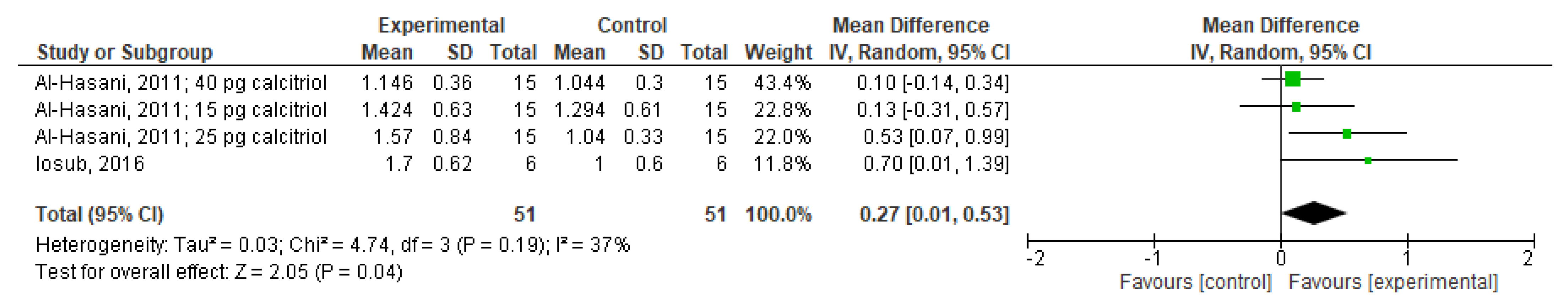
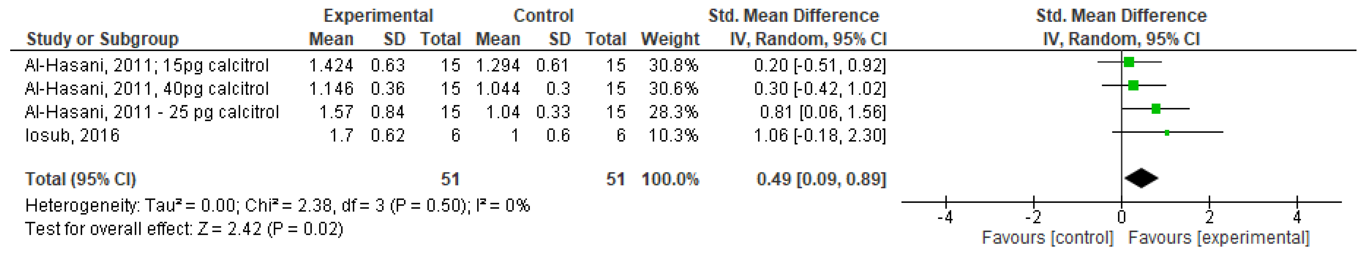
3.5. Quantitative Data Synthesis and Meta-Analysis Results

Meta-analysis enabled to quantify the results from two eligible human clinical studies and to obtain pooled data of mean difference, with 95% confidence intervals (CIs), concerning the clinical use of CTL during orthodontic treatment. Pooled combined data were calculated for various doses of CTL used. Data synthesis rendered from meta-analysis demonstrated mild discrepancies in tooth movement between control and experimental groups (mean difference = 0.27; 95% CI: 0.01–0.53, p = 0.04; std mean difference = 0.49; 95% CI: 0.09–0.89), p = 0.02) (Figure 3 and Figure 4) and moderate heterogenicity (I2 = 37%).

4. Discussion

Prolonged duration of orthodontic treatment is a crucial factor influencing outcome, quality of care, and patients’ satisfaction. The implementation of pharmacological modalities to stimulate and control OTM could potentially enhance the predictability of orthodontic therapy and be a valuable adjunctive tool, especially when used together with fixed orthodontic appliances. An ideal pharmacological agent that would promote and stimulate tooth position alterations is expected to be a valuable adjunctive tool in orthodontic treatment using especially fixed orthodontic appliances. The PROSPERO protocol revealed that the current review is the first that aimed to assess the impact of CTL on tooth movement caused by orthodontic forces. Although the assessment of quality, grading, and determination of the level of recommendation of the included studies were approached using validated and reliable tools, a possibility of subjective bias exists. Additionally, one important study was not included as this systematic review focused only on articles written in the English language, while that study was written in Spanish [19].
The literature search identified 13 animal studies, two in vitro studies, eight reviews, and two clinical trials and two cross-sectional studies, which all demonstrated a significant variability in the level of evidence, quality, and methodology. The GRADE-based evidence was low for all animal studies, however in single cases received high level of recommendation was received. A cumulative extrapolation of the data from the animal studies was complicated by differences in the research protocol, the route of CTL administration, the clinical orthodontic method used, and the period of observation.
All the studies used rats in their experiments, with the exception of one study, where cats were used [28]. In most of the studies, CTL was administered locally, except for two studies, in which a systemic route was used [23,25]. Moreover, three different techniques were used to mobilize teeth; some studies used coil springs [22,23,27], others elastomeric chains [18,48], and the third group of studies applied helical stainless steel loops [17,24,27]. The period of observation also varied as the rate of tooth movement was, in some studies, measured for two weeks [17,18,22] and, in others, for 21 days or 28 days [23]. The majority of animal studies on vitamin D3 injected 20 microliters (uL) of calcitriol locally in the site of tooth movement [24,27,28]; others used a low dose of vitamin D3 diet [25]. Differences between the study protocols used in animal studies can have a significant impact on study outcomes and conclusions, also making it difficult to compare and pool the results cumulatively. Although a number of animal studies showed CTL as positive regulator of bone metabolism and tooth movement, it is evident that these results cannot be directly applied to humans, due to obvious differences between species.
Four clinical studies were found that involved human participants and concentrated on investigating the effect of local CTL injections. Among these, two studies looked at the effect of CTL on external root resorption and the other two on tooth movement [43,44]. Both orthodontic tooth movement studies had weaknesses, including lack of randomization and undefined method of sample size calculation. In addition, canine distalization was only measured for a short period of time, either for three weeks [43] or for one month [44]; considering such a short duration, it would have been impossible to detect significant differences in the tooth movement discrepancy. This is in contrast to other studies concentrating on tooth movement that chose to measure canine traction for the minimum of three months [49,50,51].
Both human studies also lacked justification and rational evaluation for the dose of CTL injections. Al-Hasani and colleagues reported that a low dose of 25 picogram/mL produced a higher rate of tooth movement with no statistically significant difference between the study side and control sides [43]. On the other hand, Iosub Ciur and colleagues used a higher dose of 42 picogram/mL and reported 70% greater tooth movement when comparing to the contralateral control side [44]. Notably, in both studies the chosen route of calcitriol administration was by local injections that might not be compatible with daily oral medication. Both studies measured the distance between the distal surface of the canine to the mesial surface of the second premolar. Anchorage loss was not assessed, even though mesial drifting of the second premolar can occur when distalizing the canine by attaching a nickel titanium coil spring to the 1st molar rather than using absolute anchorage, such as a temporary anchorage device. Moreover, Iosub Ciur et al. utilized 3D CBCT for measuring canine movement, whereas Al-Hasani et al. used a direct-intraoral measure by digital vernier, which could be less reliable than computer-based measurements [43,44].
Although CTL was administered locally, it is possible that residue of CTL could also be transmitted from the experimental side to other intraoral control areas. Interestingly, Al-Hasani et al. found faster movement of canine even in the control side compared to the normal rate reported in other studies, that was justified by the possible effect of CTL also upon the non-experimental side, even when initially injected at the experimental side [43]. In addition, the medication bioavailability/bioabsorption was measured in neither of the studies, which seems an important factor affecting the tooth position changes but was not measured in either study.
Unlike most accelerating techniques used in orthodontics, which are based on the exacerbation of the inflammatory process by regional acceleration phenomena (RAP) [52], the active form of vitamin D (calcitriol) stimulates bone marrow to form osteoclast, so acts as an inducer of osteoclastic bone resorption [53,54,55]. However, the faster rate of tooth movement might lead to a higher rate of root resorption [56].

5. Strengths and Limitations

The current review followed a registered protocol in the International Prospective Register of Systematic Reviews (PROSPERO). The keywords search strategies used in this review were broad to all possible eligible studies based on the eligibility criteria. Two reviewers were independently involved in all stages of this review. Furthermore, this review included meta-analyses on some homogenous studies. The Oxford Level of Evidence and the GRADE approach were used to assess the included studies. Lastly, this review included human and animal studies in the data analysis. On the other hand, vitamin D3 has several generic names, which might affect the findings of this review, as some useful studies may be missed. The identification of further studies may suggest further research questions.

6. Conclusions

There is a substantial discrepancy in the level of evidence and recommendation in the effect of calcitriol on the rate of tooth movement during orthodontic treatment. Non-uniform study designs negatively impacted the obtained results in both animal and human studies. Although the preliminary evidence shows that calcitriol was found to accelerate orthodontic tooth movement, substantial difference exists in protocol and outcome methods, making it difficult to draw reliable conclusions. Despite the fact that animal studies have determined the biological effect of CTL in bone metabolism and tooth movement, insufficient sample sizes in clinical trials have resulted in lack of meaningful evidence of its impact on OTM in humans and subsequently highlight the importance for well-powered RCT studies that would aim to assess the effect of CTL on the supporting structures during orthodontic treatment with an adequate study group size and a longer follow-up period. Despite the small amount of evidence regarding the effectiveness of CTL in accelerating tooth movement, orthodontists should consider any implications of this supplement related to orthodontic treatment.

Author Contributions

Conceptualization, M.A. and A.A.-A.; methodology, M.A. and H.A.; validation, M.A., A.A.-A., M.M.A.-K. and A.D.; formal analysis, H.A. and A.D.; investigation, A.A.-A.; resources, H.A.; data curation, M.A., A.A.-A. and H.A.; writing—original draft preparation, M.A., A.A.-A., H.A. and M.M.A.-K.; writing—review and editing, A.D., M.S. and M.T.C.; visualization, M.A.; supervision, M.A. and H.A.; project administration, A.A.-A.; funding acquisition, A.D. All authors have read and agreed to the published version of the manuscript.

Funding

This research received funding was received from the Medical University of Silesia grant: KNW-1-150/N/9/I.

Conflicts of Interest

The authors declare no conflict of interest.

References

  1. Fisher, M.A.; Wenger, R.M.; Hans, M.G. Pretreatment characteristics associated with orthodontic treatment duration. Am. J. Orthod. Dentofac. Orthop. 2010, 137, 178–186. [Google Scholar] [CrossRef] [PubMed]
  2. Mavreas, D.; Athanasiou, A.E. Factors affecting the duration of orthodontic treatment: A systematic review. Eur. J. Orthod. 2008, 30, 386–395. [Google Scholar] [CrossRef] [Green Version]
  3. Geiger, A.M.; Gorelick, L.; Gwinnett, A.J.; Benson, B.J. Reducing white spot lesions in orthodontic populations with fluoride rinsing. Am. J. Orthod. Dentofac. Orthop. 1992, 101, 403–407. [Google Scholar] [CrossRef]
  4. Pandis, N.; Nasika, M.; Polychronopoulou, A.; Eliades, T. External apical root resorption in patients treated with conventional and self-ligating brackets. Am. J. Orthod. Dentofac. Orthop. 2008, 134, 646–651. [Google Scholar] [CrossRef]
  5. Long, H.; Pyakurel, U.; Wang, Y.; Liao, L.; Zhou, Y.; Lai, W. Interventions for accelerating orthodontic tooth movement: A systematic review. Angle Orthod. 2013, 83, 164–171. [Google Scholar] [CrossRef] [PubMed] [Green Version]
  6. El-Angbawi, A.; Mcintyre, G.T.; Fleming, P.S.; Bearn, D.R. Non-surgical adjunctive interventions for accelerating tooth movement in patients undergoing fixed orthodontic treatment. Cochrane Database Syst. Rev. 2015, 11, CD010887. [Google Scholar] [CrossRef] [PubMed]
  7. Sivarajan, S.; Doss, J.G.; Papageorgiou, S.N.; Cobourne, M.T.; Wey, M.C. Mini-implant supported canine retraction with micro-osteoperforation: A split-mouth randomized clinical trial. Angle Orthod. 2019, 89, 183–189. [Google Scholar] [CrossRef] [PubMed] [Green Version]
  8. Kalemaj, Z.; Debernardl, C.L.; Buti, J. Efficacy of surgical and non-surgical interventions on accelerating orthodontic tooth movement: A systematic review. Eur. J. Oral Implantol. 2015, 8, 9–24. [Google Scholar]
  9. Hoogeveen, E.J.; Jansma, J.; Ren, Y. Surgically facilitated orthodontic treatment: A systematic review. Am. J. Orthod. Dentofac. Orthop. 2014, 145, 51–64. [Google Scholar] [CrossRef]
  10. Nayak, K.; Galil, W.; Wiltshire, P. Molecular Biology of Orthodontic Tooth Movement. J. Dent. Oral Health 2013, 1, 1–6. [Google Scholar]
  11. Roodman, G.D. Role of cytokines in the regulation of bone resorption. Calcif. Tissue Int. 1993, 53, 94–98. [Google Scholar] [CrossRef] [PubMed]
  12. Younis, L.T.; Masood, M.; Ismail, F.; Bahnasi, E. The Role of Components and Molecules of Periodontal Ligament in Orthodontic Tooth Movement. Asian J. Dent. Res. 2019, 2, 1–12. [Google Scholar]
  13. Kouskoura, T.; Katsaros, C.; von Gunten, S. The potential use of pharmacological agents to modulate orthodontic tooth movement (OTM). Front. Physiol. 2017, 8, 1–9. [Google Scholar] [CrossRef] [PubMed] [Green Version]
  14. Kogawa, M.; Findlay, D.; Anderson, P.H.; Ormsby, R.; Vincent, C.; Morris, H.A.; Atkins, G.J. Osteoclastic Metabolism of 25(OH)-Vitamin D3: A Potential Mechanism for Optimization of Bone Resorption. Endocrinology 2010, 10, 4613–4625. [Google Scholar] [CrossRef] [Green Version]
  15. Reichel, H.; Koeffl, H.; Norman, A. The role of the vitamin D3 endocrine system in health and disease. N. Engl. J. Med. 1989, 320, 980–991. [Google Scholar] [CrossRef] [PubMed]
  16. Posa, F.; Di Benedetto, A.; Colaianni, G.; Cavalcanti-Adam, E.; Brunetti, G.; Porro, C.; Trotta, T.; Grano, M.; Mori, G. Vitamin D3 effects on osteoblastic differentiation of mesenchymal stem cells from dental tissues. Stem Cells Int. 2016, 2016, 1–9. [Google Scholar] [CrossRef] [PubMed]
  17. Kale, S.; Kocadereli, I.; Atilla, P.; Asan, E. Comparison of the effects of 1,25 dihydroxycholecalciferol and prostaglandin E2 on orthodontic tooth movement. Am. J. Orthod. Dentofac. Orthop. 2004, 125, 607–614. [Google Scholar] [CrossRef]
  18. Kawakami, M.; Takano-Yamamoto, T. Local injection of 1,25-dihydroxyvitamin D3 enhanced bone formation for tooth stabilization after experimental tooth movement in rats. J. Bone Miner. Metab. 2004, 22, 541–546. [Google Scholar] [CrossRef]
  19. Blanco, J.; Diaz, R.; Gross, H. Efecto de laadministración sistémica del1,25Dihidrxicolecalciferol sobrelavelocidad del movimiento ortodóncico en humanos. Estudio Clínico Revista Odontos 2001, 8, 13–24. [Google Scholar]
  20. Guyatt, G.; Oxman, A.; Vist, G.; Kunz, R.; Falck-Ytter, Y.; Alnoso-Coelle, P.; Schünemann, H.J. An emerging consensus on rating quality of evidence and strength of recommendations. Br. Med. J. 2008, 336, 924–926. [Google Scholar] [CrossRef] [Green Version]
  21. OCEBM Levels of Evidence Working Group. The Oxford 2011 Levels of Evidence. Oxford Centre for Evidence-Based Medicine. 2016. Available online: http://www.cebm.net/index.aspx?o=5653 (accessed on 23 June 2020).
  22. Ratri Nareswari, R.; Narmada, I.; Djaharu’ddin, I.; Putrani, N.; Nugrada, A. Effect of vitamin D3 administration on vascular endothelial growth factor expression and angiogenesis number in orthodontic tooth movement of pregnant Wistar rat. J. Postgrad. Med. Inst. 2019, 33, 182–188. [Google Scholar]
  23. Cui, J.; Li, J.; Wang, W.; Han, X.; Sun, J.; Feng, W.; Liu, B.; Liu, H.; Amizuka, N.; Li, M. The effect of calcitriol on high mobility group box 1 expression in periodontal ligament cells during orthodontic tooth movement in rats. J. Mol. Histol. 2016, 47, 221–228. [Google Scholar] [CrossRef]
  24. Kawakami, M. Effects of local application of 1,25 (OH)2D3 on experimental tooth movement in rats. J. Osaka 1990, 35, 128–146. [Google Scholar]
  25. Bielaczyc, A.; Golebiewska, M. Ultrastructural changes of a tooth root in young rats fed a low calcium and vitamin D3-deficient diet. Roczniki Akademii Medycznej w Bialymstoku 1990, 42, 153–158. [Google Scholar]
  26. Baran, S.; Hamamci, O.; Akalar, M. An investigation of the effects of the local use of 1:25 dihydroxycholecalciferol (1:25 D) on tension sites during experimental tooth movement in rats. J. Marmara Uni. Dent. Fac. 1996, 2, 557–561. [Google Scholar]
  27. Takano-Yamamoto, T.; Kawakami, M.; Kobayshiro, Y.; Sakuda, M. The Effect of Local Application of 1,25-Dihydroxycholecalciferol on Osteoclast Numbers in Orthodontically Treated Rats. J. Dent. Res. 1992, 71, 53–59. [Google Scholar] [CrossRef] [PubMed]
  28. Collins, M.K.; Sinclair, P.M. The local use of vitamin D3 to increase the rate of orthodontic tooth movement. Am. J. Orthod. Dentofac. Orthop. 1988, 94, 278–284. [Google Scholar] [CrossRef]
  29. Yamasaki, K. Effects of vitamin D3 on orthodontic tooth movement in rats. Kokubyo Gakkai Zasshi 1976, 43, 41–63. [Google Scholar] [CrossRef]
  30. Imazu, H. Histo-biochemical changes during tooth movement under controlled fed rat. Gifu Shika Gakkai Zasshi 1984, 11, 401–423. [Google Scholar]
  31. Kawal, H. Tooth movement in rats during a calcium and vitamin D3 deficient regimen. Gifu Shika Gakkai Zasshi 1984, 11, 380–400. [Google Scholar]
  32. Zhang, D.; Lorenzo, J. 1,25(OH)2 vitamin D33 induces IL-1 alpha mRNA expression from osteoblastic-like and bone marrow co-culture cells. Chin. J. Stomatol. 1998, 33, 375–377. [Google Scholar]
  33. Basdra, E.K.; Komposch, G. Osteoblast-like properties of human periodontal ligament cells: An in vitro analysis. Eur. J. Orthod. 1997, 19, 615–621. [Google Scholar] [CrossRef]
  34. Tehranchi, A.; Sadighnia, A.; Younessian, F.; Abdi, A.; Shirvani, A. Correlation of Vitamin D3 status and orthodontic-induced external apical root resorption. Dent. Res. J. 2017, 14, 403–411. [Google Scholar]
  35. Fontana, M.L.S.S.N.; De Souza, C.M.H.; Bernardino, J.F.; Hoette, F.; Hoette, M.L.; Thum, L.; Terumi, O.O.; Capelozza Filho, L.; Olandoski, M.; Trevilatto, P.C. Association analysis of clinical aspects and vitamin D3 receptor gene polymorphism with external apical root resorption in orthodontic patients. Am. J. Orthod. Dentofac. Orthop. 2012, 142, 339–347. [Google Scholar] [CrossRef] [PubMed]
  36. Kaklamanos, E.G.; Makrygiannakis, M.A.; Athanasiou, A.E. Does medication administration affect the rate of orthodontic tooth movement and root resorption development in humans? A systematic review. Eur. J. Orthod. 2019, 42, 407–414. [Google Scholar] [CrossRef] [PubMed]
  37. Khalaf, K.; Mando, M. Effect of Drugs on Orthodontic Tooth Movement in Human Beings: A Systematic Review of Randomized Clinical Trials. Open Dent. J. 2019, 13, 22–32. [Google Scholar] [CrossRef] [Green Version]
  38. Kacprzak, A.; Strzecki, A. Methods of accelerating orthodontic tooth movement: A review of contemporary literature. Dent. Med. Probl. 2018, 55, 197–206. [Google Scholar] [CrossRef] [Green Version]
  39. Batmaraj, U. The effects of commonly used drugs on orthodontic tooth movement: A systematic review. Asian J. Pharm. Clin. Res. 2014, 7, 10–14. [Google Scholar]
  40. Camacho, A.D. Dental movement acceleration: Literature review by an alternative scientific evidence method. World J. Methodol. 2014, 4, 151–162. [Google Scholar] [CrossRef] [PubMed]
  41. Nimeri, G.; Kau, C.; Abou-Kheir, N.; Corona, R. Acceleration of tooth movement during orthodontic treatment-A frontier in orthodontics. Prog. Orthod. 2013, 14, 42–50. [Google Scholar] [CrossRef] [Green Version]
  42. Tyrovola, J.; Spyropoulos, M. Effects of drugs and systemic factors on orthodontic treatment. Quintessence Int. 2001, 32, 365–371. [Google Scholar] [PubMed]
  43. Al-Hasani, N.R.; Al-Bustani, A.I.; Ghareeb, M.M.; Hussain, S.A. Clinical efficacy of locally injected calcitriol in orthodontic tooth movement. Int. J. Pharm. Pharm. Sci. 2011, 3, 139–143. [Google Scholar]
  44. Iosub Ciur, M.D.; Zetu, I.N.; Haba, D.; Viennot, S.; Bourgeois, D.; Andrian, S. Evaluation of the Influence of Local Administration of Vitamin D3 on the Rate of Orthodontic Tooth Movement. Rev. Med. Chir. Soc. Med. Nat. Iasi. 2016, 120, 694–699. [Google Scholar] [PubMed]
  45. Bartzela, T.; Türp, J.C.; Motschall, E.; Maltha, J.C. Medication effects on the rate of orthodontic tooth movement: A systematic literature review. Am. J. Orthod. Dentofac. Orthop. 2009, 135, 16–26. [Google Scholar] [CrossRef]
  46. Makrygiannakis, M.A.; Kaklamanos, E.G.; Athanasiou, A.E. Does common prescription medication affect the rate of orthodontic tooth movement? A systematic review. Eur. J. Orthod. 2018, 40, 649–659. [Google Scholar] [CrossRef] [PubMed]
  47. Leethanakul, C.; Kanokkulchai, S.; Pongpanich, S.; Leepong, N.; Charoemratrote, C. Interseptal bone reduction on the rate of maxillary canine retraction. Angle Orthod. 2014, 84, 839–845. [Google Scholar] [CrossRef] [PubMed] [Green Version]
  48. Takano-Yamamoto, T.; Kawakami, M.; Yamashiro, T. Effect of Age on the Rate of Tooth Movement in Combination with Local Use of 1,25(OH) 2D3 and Mechanical Force in the Rat. J. Dent. Res. 1992, 71, 1487–1492. [Google Scholar] [CrossRef]
  49. Aboul-Ela, S.M.B.E.D.; El-Beialy, A.R.; El-Sayed, K.M.F.; Selim, E.M.N.; El-Mangoury, N.H.; Mostafa, Y.A. Miniscrew implant-supported maxillary canine retraction with and without corticotomy-facilitated orthodontics. Am. J. Orthod. Dentofac. Orthop. 2011, 139, 252–259. [Google Scholar] [CrossRef]
  50. Da Silva Sousa, M.V.; Scanavini, M.A.; Sannomiya, E.K.; Velasco, L.G.; Angelieri, F. Influence of low-level laser on the speed of orthodontic movement. Photomed. Laser Surg. 2011, 29, 191–196. [Google Scholar] [CrossRef]
  51. Eltimamy, A.; El-Sharaby, F.A.; Eid, F.H.; El-Dakrory, A.E. The effect of local pharmacological agents in acceleration of orthodontic tooth movement: A systematic review. Open Access Maced. J. Med. Sci. 2019, 7, 882–886. [Google Scholar] [CrossRef] [Green Version]
  52. Frost, H.M. The regional acceleratory phenomenon. Orthop. Clin. N. Am. 1981, 12, 725–726. [Google Scholar]
  53. Roodman, G.D.; Ibbotson, K.J.; MacDonald, B.R.; Kuehl, T.J.; Mundy, G.R. 1, 25-Dihydroxyvitamin D3 causes formation of multinucleated cells with several osteoclast characteristics in cultures of primate marrow. Proc. Natl. Acad. Sci. USA 1985, 82, 8213–8217. [Google Scholar] [CrossRef] [PubMed] [Green Version]
  54. Fuller, K.; Chambers, T.J. Generation of osteoclasts in cultures of rabbit bone marrow and spleen cells. J. Cell. Physiol. 1987, 132, 441–452. [Google Scholar] [CrossRef] [PubMed]
  55. Takahashi, N.; Yamana, H.; Yoshiki, S.; Roodman, G.D.; Mundy, G.R.; Jnoes, S.J.; Boyde, A.; Suda, T. Osteoclast-like cell formation and its regulation by osteotropic hormones in mouse bone marrow cultures. Endocrinology 1988, 122, 1373–1382. [Google Scholar] [CrossRef] [PubMed]
  56. Ganesh, M.L.; Saravana Pandian, K. Acceleration of tooth movement during orthodontic treatment-A frontier in orthodontics. J. Pharm. Sci. Res. 2017, 9, 741–744. [Google Scholar]
Figure 1. The potential acceleration of orthodontic tooth movement. The mechanism of CTL biological effect.
Applsci 11 08882 g001
Figure 2. Flow diagram of the selection process.
Applsci 11 08882 g002
Figure 3. Results of meta-analysis (mean difference) and forest plot.
Applsci 11 08882 g003
Figure 4. Results of meta-analysis (std mean difference) and forest plot.
Applsci 11 08882 g004
Table 1. Main characteristics of the animal studies included in systematic review.
Author (Year)Study Design and MethodSample SizeAimsStudy DurationInterventionOrthodontic InterventionFindingsLevel of Evidence †Level of Recommendation ††
Ratri Nareswari et al. (2019) [22]In vivo experimental study, histological and immuohistochemistry.28 female Wistar ratsTo investigate the effect of vitamin D3 on vascular endothelial growth factor expression during orthodontic tooth movement.14 daysVitamin D3 (0.2 mg/kg) was given every 3 days.Coil spring (Ni-Ti) was inserted between maxillary central incisor and
1st left maxillary molar.
Vitamin D3 administration did not significantly increase VEGF expression and angiogenesis number orthodontic tooth movement.5⊕⊕⊕
Cui et al. (2016) [23]In vivo experimental study and immunohistochemistry.30 male Wistar ratsTo investigate the effect of 1,25(OH)2D3 on HMGB1 expression by periodontal ligament cells in an orthodontic rat model.28 daysAdministered with 1,25(OH)2D3 (100 ng/kg body weight) was adminstarted once every other day.NiTi coil spring on the maxillary first molarIndicates that administration of 1,25(OH)2D3 might create a favorable environment for orthodontic tooth movement.5⊕⊕⊕⊕
Kawakami and Takano-Yamamoto (1990) [24]In vivo experimental study and histomorphometric analysis16 rats
male Wistar rats
To investigate the effect local injection of vitamin D3 during tooth movement.14 days20 µL of 1,25(OH)2D3A piece of orthodontic elasticSignificant increase in mineral appositional rate associated with increased osteoblast surface value.5⊕⊕⊕
Kale et al. (2004) [17]In vivo experimental study and histological examination.37 Wistar ratsTo investigate the effect of local administrations of prostaglandin E2 and 1,25-dihydroxycholecalciferol (1,25-DHCC) on orthodontic tooth movement in rats.9 daysProstaglandin E2 (PGE2) and 1,25-dihydroxycholecalciferol (1,25-DHCC)A helical loop stainless steel springBoth Prostaglandin E2 and 1,25-DHCC enhanced the amount of tooth movement significantly.5⊕⊕
Bielaczyc and Golebiewska (1997) [25]In vivo experimental study with scanning microscopy observation.20 young Wistar ratsTo investigate the structural changes on the roots in rats fed by deficient diet with low calcium and vitamin D3.InaccessibleFeeding a low calcium and vitamin D3.InaccessibleIncreased cementolysis and decreased mineralization of cementum and dentin.5⊕⊕
Baran et al. (1996) [26]In vivo experimental study and histological examination.32 Wistar albino ratsTo assess the effects of 1:25 dihydroxychole-calciferol, one of the most potent osteoclastic activity stimulants.InaccessibleAdministration of 1:25 dihydroxychole-calciferol.InaccessibleHigher area of unilateral apposition was observed in the experimental group.5⊕⊕
Takano-Yamamoto et al. (1992)In vivo experimental study and histological examination on rats.Experiment I: 112 rats divided into two groups. Experiment II: 80 rats.To investigate the effect of local injection of 1,25-(OH)2D3
on osteoclast numbers during tooth movement.
In experiment 1: 3 days.
In experiment 2:
15 days.
20 µL of 1,25(OH)2D3 was injected locally with phosphate-buffered saline (PBS) on the control side.Elastic band was placed between the first and the second upper molars.Increased osteoclast number and bone resorption.5⊕⊕⊕⊕
Takano-Yamamoto et al. (1992) [27]In vivo experimental study and blood chemistry30 seven week-old and 30
28 week-old male Wistar rats.
To investigate the effect of local injection of vitamin D3 in young and mature rats during tooth movement.21 days20 µL of 1,25(OH)2D3
was injected locally with phosphate-buffered saline (PBS) on the control side.
A coil spring was used with a force ranging between 5 g and 30 g.No change was observed in serum calcium, phosphate, and alkaline phosphatase activity.5⊕⊕⊕
Kawakami (1990) [24]In vivo experimental study, fluorescent labeling and quantitative histology on rats.Wistar male rats
(the number was not accessible)
To investigate the effect of locally injected vitamin D3, on the rate of tooth movement in rats.20 days20 µL was locally injected.A piece of orthodontic helical spring was used to move the first molar.The rate of tooth movement was increased.5⊕⊕⊕
Collins and Sinclair (1988) [28]In vivo experimental study with histological examination and blood chemistry.10 cats,
5 cats pilot study
To investigate the effect of local administration of vitamin D3 on the rate of tooth movement.21 daysInjection of vitamin D3 metabolite into PDL.Canine retraction with light wire retraction spring.The rate of tooth movement and osteoclasts number was increased.5⊕⊕⊕
Three studies were not accessible [29,30,31], † Level of evidence according to the Oxford Level of Evidence. †† Grading: high ((⊕⊕⊕⊕)—we were very confident that the effect of the study reflects the actual effect; moderate (⊕⊕⊕)—we are quite confident that the effect in the study is close to the true effect, but it is also possible it is substantially different; low (⊕⊕)—the true effect may differ significantly from the estimate; and lastly, very low (⊕)—the true effect is likely to be substantially different from the estimated effect.
Table 2. The summary of two in vitro studies.
Authors
Years
Type of StudyActive SubstanceModelFindingsLevel of Evidence †Level of Recommendation ††
Zhang and Lorenzo (1998) [32]Histochemical and biochemical
study
Vitamin 1,25(OH) 2D3
and Interleukin-1 (IL-1)
IL-1 alpha mRNA expression in bone marrow and osteoblastic cell co-cultures by semi-quantitative RT-PCR.IL-1 involved in osteoclast formation induced by 1,25(OH)2D3 and IL-1 stimulate osteoclast progenitor proliferation.5⊕⊕⊕⊕
Basdra and Komposch (1997) [33]Histochemical and biochemical
study
1α,25-dihydroxyvitamin D3PDL fibroblasts isolated from human periodontium (the roots of healthy extracted third molar) were cultured.Human PDL fibroblast exhibit phenotypic characteristics similar to that of osteoblast-like cells and have the potential to differentiate into osteoblasts and/or cementoblast.5⊕⊕⊕⊕
† Level of evidence according to the Oxford Level of Evidence. †† Grading: high ((⊕⊕⊕⊕)—we are very confident that the effect of the study reflects the actual effect; moderate (⊕⊕⊕)—we are quite confident that the effect in the study is close to the true effect, but it is also possible it is substantially different; low (⊕⊕)—the true effect may differ significantly from the estimate; and lastly, very low (⊕)—the true effect is likely to be substantially different from the estimated effect.
Table 3. The summary of studies that assessed relation between calcitriol level and external apical root resorption (n = 2).
Authors
Years
Type of StudyActive SubstanceNumber
of Participants
GenderAge of Participants (Range)Group CharacteristicsResultsLevel of Evidence †Level of Recommendation ††
Group 1Group 2Group 3
Tehranchi et al. (2017) [34]Cross sectional studyVitamin D33423.5% males and
76.5% females
12–23 yearsClass I
13 (38.2%)
Class II
18 (53%)
Class III
3 (8%)
Vitamin D3 level is not among the clinical variables that are potential contributors for EARR.3⊕⊕⊕
Fontana et al. (2012) [35]Cross sectional studyVitamin D337744.8% males and 55.2% females8–21 years160 (42%) with EARR ≤1.43 mm179(47%) with EARR>1.43 mm38(11%) untreated subjectsVitamin D3 receptor polymorphism was associated with EARR in orthodontic patients2⊕⊕⊕⊕
EARR: External apical root resorption. † Level of evidence according to the Oxford Level of Evidence. †† Grading: high ((⊕⊕⊕⊕)—we are very confident that the effect of the study reflects the actual effect; moderate (⊕⊕⊕)—we are quite confident that the effect in the study is close to the true effect, but it is also possible it is substantially different; low (⊕⊕)—the true effect may differ significantly from the estimate; and lastly very low (⊕)—the true effect is likely to be substantially different from the estimated effect).
Table 4. The summary of review concerning papers on the relation between calcitriol and orthodontic treatment.
Authors/YearsFindings
Kaklamanos et al. (2019) [36]The rate of orthodontic tooth movement may be affected by consuming specific analgesics for a few days
Khalaf and Mando (2019) [37]The rate of tooth movement was reduced by acetylsalicylic acid and ibuprofen whereas no impact on orthodontic tooth movement was observed when paracetamol, Rofecoxib, and tenoxicam were consumed.
Kacprzak and Strzecki (2018) [38]In adults, corticotomy and its modifications might prove to be a useful in decreasing the duration of orthodontic treatment
Kouskoura et al. (2017) [13]Many drugs can and will influence both bone metabolism and can influence cellular functions
Batmaraj (2014) [39]Beneficial and harmful effects and clinical efficacy of new drugs must be tested and investigated.
Camacho (2014) [40]Level of evidence was as follows: surgery first followed by low level laser beam application, corticotomy, and periodontal distraction
Nimeri et al. (2013) [41]Piezocision technique is an effective approach to accelerate tooth movement
Tyrovola and Spyropoulos (2001) [42]Hormones and drugs such as estrogen, androgen, bisphosphonates, vitamin D3, and calcitonin could delay orthodontic tooth movement, whereas thyroid hormones and corticosteroids might enhance orthodontic tooth movement.
Table 5. The characteristics of clinical trials included in the systematic review.
Authors/YearStudy DesignCountryAim (s)Sample SizeGenderAge (Range)Level of Evidence †Level of Recommendation ††
Al-Hasani et al. (2011) [43]RCT (for dosage)
Split mouth
IraqTo investigate the effect of locally injected vitamin D3 (calcitriol) on the rate of orthodontic tooth movement.15Not
reported
17–28 years1⊕⊕⊕⊕
Iosub Ciur et al. (2016) [44]RCT (for dosage)
Split mouth
FranceTo investigate the effect of local administration of vitamin D3 on the dental roots and the rate of orthodontic tooth movement.4M:2(50%)
F:2(50%)
13–34 years1⊕⊕⊕⊕
† Level of evidence according to the Oxford Level of Evidence. †† Grading: high ((⊕⊕⊕⊕)—we are very confident that the effect of the study reflects the actual effect; moderate (⊕⊕⊕):—we are quite confident that the effect in the study is close to the true effect, but it is also possible it is substantially different; low (⊕⊕)—the true effect may differ significantly from the estimate; and lastly, very low (⊕)—the true effect is likely to be substantially different from the estimated effect).
Publisher’s Note: MDPI stays neutral with regard to jurisdictional claims in published maps and institutional affiliations.

