Insights in the Role of Lipids, Oxidative Stress and Inflammation in Rheumatoid Arthritis Unveiled by New Trends in Lipidomic Investigations

 ,
, 
Abstract
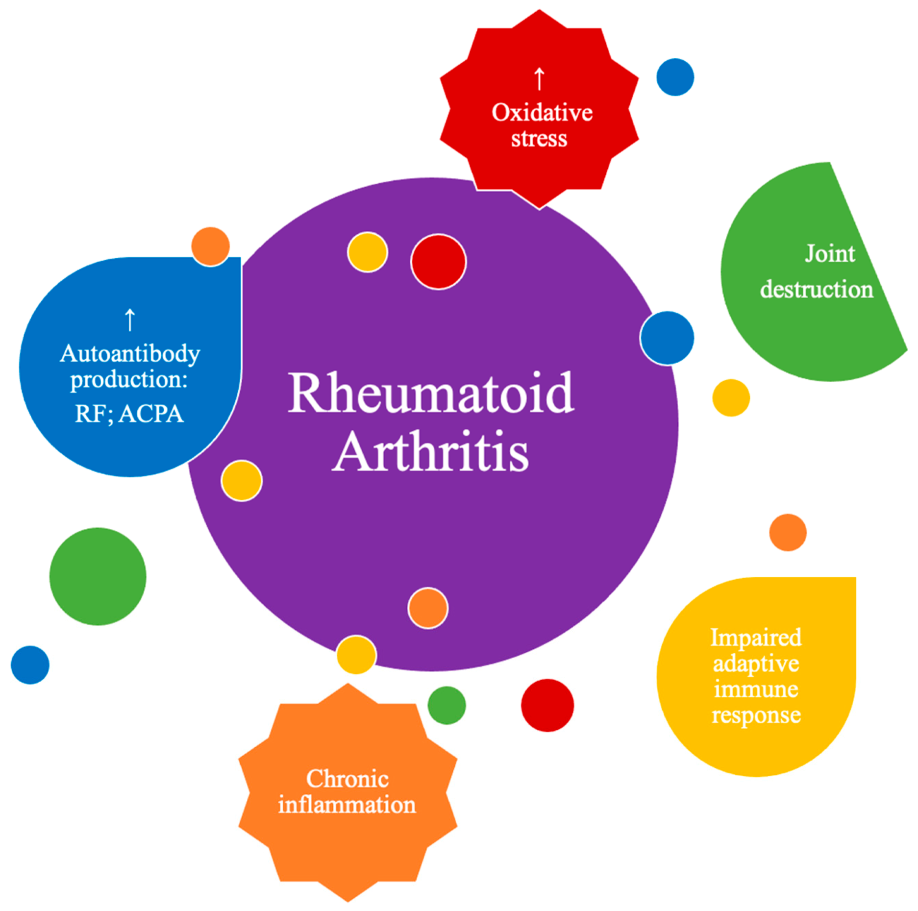
:1. Rheumatoid Arthritis
2. Oxidative Stress in Rheumatoid Arthritis
3. Lipids Are Key Players in Rheumatoid Arthritis
Dyslipidemia as an Important Contributor to RA Pathogenesis
4. Lipidomic Studies in Rheumatoid Arthritis
4.1. Lipidomic Analysis of Serum/Plasma in Rheumatoid Arthritis Patients
4.1.1. Fatty Acid Profiling Analysis
4.1.2. FA Analysis after a Fasting Period
4.1.3. FA Analysis after Supplementation Intake
4.1.4. Phospholipidomic Profiling Analysis by LC-MS
4.1.5. Identification of Lipid Peroxidation Products by LC-MS

| Reference | Analytical Method | Lipid Extraction Method | N° and Age (Years) of RA Patients | Extra Details of the Study | Molecular Specie | Results | |
|---|---|---|---|---|---|---|---|
| ↓ Decrease | ↑ Increase | ||||||
| FA profiling analysis | |||||||
| Bruderlein et al. [98] | GC-MS | Gloster and Fletcher method | 20 RA Age: 25–66 | NSAID treatment. | FA esterified to PL | 16:0; 18:2 ω-6; 18:3 ω-3 | 18:1 ω-9; 20:3 ω-6; 20:3 ω-9; (20:3 + 20:4) ω-6/18:2 ω-6 ratio |
| Jacobsson et al. [97] | GC-MS | INF | Two groups: 21 RA Age: 25–78 21 RA Age: 3–43 | NSAID treatment. | FA esterified to PC | 16:1; 18:0; 18:2 ω-6; 18:3 ω-3; 20:5 ω-3; 22:5 ω-3; 18:2/20:4 ratio; Σ ω-6 | 14:0; 16:0; 18:1; Σ saturated FA |
| Suryaprabha et al. [99] | GC-MS | Methanol:Chloroform (1:2, v/v) | 14 RA Mean age: 33 ± 9 | Not on drugs when sample collection. | FA esterified to PL | 18:0; 18:3 ω-6; 18:3 ω-3; 20:3 ω-6; 20:5 ω-3; 22:6 ω-3 | - |
| Rodríguez-Carrio et al. [93] | LC-MS/MS | MTBE | 124 RA Mean age: 52.47 ± 12.76 | 43 patients with smoking habits. | Plasma FFA | 16:0; 16:1 ω-7; 18:1 ω-9; 20:4 ω-6; 20:5 ω-3; 22:6 ω-3 | - |
| Analysis after a fasting period | |||||||
| Hafström et al. [100] | GC-MS | Methanol:Chloroform (2:1, v/v) | 14 RA Age: 34–65 | No steroids/NSAID treatment for the previous 3 months. 7 days of fasting. | FA esterified to PL | 20:3 ω-6 | 20:4 ω-6; 20:5 ω-3 |
| Haugen et al. [101] | GC-MS | n-butanol (butyl alcohol) | 53 RA Mean age: 51 | Fasting period of 7–10 days followed by 1-year vegetarian diet. | FA esterified to PL | After vegan diet 14:0; 16:0; 16:1; 18:0; 18:1; 18:3 ω-3; 20:1; 20:3 ω-9; 20:3 ω-6; 20:4 ω-6; 20:5 ω-3; 22:0; 22:1 ω-11; 22:4 ω-6; 22:5 ω-6; 22:5 ω-3; 22:6 ω-3; Σ ω-6; Σ ω-3; Σ saturated FA | - |
| Fraser et al. [94] | GC-MS | n-hexane | 9 RA Age: 31–65 | Steroids/NSAID treatment. 7 days of fasting. | Plasma FFA | - | 14:0; 16:0; 16:1 ω-7; 18:0; 18:1 ω-9; 18:2 ω-6; 18:3 ω-3; Σ FFA |
| Analysis after supplementation intake | |||||||
| Kremer et al. [95] | GC-MS | INF | 37 RA Age: INF | NSAID treatment. EPA supplementation. | Plasma FFA | - | With supplementation 20:5 |
| Remans et al. [102] | GC-MS | Bligh & Dyer method | 26 RA Mean age: 59.5 ± 11.0 | Steroids treatment. Nutritional supplement with PUFA and micronutrients. | FA esterified to PL | With supplementation 20:4 | With supplementation Σ ω-3 PUFA (20:5; 22:5; 22:6) |
| Jäntti et al. [96] | GC-MS | Methanol:Chloroform (1:1, v/v) | Two groups: 10 RA Mean age: 50 10 RA Mean age: 38 | No NSAID treatment for the previous 7 days. Evening primrose oil/olive oil supplementation. | Plasma FFA | With supplementation 18:1; 20:5 | With supplementation 18:2; 18:3; 20:3; 20:4 |
| Phospholipidomic profiling analysis by LC-MS | |||||||
| Łuczaj et al. [113] | LC-MS | Modified Folch method | 9 RA Age: 23–79 | No steroids/NSAID treatment. Excluded heavy smokers. | Plasma PL | PC(40:2); PC(40:3); PC(42:3) | LPC(16:1); LPC(24:3); PC(34:3); PC(36:3); PC(38:2); PC(38:3); PC(38:4); LPE(16:0); LPE(18:0); PE(30:1); PI(36:1); PI(36:2); PI(36:3); PI(36:4); PI(38:3); PI(38:4); SM(d34:2); SM(d38:1); SM(d40:1); SM(d40:2) |
| Identification of lipid peroxidation products by LC-MS | |||||||
| Charles-Schoeman et al. [118] | LC-MS/MS | Methanol/water | 10 RA Mean age: 49.6 ± 11.8 | 10% of the patients with smoking habits. | Plasma HDL + LDL | - | 5-HETE; 15-HETE; 9-HODE; 13-HODE |
4.2. Lipidomic Analysis of Other Types of Samples of Rheumatoid Arthritis Patients
| Reference | Analytical Method | Lipid Extraction Method | N° and Age (Years) of RA Patients | Extra Details of the Study | Molecular Specie | Results | |
|---|---|---|---|---|---|---|---|
| ↓ Decrease | ↑ Increase | ||||||
| Kim et al. [122] | GC-MS | Folch method | 10 RA Age: INF | - | FFA | - | - |
| Hafström et al. [100] | GC-MS | Methanol:Chloroform (2:1, v/v) | 14 RA Age: 34–65 | No steroids/NSAID treatment for the previous 3 months. 7 days of fasting. | Membrane FA | Neutrophils: NSD Platelets: 20:3 ω-6 | Neutrophils: NSD Platelets: 20:4 ω-6; 20:5 ω-3 |
| Masoom-Yasinzai [126] | GC-MS | Chloroform:Methanol (2:1, v/v) | 15 RA Age: INF | No steroids/hypolipidemic treatment. | Membrane FA | 20:5 ω-3 | - |
| Lee et al. [127] | GC-MS | INF | 100 RA Mean age: 48.39 ± 9.69 | Steroids/NSAID treatment. 13% of the patients with smoking habits. | Membrane FA | 18:1 ω-9; 18:2 ω-6; 18:3 ω-3; 20:5 ω-3; ω-3 index; 20:5/20:4 ratio | 14:0; 16:0; 16:1 ω-7; 18:0; 18:3 ω-6 |
| Park et al. [128] | GC-MS | INF | Two groups: 41 RA Mean age: 49.24 ± 10.46 40 RA Mean age: 47.63 ± 8.78 | Steroids/NSAID treatment. 15 patients with smoking habits. ω-3 PUFA supplementation. | Membrane FA | With supplementation 18:1 ω-9; 18:2 ω-6; 20: 4 ω-6; Σ ω-6 PUFA; ω-6/ω-3 ratio | With supplementation 20:5 ω-3; Σ ω-3 PUFA |
| Fraser et al. [94] | GC-MS | Benzene | 9 RA Age: 31–65 | Steroids/NSAID treatment. 7 days of fasting. | Membrane FA | NSD | NSD |
| Jacobsson et al. [97] | GC-MS | 2% Sulfuric acid in Methanol:Toluene (1:1, v/v) | Two groups: 21 RA Age: 25–78 21 RA Age: 3–43 | NSAID treatment. | FA | 18:2 ω-6; 18:3 ω-3; Σ ω-6 | - |
4.3. Lipidomic Analysis of Erythrocytes from Pre-Clinical Rheumatoid Arthritis Subjects
| Reference | Analytical Method | Lipid Extraction Method | N° and Age (Years) of RA Patients | Extra Details of the Study | Results | |
|---|---|---|---|---|---|---|
| ↓ Decrease | ↑ Increase | |||||
| Gan et al. [135] | GC-MS | INF | 30 pre-RA Mean age: 45.6 ± 16.5 | 2 patients with smoking habits. | Anti-CCP2 + 22:6; Σ ω-3 FA%; 20:5 + 22:6 | - |
| Gan et al. [136] | GC-MS | INF | Two groups: 40 pre-RA Mean age: 43.7 ± 15.4 27 pre-RA Mean age: 48.1 ± 13.2 | 5 patients with smoking habits. | RF + and SE + 22:5 ω-3; 22:6 ω-3; Σ ω-3 FA%; 20:5 + 22:6 Anti-CCP2 + and SE + 20:5 ω-3; 22:6 ω-3; Σ ω-3 FA%; 20:5 + 22:6 | - |
| Pablo et al. [137] | GC-MS | Chloroform:Methanol (2:1, v/v) | 96 pre-RA Mean age: 51 ± 7.56 | 29 patients with smoking habits. | - | 20:3 ω-6; 22:5 ω-3 |
5. Concluding Remarks and Future Perspectives
Funding
Institutional Review Board Statement
Informed Consent Statement
Data Availability Statement
Conflicts of Interest
References
- Alamanos, Y.; Voulgari, P.V.; Drosos, A.A. Incidence and prevalence of rheumatoid arthritis, based on the 1987 American College of Rheumatology criteria: A systemic review. Sem. Arthritis Rheum. 2006, 36, 182–188. [Google Scholar] [CrossRef] [PubMed]
- Sandoughi, M.; Kaykhaei, M.A.; Shahrakipoor, M.; Darvishzadeh, R.; Nikbakht, M.; Shahbakhsh, S.; Zakeri, Z. Clinical manifestations and disease activity score of rheumatoid arthritis in southeast of Iran. Rheumatol. Res. 2017, 2, 61–64. [Google Scholar] [CrossRef] [Green Version]
- Jung, N.; Bueb, J.L.; Tolle, F.; Bréchard, S. Regulation of neutrophil pro-inflammatory functions sheds new light on the pathogenesis of rheumatoid arthritis. Biochem. Pharmacol. 2019, 165, 170–180. [Google Scholar] [CrossRef] [PubMed]
- Aletaha, D.; Blüml, S. Therapeutic implications of autoantibodies in rheumatoid arthritis. RMD Open 2016, 2, e000009. [Google Scholar] [CrossRef] [Green Version]
- Aggarwal, R.; Liao, K.; Nair, R.; Ringold, S.; Costenbader, K.H. Anti-citrullinated peptide antibody assays and their role in the diagnosis of rheumatoid arthritis. Arthritis Rheum. 2009, 61, 1472–1483. [Google Scholar] [CrossRef] [Green Version]
- Aletaha, D.; Neogi, T.; Silman, A.J.; Funovits, J.; Felson, D.T.; Bingham, C.O.; Birnbaum, N.S.; Burmester, G.R.; Bykerk, V.P.; Cohen, M.D.; et al. 2010 Rheumatoid arthritis classification criteria: An American College of Rheumatology/European League Against Rheumatism collaborative initiative. Arthritis Rheum. 2010, 62, 2569–2581. [Google Scholar] [CrossRef]
- Bartok, B.; Firestein, G.S. Fibroblast-like synoviocytes: Key effector cells in rheumatoid arthritis. Immunol. Rev. 2010, 233, 233–255. [Google Scholar] [CrossRef]
- Marston, B.; Palanichamy, A.; Anolik, J.H. B cells in the pathogenesis and treatment of rheumatoid arthritis. Curr. Opin. Rheumatol. 2010, 22, 307–315. [Google Scholar] [CrossRef]
- Su, Z.; Yang, R.; Zhang, W.; Xu, L.; Zhong, Y.; Yin, Y.; Cen, J.; DeWitt, J.P.; Wei, Q. The synergistic interaction between the calcineurin B subunit and IFN-γ enhances macrophage antitumor activity. Cell Death Dis. 2015, 6, e1740. [Google Scholar] [CrossRef]
- Tolboom, T.C.; Pieterman, E.; Van Der Laan, W.H.; Toes, R.E.; Huidekoper, A.L.; Nelissen, R.G.; Breedveld, F.C.; Huizinga, T.W. Invasive properties of fibroblast-like synoviocytes: Correlation with growth characteristics and expression of MMP-1, MMP-3, and MMP-10. Ann. Rheum. Dis. 2002, 61, 975–980. [Google Scholar] [CrossRef] [Green Version]
- Sabeh, F.; Fox, D.; Weiss, S.J. Membrane-type I matrix metalloproteinase-dependent regulation of rheumatoid arthritis synoviocyte function. J. Immunol. 2010, 184, 6396–6406. [Google Scholar] [CrossRef] [PubMed]
- Smiljanovic, B.; Radzikowska, A.; Kuca-Warnawin, E.; Kurowska, W.; Grün, J.R.; Stuhlmüller, B.; Bonin, M.; Schulte-Wrede, U.; Sörensen, T.; Kyogoku, C.; et al. Monocyte alterations in rheumatoid arthritis are dominated by preterm release from bone marrow and prominent triggering in the joint. Ann. Rheum. Dis. 2018, 77, 300–308. [Google Scholar] [CrossRef] [PubMed]
- Cascão, R.; Moura, R.A.; Perpétuo, I.; Canhão, H.; Vieira-Sousa, E.; Mourão, A.F.; Rodrigues, A.M.; Polido-Pereira, J.; Queiroz, M.V.; Rosário, H.S.; et al. Identification of a cytokine network sustaining neutrophil and Th17 activation in untreated early rheumatoid arthritis. Arthritis Res. Ther. 2010, 12, R196. [Google Scholar] [CrossRef] [PubMed] [Green Version]
- Wright, H.L.; Moots, R.J.; Bucknall, R.C.; Edwards, S.W. Neutrophil function in inflammation and inflammatory diseases. Rheumatology 2010, 49, 1618–1631. [Google Scholar] [CrossRef] [Green Version]
- Larbre, J.P.; Moore, A.R.; Da Silva, J.A.; Iwamura, H.; Ioannou, Y.; Willoughby, D.A. Direct degradation of articular cartilage by rheumatoid synovial fluid: Contribution of proteolytic enzymes. J. Rheumatol. 1994, 21, 1796–1801. [Google Scholar] [PubMed]
- Khandpur, R.; Carmona-Rivera, C.; Vivekanandan-Giri, A.; Gizinski, A.; Yalavarthi, S.; Knight, J.S.; Friday, S.; Li, S.; Patel, R.M.; Subramanian, V.; et al. NETs are a source of citrullinated autoantigens and stimulate inflammatory responses in rheumatoid arthritis. Sci. Transl. Med. 2013, 5, 178ra40. [Google Scholar] [CrossRef] [Green Version]
- Bala, A.; Mondal, C.; Haldar, P.K.; Khandelwal, B. Oxidative stress in inflammatory cells of patient with rheumatoid arthritis: Clinical efficacy of dietary antioxidants. Inflammopharmacology 2017, 25, 595–607. [Google Scholar] [CrossRef]
- Karami, J.; Aslani, S.; Jamshidi, A.; Garshasbi, M.; Mahmoudi, M. Genetic implications in the pathogenesis of rheumatoid arthritis; an updated review. Gene 2019, 702, 8–16. [Google Scholar] [CrossRef]
- Takeno, M.; Kitagawa, S.; Yamanaka, J.; Teramoto, M.; Tomita, H.; Shirai, N.; Itoh, S.; Hida, S.; Hayakawa, K.; Onozaki, K.; et al. 5-Hydroxy-2-methylpyridine isolated from cigarette smoke condensate aggravates collagen-induced arthritis in mice. Biol. Pharm. Bull. 2018, 41, 877–884. [Google Scholar] [CrossRef]
- Croia, C.; Bursi, R.; Sutera, D.; Petrelli, F.; Alunno, A.; Puxeddu, I. One year in review 2019: Pathogenesis of rheumatoid arthritis. Clin. Exp. Rheumatol. 2019, 37, 347–357. [Google Scholar]
- Knevel, R.; De Rooy, D.P.; Saxne, T.; Lindqvist, E.; Leijsma, M.K.; Daha, N.A.; Koeleman, B.P.; Tsonaka, R.; Houwing-Duistermaat, J.J.; Schonkeren, J.J. A genetic variant in osteoprotegerin is associated with progression of joint destruction in rheumatoid arthritis. Arthritis Res. Ther. 2014, 16, R108. [Google Scholar] [CrossRef] [PubMed] [Green Version]
- De Rooy, D.P.; Tsonaka, R.; Andersson, M.L.; Forslind, K.; Zhernakova, A.; Frank-Bertoncelj, M.; de Kovel, C.G.; Koeleman, B.P.; van der Heijde, D.M.; Huizinga, T.W. Genetic factors for the severity of ACPA-negative rheumatoid arthritis in 2 cohorts of early disease: A genome-wide study. J. Rheumatol. 2015, 42, 1383–1391. [Google Scholar] [CrossRef] [PubMed]
- Van Vollenhoven, R.F. Progress in RA genetics, pathology and therapy. Nat. Rev. Rheumatol. 2013, 9, 70–72. [Google Scholar] [CrossRef] [PubMed]
- Hanaoka, B.Y.; Ithurburn, M.P.; Rigsbee, C.A.; Bridges, S.L.; Moellering, D.R.; Gower, B.; Bamman, M. Chronic inflammation in RA: Mediator of skeletal muscle pathology and physical impairment. Arthritis Care Res. 2020, 71, 173–177. [Google Scholar] [CrossRef] [PubMed] [Green Version]
- Huffman, K.M.; Jessee, R.; Andonian, B.; Davis, B.N.; Narowski, R.; Huebner, J.L.; Kraus, V.B.; McCracken, J.; Gilmore, B.F.; Tune, K.N.; et al. Molecular alterations in skeletal muscle in rheumatoid arthritis are related to disease activity, physical inactivity, and disability. Arthritis Res. Ther. 2017, 19, 12. [Google Scholar] [CrossRef] [PubMed] [Green Version]
- VanderVeen, B.N.; Fix, D.K.; Carson, J.A. Disrupted skeletal muscle mitochondrial dynamics, mitophagy, and biogenesis during cancer cachexia: A role for inflammation. Oxid. Med. Cell. Longev. 2017, 2017, 3292087. [Google Scholar] [CrossRef] [PubMed]
- Petersen, K.F.; Dufour, S.; Befroy, D.; Garcia, R.; Shulman, G.I. Impaired mitochondrial activity in the insulin-resistant offspring of patients with type 2 diabetes. N. Engl. J. Med. 2004, 350, 664–671. [Google Scholar] [CrossRef] [Green Version]
- Giles, J.T.; Danielides, S.; Szklo, M.; Post, W.S.; Blumenthal, R.S.; Petri, M.; Schreiner, P.J.; Budoff, M.; Detrano, R.; Bathon, J.M. Insulin resistance in rheumatoid arthritis: Disease-related indicators and associations with the presence and progression of subclinical atherosclerosis. Arthritis Rheumatol. 2015, 67, 626–636. [Google Scholar] [CrossRef] [Green Version]
- Lanchais, K.; Capel, F.; Tournadre, A. Could omega 3 fatty acids preserve muscle health in rheumatoid arthritis? Nutrients 2020, 12, 223. [Google Scholar] [CrossRef] [Green Version]
- Baker, J.F.; Mostoufi-Moab, S.; Long, J.; Zemel, B.; Ibrahim, S.; Taratuta, E.; Leonard, M.B. Intramuscular fat accumulation and associations with body composition, strength, and physical functioning in patients with rheumatoid arthritis. Arthritis Care Res. 2018, 70, 1727–1734. [Google Scholar] [CrossRef] [Green Version]
- Filippin, L.I.; Vercelino, R.; Marroni, N.P.; Xavier, R.M. Redox signalling and the inflammatory response in rheumatoid arthritis. Clin. Exp. Immunol. 2008, 152, 415–422. [Google Scholar] [CrossRef] [PubMed]
- Quinonez-Flores, C.M.; Gonzalez-Chavez, S.A.; Del Rio Najera, D.; Pacheco-Tena, C. Oxidative stress relevance in the pathogenesis of the rheumatoid arthritis: A systematic review. Biomed Res. Int. 2016, 2016. [Google Scholar] [CrossRef] [PubMed] [Green Version]
- Das, D.C.; Jahan, I.; Uddin, M.G.; Hossain, M.M.; Chowdhury, M.A.Z.; Fardous, Z.; Rahman, M.M.; Kabir, A.K.M.H.; Deb, S.R.; Siddique, M.A.B.; et al. Serum CRP, MDA, Vitamin C, and trace elements in Bangladeshi patients with rheumatoid arthritis. Biol. Trace Elem. Res. 2021, 199, 76–84. [Google Scholar] [CrossRef] [PubMed]
- Aryaeian, N.; Djalali, M.; Shahram, F.; Jazayeri, S.H.; Chamari, M.; Nazari, S.A. Beta-carotene, vitamin E, MDA, glutathione reductase and arylesterase activity levels in patients with active rheumatoid arthritis. Iran. J. Public Health 2011, 40, 102–109. [Google Scholar]
- Hassan, M.Q.; Hadi, R.A.; Al-Rawi, Z.S.; Padron, V.A.; Stohs, S.J. The glutathione defense system in the pathogenesis of rheumatoid arthritis. J. Appl. Toxicol. 2001, 21, 69–73. [Google Scholar] [CrossRef]
- Kalpakcioglu, B.; Senel, K. The interrelation of glutathione reductase, catalase, glutathione peroxidase, superoxide dismutase, and glucose-6-phosphate in the pathogenesis of rheumatoid arthritis. Clin. Rheumatol. 2008, 27, 141–145. [Google Scholar] [CrossRef]
- Veselinovic, M.; Barudzic, N.; Vuletic, M.; Zivkovic, V.; Tomic-Lucic, A.; Djuric, D.; Jakovljevic, V. Oxidative stress in rheumatoid arthritis patients: Relationship to diseases activity. Mol. Cell. Biochem. 2014, 391, 225–232. [Google Scholar] [CrossRef]
- Hansson, G.K.; Libby, P. The immune response in atherosclerosis: A double-edged sword. Nat. Rev. Immunol. 2006, 6, 508–519. [Google Scholar] [CrossRef]
- Hassan, S.Z.; Gheita, T.A.; Kenawy, S.A.; Fahim, A.T.; Sorougy, I.M.E.-; Abdou, M.S. Oxidative stress in systemic lupus erythematosus and rheumatoid arthritis patients: Relationship to disease manifestations and activity. Int. J. Rheum. Dis. 2011, 14, 325–331. [Google Scholar] [CrossRef]
- Chowdhury, C.S.; Giaglis, S.; Walker, U.A.; Buser, A.; Hahn, S.; Hasler, P. Enhanced neutrophil extracellular trap generation in rheumatoid arthritis: Analysis of underlying signal transduction pathways and potential diagnostic utility. Arthritis Res. Ther. 2014, 16, R122. [Google Scholar] [CrossRef] [Green Version]
- Pérez-Sánchez, C.; Ruiz-Limón, P.; Aguirre, M.A.; Jiménez-Gómez, Y.; Arias-De La Rosa, I.; Ábalos-Aguilera, M.C.; Rodriguez-Ariza, A.; Castro-Villegas, M.C.; Ortega-Castro, R.; Segui, P.; et al. Diagnostic potential of NETosis-derived products for disease activity, atherosclerosis and therapeutic effectiveness in Rheumatoid Arthritis patients. J. Autoimmun. 2017, 82, 31–40. [Google Scholar] [CrossRef] [PubMed]
- Karaman, A.; Binici, D.N.; Melikoğlu, M.A. Comet assay and analysis of micronucleus formation in patients with rheumatoid arthritis. Mutat. Res. Toxicol. Environ. Mutagen. 2011, 721, 1–5. [Google Scholar] [CrossRef] [PubMed]
- Hajizadeh, S.; DeGroot, J.; TeKoppele, J.M.; Tarkowski, A.; Collins, L.V. Extracellular mitochondrial DNA and oxidatively damaged DNA in synovial fluid of patients with rheumatoid arthritis. Arthritis Res. Ther. 2003, 5, R234–R240. [Google Scholar] [CrossRef] [PubMed] [Green Version]
- Stamp, L.K.; Khalilova, I.; Tarr, J.M.; Senthilmohan, R.; Turner, R.; Haigh, R.C.; Winyard, P.G.; Kettle, A.J. Myeloperoxidase and oxidative stress in rheumatoid arthritis. Rheumatology 2012, 51, 1796–1803. [Google Scholar] [CrossRef] [PubMed] [Green Version]
- Phull, A.-R.; Nasir, B.; ul Haq, I.; Kim, S.J. Oxidative stress, consequences and ROS mediated cellular signaling in rheumatoid arthritis. Chem. Biol. Interact. 2018, 281, 121–136. [Google Scholar] [CrossRef]
- García-González, A.; Gaxiola-Robles, R.; Zenteno-Savín, T. Oxidative stress in patients with rheumatoid arthritis. Rev. Investig. Clin. 2015, 67, 46–53. [Google Scholar]
- Tetik, S.; Ahmad, S.; Alturfan, A.A.; Fresko, I.; Disbudak, M.; Sahin, Y.; Aksoy, H.; Yardimci, K.T. Determination of oxidant stress in plasma of rheumatoid arthritis and primary osteoarthritis patients. Indian J. Biochem. Biophys. 2010, 47, 353–358. [Google Scholar]
- Toukap, A.N.; Delporte, C.; Noyon, C.; Franck, T.; Rousseau, A.; Serteyn, D.; Raes, M.; Vanhaeverbeek, M.; Moguilevsky, N.; Nève, J.; et al. Myeloperoxidase and its products in synovial fluid of patients with treated or untreated rheumatoid arthritis. Free Radic. Res. 2014, 48, 461–465. [Google Scholar] [CrossRef] [Green Version]
- Datta, S.; Kundu, S.; Ghosh, P.; De, S.; Ghosh, A.; Chatterjee, M. Correlation of oxidant status with oxidative tissue damage in patients with rheumatoid arthritis. Clin. Rheumatol. 2014, 33, 1557–1564. [Google Scholar] [CrossRef]
- Dalle-Donne, I.; Rossi, R.; Colombo, R.; Giustarini, D.; Milzani, A. Biomarkers of oxidative damage in human disease. Clin. Chem. 2006, 52, 601–623. [Google Scholar] [CrossRef]
- Ersoy, Y.; Ozerol, E.; Baysal, O.; Temel, I.; MacWalter, R.S.; Meral, U.; Altay, Z.E. Serum nitrate and nitrite levels in patients with rheumatoid arthritis, ankylosing spondylitis, and osteoarthritis. Ann. Rheum. Dis. 2002, 61, 76–78. [Google Scholar] [CrossRef] [PubMed] [Green Version]
- Nicholls, S.J.; Hazen, S.L. Myeloperoxidase, modified lipoproteins, and atherogenesis. J. Lipid Res. 2009, 50, S346–S351. [Google Scholar] [CrossRef] [PubMed] [Green Version]
- Šoltés, L.; Mendichi, R.; Kogan, G.; Schiller, J.; Stankovská, M.; Arnhold, J. Degradative action of reactive oxygen species on hyaluronan. Biomacromolecules 2006, 7, 659–668. [Google Scholar] [CrossRef] [PubMed]
- Bello, H.; Dandare, A.; Danmaliki, G. Effects of cigarette smoking on lipid peroxidation and serum antioxidant vitamins. IOSR J. Pharm. Biol. Sci. 2017, 12, 40–44. [Google Scholar] [CrossRef]
- Gornati, R.; Colombo, G.; Clerici, M.; Rossi, F.; Gagliano, N.; Riva, C.; Colombo, R.; Dalle-Donne, I.; Bernardini, G.; Milzani, A. Protein carbonylation in human endothelial cells exposed to cigarette smoke extract. Toxicol Lett. 2013, 218, 118–128. [Google Scholar] [CrossRef] [PubMed]
- Colombo, G.; Garavaglia, M.L.; Astori, E.; Giustarini, D.; Rossi, R.; Milzani, A.; Dalle-Donne, I. Protein carbonylation in human bronchial epithelial cells exposed to cigarette smoke extract. Cell Biol. Toxicol. 2019, 35, 345–360. [Google Scholar] [CrossRef] [PubMed]
- Bochkov, V.N.; Oskolkova, O.V.; Birukov, K.G.; Levonen, A.-L.; Binder, C.J.; Stöckl, J. Generation and biological activities of oxidized phospholipids. Antioxid. Redox Signal. 2009, 12, 1009–1059. [Google Scholar] [CrossRef] [Green Version]
- Phaniendra, A.; Jestadi, D.B.; Periyasamy, L. Free radicals: Properties, sources, targets, and their implication in various diseases. Indian J. Clin. Biochem. 2015, 30, 11–26. [Google Scholar] [CrossRef] [Green Version]
- Ediz, L.; Hiz, O.; Ozkol, H.; Gulcu, E.; Toprak, M.; Ceylan, M.F. Relationship between anti-CCP antibodies and oxidant and anti-oxidant activity in patients with rheumatoid arthritis. Int. J. Med. Sci. 2011, 8, 139–147. [Google Scholar] [CrossRef] [Green Version]
- Mishra, R.; Singh, A.; Chandra, V.; Negi, M.P.S.; Tripathy, B.C.; Prakash, J.; Gupta, V. A comparative analysis of serological parameters and oxidative stress in osteoarthritis and Rheumatoid arthritis. Rheumatol. Int. 2012, 32, 2377–2382. [Google Scholar] [CrossRef]
- Alver, A.; Şentürk, A.; Çakirbay, H.; Menteşe, A.; Gökmen, F.; Keha, E.E.; Uçar, F. Carbonic anhydrase II autoantibody and oxidative stress in rheumatoid arthritis. Clin. Biochem. 2011, 44, 1385–1389. [Google Scholar] [CrossRef]
- Shah, D.; Wanchu, A.; Bhatnagar, A. Interaction between oxidative stress and chemokines: Possible pathogenic role in systemic lupus erythematosus and rheumatoid arthritis. Immunobiology 2011, 216, 1010–1017. [Google Scholar] [CrossRef] [PubMed]
- Desai, P.B.; Manjunath, S.; Kadi, S.; Chetana, K.; Vanishree, J. Oxidative stress and enzymatic antioxidant status in rheumatoid arthritis: A case control study. Eur. Rev. Med. Pharmacol. Sci. 2010, 14, 959–967. [Google Scholar] [PubMed]
- Łuczaj, W.; Gindzienska-Sieskiewicz, E.; Jarocka-Karpowicz, I.; Andrisic, L.; Sierakowski, S.; Zarkovic, N.; Waeg, G.; Skrzydlewska, E. The onset of lipid peroxidation in rheumatoid arthritis: Consequences and monitoring. Free Radic. Res. 2016, 50, 304–313. [Google Scholar] [CrossRef]
- Thiele, G.M.; Duryee, M.J.; Anderson, D.R.; Klassen, L.W.; Mohring, S.M.; Young, K.A.; Benissan-Messan, D.; Sayles, H.; Dusad, A.; Hunter, C.D.; et al. Malondialdehyde-acetaldehyde adducts and anti-malondialdehyde-acetaldehyde antibodies in rheumatoid arthritis. Arthritis Rheumatol. 2015, 67, 645–655. [Google Scholar] [CrossRef] [PubMed] [Green Version]
- Staro´n, A.; Mąkosa, G.; Koter-Michalak, M. Oxidative stress in erythrocytes from patients with rheumatoid arthritis. Rheumatol. Int. 2012, 32, 331–334. [Google Scholar]
- Rho, Y.H.; Chung, C.P.; Oeser, A.; Solus, J.F.; Gebretsadik, T.; Shintani, A.; Raggi, P.; Milne, G.L.; Stein, C.M. Interaction between oxidative stress and high-density lipoprotein cholesterol is associated with severity of coronary artery calcification in rheumatoid arthritis. Arthritis Care Res. 2010, 62, 1473–1480. [Google Scholar] [CrossRef] [Green Version]
- Kwaśny-Krochin, B.; Głuszko, P.; Undas, A. Plasma asymmetric dimethylarginine in active rheumatoid arthritis: Links with oxidative stress and inflammation. Pol. Arch. Med. Wewn. 2012, 122, 270–276. [Google Scholar] [CrossRef] [Green Version]
- Calder, P.C. n-3 polyunsaturated fatty acids, inflammation, and inflammatory diseases. Am. J. Clin. Nutr. 2006, 83, 1505S–1519S. [Google Scholar] [CrossRef]
- Ruggiero, C.; Lattanzio, F.; Lauretani, F.; Gasperini, B.; Andres-Lacueva, C.; Cherubini, A. n-3 polyunsaturated fatty acids and immune-mediated diseases: Inflammatory bowel disease and rheumatoid arthritis. Curr. Pharm. Des. 2009, 15, 4135–4148. [Google Scholar] [CrossRef]
- Cleland, L.G.; James, M.J. Rheumatoid arthritis and the balance of dietary n-6 and n-3 essential fatty acids. Br. J. Rheumatol. 1997, 36, 513–514. [Google Scholar] [CrossRef] [PubMed]
- Kromann, N.; Green, A. Epidemiological studies in the Upernavik district, Greenland. Incidence of some chronic diseases 1950-74. Acta Med 1980, 208, 401–406. [Google Scholar]
- Linos, A.; Kaklamani, V.G.; Kaklamani, E.; Koumantaki, Y.; Giziaki, E.; Papazoglou, S.; Mantzoros, C.S. Dietary factors in relation to rheumatoid arthritis: A role for olive oil and cooked vegetables? Am. J. Clin. Nutr. 1999, 70, 1077–1082. [Google Scholar] [CrossRef] [PubMed] [Green Version]
- Linos, A.; Kaklamanis, E.; Kontomerkos, A.; Koumantaki, Y.; Gazi, S.; Vaiopoulos, G.; Tsokos, G.C.; Kaklamanis, P. The effect of olive oil and fish consumption on rheumatoid arthritis—A case control study. Scand. J. Rheumatol. 1991, 20, 419–426. [Google Scholar] [CrossRef] [PubMed]
- Kavanaugh, A. Dyslipoproteinaemia in a subset of patients with rheumatoid arthritis. Ann. Rheum. Dis. 1994, 53, 551–552. [Google Scholar] [CrossRef] [PubMed] [Green Version]
- Dessein, P.H.; Joffe, B.I.; Veller, M.G.; Stevens, B.A.; Tobias, M.; Reddi, K.; Stanwix, A.E. Traditional and nontraditional cardiovascular risk factors are associated with atherosclerosis in rheumatoid arthritis. J. Rheumatol. 2005, 32, 435–442. [Google Scholar]
- Curtis, J.R.; John, A.; Baser, O. Dyslipidemia and changes in lipid profiles associated with rheumatoid arthritis and initiation of anti-tumor necrosis factor therapy. Arthritis Care Res. 2012, 64, 1282–1291. [Google Scholar] [CrossRef] [Green Version]
- Bag-Ozbek, A.; Giles, J.T. Inflammation, adiposity, and atherogenic dyslipidemia in rheumatoid arthritis: Is there a paradoxical relationship? Curr. Allergy Asthma Rep. 2015, 15, 1–10. [Google Scholar] [CrossRef]
- Park, Y.B.; Lee, S.K.; Lee, W.K.; Suh, C.H.; Lee, C.W.; Lee, C.H.; Song, C.H.; Lee, J. Lipid profiles in untreated patients with rheumatoid arthritis. J. Rheumatol. 1999, 26, 1701–1704. [Google Scholar]
- Kowsalya, R.; Sreekantha; Chandra, V. Remya Dyslipidemia with altered oxidant– antioxidant status in rheumatoid arthritis. Int. J. Pharma Bio Sci. 2011, 2, 424–428. [Google Scholar]
- Boers, M.; Nurmohamed, M.T.; Doelman, C.J.A.; Lard, L.R.; Verhoeven, A.C.; Voskuyl, A.E.; Huizinga, T.W.J.; Stadt, R.J.; Dijkmans, B.A.C.; Linden, S. Influence of glucocorticoid and disease activity on total and high density lipoprotein cholesterol in patients with rheumatoid arthritis. Ann. Rheum. Dis. 2003, 62, 842–845. [Google Scholar] [CrossRef] [PubMed] [Green Version]
- Charles-schoeman, C.; Fleischmann, R.; Davignon, J.; Schwartz, H.; Turner, S.M.; Beysen, C.; Milad, M.; Hellerstein, M.K.; Luo, Z.; Kaplan, I.V.; et al. Potential mechanisms leading to the abnormal lipid profile in patients with rheumatoid arthritis versus healthy volunteers and reversal by Tofacitinib. Arthritis Rheumatol. 2015, 67, 616–625. [Google Scholar] [CrossRef] [PubMed]
- Singh, H.V.; Shrivastava, A.K.; Raizada, A.; Singh, S.K.; Pandey, A.; Singh, N.; Yadav, D.; Sharma, H. Atherogenic lipid profile and high sensitive C-reactive protein in patients with rheumatoid arthritis. Clin. Biochem. 2013, 46, 1007–1012. [Google Scholar] [CrossRef] [PubMed]
- Cacciapaglia, F.; Anelli, M.G.; Rinaldi, A.; Serafino, L.; Covelli, M.; Scioscia, C.; Iannone, F.; Lapadula, G. Lipid profile of rheumatoid arthritis patients treated with anti-tumor necrosis factor-alpha drugs changes according to disease activity and predicts clinical response. Drug Dev. Res. 2014, 75, S77–S80. [Google Scholar] [CrossRef] [PubMed]
- Myasoedova, E.; Crowson, C.S.; Kremers, H.M.; Roger, V.L.; Fitz-gibbon, P.D.; Therneau, T.M.; Gabriel, S.E. Lipid paradox in rheumatiod arthritis: The impact of serum lipid measures and systemic inflammation on the risk of cardiovascular disease. Ann. Rheum. Dis. 2012, 70, 482–487. [Google Scholar] [CrossRef] [PubMed] [Green Version]
- Carmena, R.; Duriez, P.; Fruchart, J.C. Atherogenic lipoprotein particles in atherosclerosis. Circulation 2004, 109, III-2–III-7. [Google Scholar] [CrossRef] [Green Version]
- Nowak, B.; Madej, M.; Łuczak, A.; Małecki, R.; Wiland, P. Disease activity, oxidized-LDL fraction and anti-oxidized LDL antibodies influence cardiovascular risk in rheumatoid arthritis. Adv. Clin. Exp. Med. 2016, 25, 43–50. [Google Scholar] [CrossRef] [Green Version]
- Ajeganova, S.; de Faire, U.; Jogestrand, T.; Frostegård, J.; Hafström, I. Carotid atherosclerosis, disease measures, oxidized low-density lipoproteins, and atheroprotective natural antibodies for cardiovascular disease in early rheumatoid arthritis—An inception cohort study. J. Rheumatol. 2012, 39, 1146–1154. [Google Scholar] [CrossRef] [Green Version]
- Hitchon, C.A.; El-Gabalawy, H.S. Oxidation in rheumatoid arthritis. Arthritis Res. Ther. 2004, 6, 265–278. [Google Scholar] [CrossRef] [Green Version]
- Charles-Schoeman, C.; Watanabe, J.; Lee, Y.Y.; Furst, D.E.; Amjadi, S.; Elashoff, D.; Park, G.; McMahon, M.; Paulus, H.E.; Fogelman, A.M.; et al. Abnormal function of high-density lipoprotein is associated with poor disease control and an altered protein cargo in rheumatoid arthritis. Arthritis Rheum. 2009, 60, 2870–2879. [Google Scholar] [CrossRef] [Green Version]
- Zhao, Y.Y.; Cheng, X.L.; Lin, R.C. Lipidomics Applications for Discovering Biomarkers of Diseases in Clinical Chemistry. Int. Rev. Cell Mol. Biol. 2014, 313, 1–26. [Google Scholar] [PubMed]
- Lv, J.; Zhang, L.; Yan, F.; Wang, X. Clinical lipidomics: A new way to diagnose human diseases. Clin. Transl. Med. 2018, 7, 10–12. [Google Scholar] [CrossRef] [PubMed] [Green Version]
- Rodríguez-Carrio, J.; Alperi-López, M.; López, P.; Ballina-García, F.J.; Suárez, A. Non-esterified fatty acids profiling in rheumatoid arthritis: Associations with clinical features and Th1 response. PLoS ONE 2016, 11, 1–17. [Google Scholar] [CrossRef] [PubMed] [Green Version]
- Fraser, D.A.; Thoen, J.; Rustan, A.C.; Førre, Ø.; Kjeldsen-Kragh, J. Changes in plasma free fatty acid concentrations in rheumatoid arthritis patients during fasting and their effects upon T-lymphocyte proliferation. Rheumatology 1999, 38, 948–952. [Google Scholar] [CrossRef] [Green Version]
- Kremer, J.M.; Michalek, A.V.; Lininger, L.; Huyck, C.; Bigauoette, J.; Timchalk, M.A.; Rynes, R.I.; Zieminski, J.; Bartholomew, L.E. Effects of manipulation of dietary fatty acids on clinical manifestations of rheumatoid arthritis. Lancet 1985, 1, 184–187. [Google Scholar] [CrossRef]
- Jäntti, J.; Nikkari, T.; Solakivi, T.; Vapaatalo, H.; Isomaki, H. Evening primrose oil in rheumatoid arthritis: Changes in serum lipids and fatty acids. Ann. Rheum. Dis. 1989, 48, 124–127. [Google Scholar] [CrossRef] [Green Version]
- Jacobsson, L.; Lindgarde, F.; Manthorpe, R.; Akesson, B. Correlation of fatty acid composition of adipose tissue lipids and serum phosphatidylcholine and serum concentrations of micronutrients with disease duration in rheumatoid arthritis. Ann. Rheum. Dis. 1990, 49, 901–905. [Google Scholar] [CrossRef] [Green Version]
- Bruderlein, H.; Daniel, R.; Boismenu, D.; Julien, N.; Couture, F. Fatty acid profiles of serum phospholipids in patients suffering rheumatoid arthritis. Prog. Lipid Res. 1981, 20, 625–631. [Google Scholar] [CrossRef]
- Suryaprabha, P.; Das, U.N.; Ramesh, G.; Kumar, K.V.; Kumar, G.S. Reactive oxygen species, lipid peroxides and essential fatty acids in patients with rheumatoid arthritis and systemic lupus erythematosus. Prostaglandins Leukot. Essent. Fat. Acids 1991, 43, 251–255. [Google Scholar] [CrossRef]
- Hafström, I.; Ringertz, B.; Gyllenhammar, H.; Palmblad, J.; Harms-Ringdahl, M. Effects of fasting on disease activity, neutrophil function, fatty acid composition, and leukotriene biosynthesis in patients with rheumatoid arthritis. Arthritis Rheum. 1988, 31, 585–592. [Google Scholar] [CrossRef]
- Haugen, M.A.; Kjeldsen-Kragh, J.; Bjervea, K.S.; Høstmark, A.T.; Førre, Ø. Changes in plasma phospholipid fatty acids and their relationship to disease activity in rheumatoid arthritis patients treated with a vegetarian diet. Br. J. Nutr. 1994, 72, 555–566. [Google Scholar] [CrossRef] [PubMed] [Green Version]
- Remans, P.H.J.; Sont, J.K.; Wagenaar, L.W.; Wouters-Wesseling, W.; Zuijderduin, W.M.; Jongma, A.; Breedveld, F.C.; van Laar, J.M. Nutrient supplementation with polyunsaturated fatty acids and micronutrients in rheumatoid arthritis: Clinical and biochemical effects. Eur. J. Clin. Nutr. 2004, 58, 839–845. [Google Scholar] [CrossRef] [PubMed]
- Ferber, E.; De Pasquale, G.G.; Resch, K. Phospholipid metabolism of stimulated lymphocytes. Composition of phospholipd fatty acids. Biochim. Biophys. Acta. 1975, 398, 364–376. [Google Scholar] [CrossRef]
- Svensson, K.; Lundqvist, G.; Wide, L.; Hallgren, R. Impaired glucose handling in active rheumatoid arthritis: Relationship to the secretion of insulin and counter-regulatory hormones. Metabolism 1987, 36, 944–948. [Google Scholar] [CrossRef]
- Brenner, R. Nutritional and hormonal factors influencing desaturation of essential fatty acids. Prog. Lipid Res. 1982, 20, 41–48. [Google Scholar] [CrossRef]
- Santoli, D.; Phillips, P.D.; Colt, T.L.; Zurier, R.B. Suppression of interleukin-2 dependent human T cell growth in vitro by Prostaglandin E and their precursor fatty acids. J. Clin. Investig. 1990, 85, 424–432. [Google Scholar] [CrossRef] [PubMed] [Green Version]
- Athanassiou, P.; Athanassiou, L.; Kostoglou-Athanassiou, I. Nutritional pearls: Diet and rheumatoid arthritis. Mediterr. J. Rheumatol. 2020, 31, 319–324. [Google Scholar] [CrossRef]
- Rossetti, R.; Seiler, C.; DeLuca, P.; Laposata, M.; Zurier, R. Oral administration of unsaturated fatty acids: Effects on human peripheral blood T lymphochyte proliferation. J. Leukoc. Biol. 1997, 62, 438–443. [Google Scholar] [CrossRef]
- Purasiri, P.; McKechnie, A.; Heys, S.; Eremin, O. Modulation in vitro of human natural cytotoxicity, lymphocyte proliferative response to mitogens and cytokine production by essential fatty acids. Immunology 1997, 92, 166–172. [Google Scholar] [CrossRef]
- Søyland, E.; Nenseter, M.; Braathen, L.; Drevon, C. Very long chain n-3 and n-6 polyunsaturated fatty acids inhibit proliferation of human T-lymphocytes in vitro. Eur. J. Clin. Investig. 1993, 23, 112–121. [Google Scholar] [CrossRef]
- Proudman, S.M.; Cleland, L.G.; Metcalf, R.G.; Sullivan, T.R.; Spargo, L.D.; James, M.J. Plasma n-3 fatty acids and clinical outcomes in recent-onset rheumatoid arthritis. Br. J. Nutr. 2015, 114, 885–890. [Google Scholar] [CrossRef] [PubMed] [Green Version]
- Kremer, J.M. n-3 fatty acid supplements in rheumatoid arthritis. Am. J. Clin. Nutr. 2000, 71, 349S–351S. [Google Scholar] [CrossRef] [PubMed]
- Łuczaj, W.; Moniuszko-Malinowska, A.; Domingues, P.; Domingues, M.R.; Gindzienska-Sieskiewicz, E.; Skrzydlewska, E. Plasma lipidomic profile signature of rheumatoid arthritis versus Lyme arthritis patients. Arch. Biochem. Biophys. 2018, 654, 105–114. [Google Scholar] [CrossRef] [PubMed]
- Fuchs, B.; Schiller, J.; Wagner, U.; Häntzschel, H.; Arnold, K. The phosphatidylcholine/lysophosphatidylcholine ratio in human plasma is an indicator of the severity of rheumatoid arthritis: Investigations by 31 P NMR and MALDI-TOF MS. Clin. Biochem. 2005, 38, 925–933. [Google Scholar] [CrossRef]
- Rikitake, Y.; Hirata, K.; Kawashima, S.; Takeuchi, S.; Shimokawa, Y.; Kojima, Y.; Inoue, N.; Yokoyama, M. Signaling mechanism underlying COX-2 induction by lysophosphatidylcholine. Biochem. Biophys. Res. Commun. 2001, 281, 1291–1297. [Google Scholar] [CrossRef]
- Domingues, M.R.M.; Reis, A.; Domingues, P. Mass spectrometry analysis of oxidized phospholipids. Chem. Phys. Lipids 2008, 156, 1–12. [Google Scholar] [CrossRef]
- Miltenberger-Miltenyi, G.; Cruz-Machado, A.R.; Saville, J.; Conceição, V.A.; Calado, Â.; Lopes, I.; Fuller, M.; Fonseca, J.E. Increased monohexosylceramide levels in the serum of established rheumatoid arthritis patients. Rheumatology 2020, 59, 2085–2089. [Google Scholar] [CrossRef]
- Charles-Schoeman, C.; Meriwether, D.; Lee, Y.Y.; Shahbazian, A.; Reddy, S.T. High levels of oxidized fatty acids in HDL are associated with impaired HDL function in patients with active rheumatoid arthritis. Clin. Rheumatol. 2018, 37, 615–622. [Google Scholar] [CrossRef]
- Morgantini, C.; Natali, A.; Boldrini, B.; Imaizumi, S.; Navab, M.; Fogelman, A.M.; Ferrannini, E.; Reddy, S.T. Anti-inflammatory and antioxidant properties of HDLs are impaired in type 2 diabetes. Diabetes 2011, 60, 2617–2623. [Google Scholar] [CrossRef] [Green Version]
- Imaizumi, S.; Grijalva, V.; Navab, M.; Van Lenten, B.J.; Wagner, A.C.; Anantharamaiah, G.M.; Fogelman, A.M.; Reddy, S.T. L-4F differentially alters plasma levels of oxidized fatty acids resulting in more anti-inflammatory HDL in mice. Drug. Metab. Lett. 2010, 4, 139–148. [Google Scholar] [CrossRef]
- Seidman, A.J.; Limaiem, F. Synovial Fluid Analysis. In StatPearls; StatPearls Publishing: Treasure Island, FL, USA, 2020. [Google Scholar]
- Kim, I.C.; Cohen, A.S. Synovial fluid fatty acid composition in patients with rheumatoid arthritis, gout and degenerative joint disease. Proc. Soc. Exp. Biol. Med. 1966, 123, 77–80. [Google Scholar] [CrossRef] [PubMed]
- Newcombe, D.S.; Cohen, A.S. Chylous synovial effusion in rheumatoid arthritis: Clinical and pathogenetic significance. Am. J. Med. 1965, 38, 156–164. [Google Scholar] [CrossRef]
- Harifi, G.; Sibilia, J. Pathogenic role of platelets in rheumatoid arthritis and systemic autoimmune diseases: Perspectives and therapeutic aspects. Saudi Med. J. 2016, 37, 354–360. [Google Scholar] [CrossRef] [PubMed]
- Olumuyiwa-Akeredolu, O.-O.O.; Pretorius, E. Platelet and red blood cell interactions and their role in rheumatoid arthritis. Rheumatol. Int. 2015, 35, 1955–1964. [Google Scholar] [CrossRef] [PubMed] [Green Version]
- Masoom-Yasinzai, M. Altered fatty acid, cholesterol and Na+/K+ ATPase activity in erythrocyte membrane of rheumatoid arthritis patients. Z. Nat. C. J. Biosci. 1996, 51, 401–403. [Google Scholar] [CrossRef] [PubMed]
- Lee, A.L.; Park, Y. The association between n-3 polyunsaturated fatty acid levels in erythrocytes and the risk of rheumatoid arthritis in Korean Women. Ann. Nutr. Metab. 2013, 63, 88–95. [Google Scholar] [CrossRef] [PubMed]
- Park, Y.; Lee, A.R.; Shim, S.C.; Lee, J.H.; Choe, J.Y.; Ahn, H.; Choi, C.B.; Sung, Y.K.; Bae, S.C. Effect of n-3 polyunsaturated fatty acid supplementation in patients with rheumatoid arthritis: A 16-week randomized, double-blind, placebo-controlled, parallel-design multicenter study in Korea. J. Nutr. Biochem. 2013, 24, 1367–1372. [Google Scholar] [CrossRef]
- Calder, P.C. Immunomodulation by omega-3 fatty acids. Prostaglandins Leukot Essent Fat. Acids 2007, 77, 327–335. [Google Scholar] [CrossRef]
- Gu, Y.; Lu, C.; Zha, Q.; Kong, H.; Lu, X.; Lu, A.; Xu, G. Plasma metabonomics study of rheumatoid arthritis and its Chinese medicine subtypes by using liquid chromatography and gas chromatography coupled with mass spectrometry. Mol. Biosyst. 2012, 8, 1535–1543. [Google Scholar] [CrossRef]
- Deane, K.D.; Norris, J.M.; Holers, V.M. Preclinical rheumatoid arthritis: Identification, evaluation, and future directions for investigation. Rheum. Dis. Clin. N. Am. 2010, 36, 213–241. [Google Scholar] [CrossRef] [Green Version]
- Deane, K.D.; El-Gabalawy, H. Pathogenesis and prevention of rheumatic disease: Focus on preclinical RA and SLE. Nat. Rev. Rheumatol. 2014, 10, 212–228. [Google Scholar] [CrossRef] [PubMed] [Green Version]
- Deane, K.D.; O’Donnell, C.I.; Hueber, W.; Majka, D.S.; Lazar, A.A.; Derber, L.A.; Gilliland, W.R.; Edison, J.D.; Norris, J.M.; Robinson, W.H.; et al. The number of elevated cytokines and chemokines in preclinical sero-positive rheumatoid arthritis predicts time to diagnosis in an age-dependent manner. Arthritis Rheum. 2010, 62, 3161–3172. [Google Scholar] [CrossRef] [Green Version]
- Greenblatt, H.K.; Kim, H.A.; Bettner, L.F.; Deane, K.D. Preclinical rheumatoid arthritis and rheumatoid arthritis prevention. Curr. Opin. Rheumatol. 2020, 32, 289–296. [Google Scholar] [CrossRef] [PubMed]
- Gan, R.W.; Young, K.A.; Zerbe, G.O.; Kristen Demoruelle, M.; Weisman, M.H.; Buckner, J.H.; Gregersen, P.K.; Mikuls, T.R.; O’Dell, J.R.; Keating, R.M.; et al. Lower omega-3 fatty acids are associated with the presence of anti-cyclic citrullinated peptide autoantibodies in a population at risk for future rheumatoid arthritis: A nested case-control study. Rheumatology 2016, 55, 367–376. [Google Scholar] [CrossRef] [PubMed] [Green Version]
- Gan, R.W.; Demoruelle, M.K.; Deane, K.D.; Weisman, M.H.; Buckner, J.H.; Gregersen, P.K.; Mikuls, T.R.; O’Dell, J.R.; Keating, R.M.; Fingerlin, T.E.; et al. Omega-3 fatty acids are associated with a lower prevalence of autoantibodies in shared epitope-positive subjects at risk for rheumatoid arthritis. Ann. Rheum. Dis. 2017, 76, 147–152. [Google Scholar] [CrossRef] [Green Version]
- De Pablo, P.; Romaguera, D.; Fisk, H.L.; Calder, P.C.; Quirke, A.M.; Cartwright, A.J.; Panico, S.; Mattiello, A.; Gavrila, D.; Navarro, C.; et al. High erythrocyte levels of the n-6 polyunsaturated fatty acid linoleic acid are associated with lower risk of subsequent rheumatoid arthritis in a Southern European nested case–control study. Ann. Rheum. Dis. 2018, 77, 981–987. [Google Scholar] [CrossRef]
- McGrath, C.M.; Young, S.P. Lipid and metabolic changes in rheumatoid arthritis. Curr. Rheumatol. Rep. 2015, 17, 57. [Google Scholar] [CrossRef]
- Cas, M.D.; Roda, G.; Li, F.; Secundo, F. Functional lipids in autoimmune inflammatory diseases. Int. J. Mol. Sci. 2020, 21, 3074. [Google Scholar] [CrossRef]


Publisher’s Note: MDPI stays neutral with regard to jurisdictional claims in published maps and institutional affiliations. |
© 2021 by the authors. Licensee MDPI, Basel, Switzerland. This article is an open access article distributed under the terms and conditions of the Creative Commons Attribution (CC BY) license (http://creativecommons.org/licenses/by/4.0/).
Share and Cite
Ferreira, H.B.; Melo, T.; Paiva, A.; Domingues, M.d.R. Insights in the Role of Lipids, Oxidative Stress and Inflammation in Rheumatoid Arthritis Unveiled by New Trends in Lipidomic Investigations. Antioxidants 2021, 10, 45. https://doi.org/10.3390/antiox10010045
Ferreira HB, Melo T, Paiva A, Domingues MdR. Insights in the Role of Lipids, Oxidative Stress and Inflammation in Rheumatoid Arthritis Unveiled by New Trends in Lipidomic Investigations. Antioxidants. 2021; 10(1):45. https://doi.org/10.3390/antiox10010045
Chicago/Turabian StyleFerreira, Helena Beatriz, Tânia Melo, Artur Paiva, and Maria do Rosário Domingues. 2021. "Insights in the Role of Lipids, Oxidative Stress and Inflammation in Rheumatoid Arthritis Unveiled by New Trends in Lipidomic Investigations" Antioxidants 10, no. 1: 45. https://doi.org/10.3390/antiox10010045