Article

The Home Environment Is a Reservoir for Methicillin-Resistant Coagulase-Negative Staphylococci and Mammaliicocci

1
Department of Paraclinical Sciences, Faculty of Veterinary Medicine, Norwegian University of Life Sciences, 1433 Ås, Norway
2
Institute of Microbiology, Norwegian Armed Forces Joint Medical Services, 2027 Kjeller, Norway
3
Previwo AS, 0454 Oslo, Norway
4
Department of Companion Animal Clinical Sciences, Faculty of Veterinary Medicine, Norwegian University of Life Sciences, 1433 Ås, Norway
5
Regulations and Control Department, Animal Health, 0304 Oslo, Norway
6
Department of Bacteriology, Norwegian Institute of Public Health, 0213 Oslo, Norway
*
Author to whom correspondence should be addressed.
Antibiotics 2024, 13(3), 279; https://doi.org/10.3390/antibiotics13030279
Submission received: 13 February 2024 / Revised: 8 March 2024 / Accepted: 15 March 2024 / Published: 19 March 2024
(This article belongs to the Special Issue Antibiotics Resistance in Animals and the Environment)

Abstract

:
Coagulase-negative staphylococci (CoNS) and mammaliicocci are opportunistic human and animal pathogens, often resistant to multiple antimicrobials, including methicillin. Methicillin-resistant CoNS (MRCoNS) have traditionally been linked to hospitals and healthcare facilities, where they are significant contributors to nosocomial infections. However, screenings of non-hospital environments have linked MRCoNS and methicillin-resistant mammaliicocci (MRM) to other ecological niches. The aim of this study was to explore the home environment as a reservoir for MRCoNS and MRM. A total of 33 households, including households with a dog with a methicillin-resistant staphylococcal infection, households with healthy dogs or cats and households without pets, were screened for MRCoNS and MRM by sampling one human, one pet (if present) and the environment. Samples were analyzed by a selective culture-based method, and bacterial species were identified by MALDI-TOF MS and tested for antibiotic susceptibility by the agar disk diffusion method. Following whole-genome sequencing, a large diversity of SCCmec elements and sequence types was revealed, which did not indicate any clonal dissemination of specific strains. Virulome and mobilome analyses indicated a high degree of species specificity. Altogether, this study documents that the home environment is a reservoir for a variety of MRCoNS and MRM regardless of the type of household.

1. Introduction

Coagulase-negative staphylococci (CoNS) and Mammaliicoccus spp. (formerly known as the Staphylococcus sciuri group) are a heterogeneous group of skin and mucous membrane commensals and are also opportunistic pathogens responsible for various infections in humans and animals [1,2]. They are considered to have a lower pathogenic potential than the more virulent Staphylococcus aureus and Staphylococcus pseudintermedius. Still, CoNS cause a substantial number of infections in immunocompromised as well as in otherwise healthy patients [3]. Staphylococcus epidermidis, Staphylococcus hominis and Staphylococcus haemolyticus are significant contributors to septicemia in neonates, while Staphylococcus saprophyticus is one of the most common causative agents in urinary tract infections [4,5,6].
In addition to their opportunistic pathogenic potential, CoNS and mammaliicocci are thought to be important reservoirs for antimicrobial resistance genes (ARGs), including the mecA gene responsible for methicillin resistance, and mobile genetic elements associated with ARGs [1,7]. Thus, the potential for horizontal gene transfer of these genes to more pathogenic bacteria, such as S. aureus, is present.
As opposed to human and animal infections caused by either methicillin-resistant S. aureus (MRSA) or methicillin-resistant S. pseudintermedius (MRSP), methicillin-resistant MRCoNS infections are not monitored in Norway. Hence, the knowledge of the occurrence of MRCoNS is largely unknown. Based on reports of high rates of MRCoNS in cases of neonatal septicemia and MRCoNS-carrying health care personnel, we can assume that Norwegian healthcare facilities make up important reservoirs for MRCoNS [6,8]. However, non-hospital environments may also serve as reservoirs for these bacteria [9]. High rates of MRCoNS have been reported in public transportation systems and humans without previous exposure to health care systems [10,11]. In a previous study on the transmission of MR Staphylococcus spp. (MRS) from infected dogs to their owners, we found several species of MRCoNS and MR Mammaliicoccus spp. (MRM) in the same home environments [12]. These findings led us to wonder if there was an association between the dogs’ infection status and the existence of MRCoNS and MRM in their homes, or if MRCoNS was a standard feature in all kinds of homes.
Thus, this study aims to gain more insight into the home environment’s role as a reservoir for MRCoNS by screening the environment, humans and pets in different households for MR bacteria. Furthermore, we examine the distribution of MRCoNS and MRM by identifying the species, sequence types and SCCmec elements in the home environment. Finally, we screen the bacteria for virulence genes, ARGs and mobile genetic elements.

2. Results

2.1. Isolates

A total of 117 verified MRCoNS and MRM isolates constituted the sample material for the occurrence analysis. Of these, 103 isolates were submitted for whole-genome sequencing, of which 75 are presented in the resistome-, virulence- and SCCmec results. Thirty-nine S. epidermidis, S. haemolyticus and S. hominis isolates were included in the in silico MLST analysis, while 57 S. epidermidis, S. haemolyticus, S. hominis and S. saprophyticus isolates are presented in the mobilome analysis.

2.2. Occurrence of MRCoNS and MRM

All but three households (30/33) tested positive for a minimum of one species of MRCoNS or MRM (Table 1). The majority of the studied MRCoNS/MRM isolates were detected in the home environments (n = 107/117). Overall, MR S. saprophyticus, S. haemolyticus, S. hominis and S. epidermidis were the most prevalent species in the households. Additionally, some species appeared to be more prevalent in specific households, as follows: MR S. epidermidis (MRSE) in homes with infected dogs or healthy cats and MR S. hominis in homes with infected and/or healthy dogs. Households with infected dogs had a higher mean number of different species (3.13) compared to those with healthy pets (1.79) and without pets (1.55). A one-way ANOVA revealed that there was a statistically significant difference in the mean numbers of species between the household groups (F (2.30) = (3.487) p = 0.04). The Tukey’s HSD test found that this difference was between the homes with infected pets and those without pets (p = 0.03).
As shown in Table 1, six humans and three dogs carried MRCoNS. Two of these dogs suffered from MRSP infections, in addition to carrying MRSE, while the third dog (household H) had an MRSE infection. Their three owners tested positive for MRSE. Two owners carried MR S. haemolyticus, one owner of an infected dog and one owner of a healthy cat. One owner of a healthy dog carried MR S. warneri. None of the humans in the household without pets tested positive for MRCoNS, while all but one of the eleven home environments tested positive for at least one species of MRCoNS.

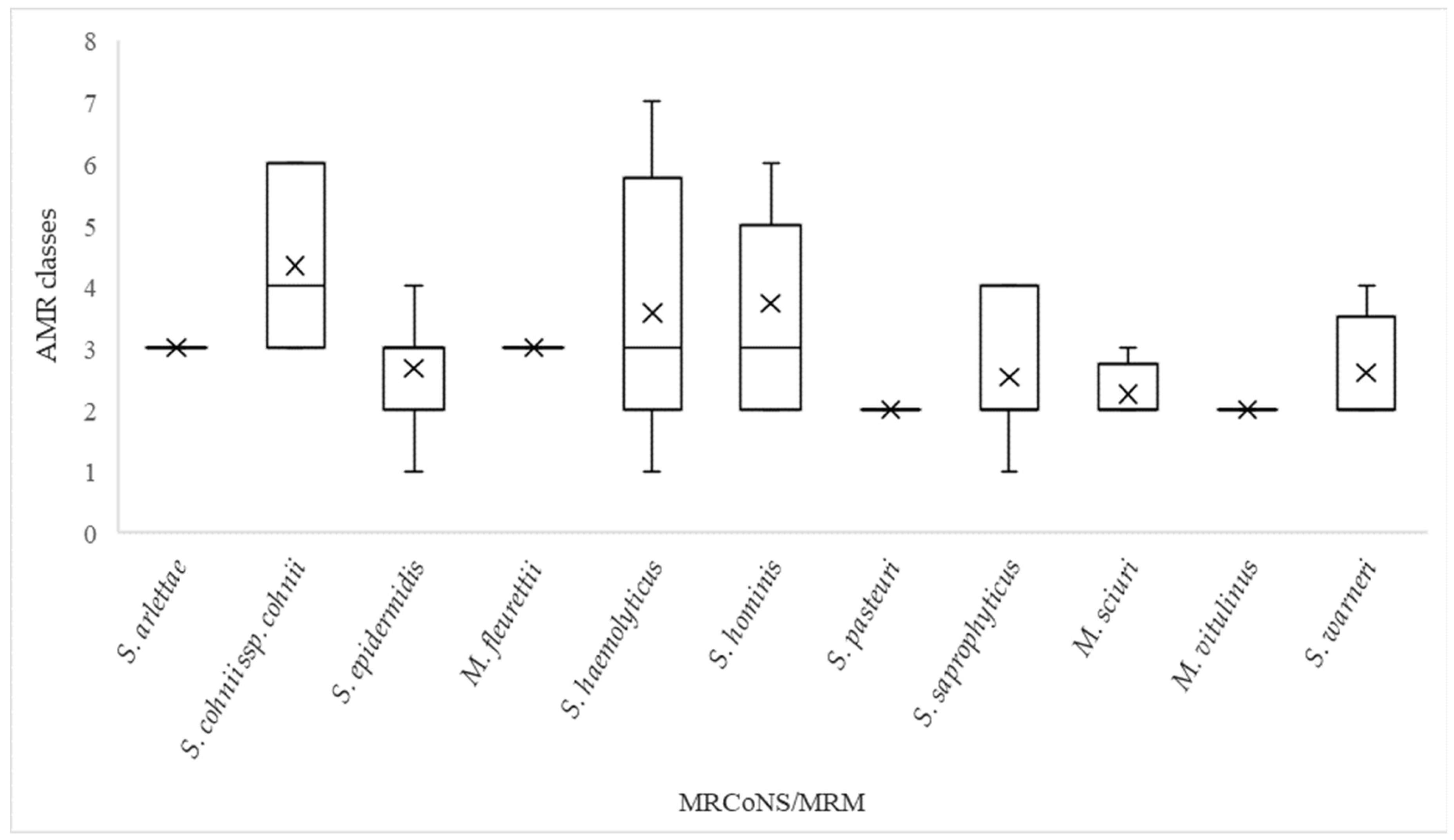
2.3. Antimicrobial Resistance

Forty-one of the seventy-five isolates were multi-resistant, expressing resistance to three or more classes of antimicrobials. S. cohnii ssp. cohnii, S. hominis and S. haemolyticus were the most resistant species, expressing resistance to means of 4.3, 3.7 and 3.6 classes, respectively (Figure 1). Following resistance to anti-staphylococcal beta-lactams, resistance to macrolides (erythromycin) and fusidanes (fusidic acid) was most frequently observed (Table 2). Worth noticing is the large proportion of S. hominis isolates that expressed no phenotypic resistance to cefoxitin and amoxicillin clavulanic acid. The resistome analysis supported the phenotypical resistance profile, displaying a high prevalence of genes conferring resistance to erythromycin and fusidic acid among the MRCoNS and MRM (Table 3) and further confirmed that all sequenced MRCoNS and MRM possessed the mecA gene. Despite the presence of mecA, we observed a variable phenotypic expression of resistance to beta-lactams among the isolates (Table 2). This was particularly evident for S. hominis, with 1/11 isolates being phenotypically susceptible to oxacillin and 5/11 isolates being susceptible to cefoxitin. Furthermore, we observed phenotypic susceptibility to amoxicillin–clavulanic acid among six MRCoNS and MRM species: S. cohnii ssp. cohnii (2/3), S. epidermidis (8/12), S. haemolyticus (2/16), S. hominis (6/11), S. warneri (1/5) and M. vitulinus (n = 1). In addition to mecA, the beta-lactamase-encoding blaZ gene was present in 16 of the 20 amoxicillin–clavulanic-acid-susceptible isolates.

2.4. SCCmec Cassettes and Sequence Types

Table 4 shows the predicted SCCmec cassettes based on detected genes and best homology in the sequenced MRCoNS and MRM isolates. For four isolates (one S. epidermidis, one S. haemolyticus and two S. hominis), the SCCmec prediction based on genes deviated from the prediction based on homology. In these cases, the SCCmec cassettes were reported based on the prediction of the genes. The S. epidermidis isolates were assigned three types (II (2a), III (3A) and IV (2b)) in addition to one non-typeable (NT) isolate. In just one household, the same SCCmec cassette was predicted in isolates of different species: an S. epidermidis and an S. saprophyticus isolate of type III (3A). The SCCmec elements of a substantial number of isolates (43/75) were non-typeable. Eleven of the fourteen NT S. haemolyticus isolates showed best homology with SCCmec type V but missed either the ccrC1 or mec class C2, or both. Three of the NT S. saprophyticus isolates had the best predicted homology to SCCmec III (3A) but missed either ccrA3 or both ccrA3 and ccrB3, while two additional S. saprophyticus isolates shared the best homology with SCCmec I (1B) but missed ccrA1 and ccrB1.
The sequence types (STs) were predicted by in silico MLST for 25 of the 39 S. epidermidis, S. haemolyticus and S. hominis isolates. The 12 S. epidermidis isolates were assigned to 10 different STs (5, 35, 57, 81, 130, 218, 224, 332, 640 (n = 2) and 679) in addition to 1 non-typeable isolate. The two MRSE ST640s were from different households. Nine of sixteen S. haemolyticus were typed to seven STs (1, 3 (n = 2), 30, 42, 49, 52 (n = 2) and 56). Isolates with identical STs were from different households. The remaining S. haemolyticus isolates had combinations of allelic profiles not reported earlier. This was also the case for 6 of the 11 S. hominis isolates, while the remaining 5 were typed to two different STs (1 and 18), all of which were from different households.

2.5. Household Analysis of Human and Pet Isolates

In households with infected pets (C, F and H), the dogs and owners concurrently tested positive for MRSE (Table 1). In household H, the dog, owner and home environment tested positive for MRSE ST640 with identical susceptibility profiles, resistance genes and SCCmec elements [12]. In contrast, the dog and owner in household C carried two different STs (MRSE ST679 and ST130), presenting different resistance genes and SCCmec types (Supplementary Materials Table S1). MRSE isolates with identical susceptibility profiles to the dog isolate were detected in the bathroom and kitchen. However, no isolates similar to the MRSE found on the owner were recovered from the home environment. We observed similar findings in household F, in which the owner and dog carried MRSE with different STs (ST218 and ST5) and SCCmec cassettes (III (3A) and IVa (2B)). Contrary to household C, we recovered only isolates with identical susceptibility profiles to the human isolate from the home environment. In household D, MR S. haemolyticus ST42 and ST1 were recovered from the owner, while the dog tested negative for these. Only the MR S. haemolyticus ST42 was detected in the home environment.
Two owners of healthy pets carried MRCoNS. The first owner tested positive for a MR S. warneri possessing a SCCmec cassette V (5C2 and 5). An MR S. warneri with an identical SCCmec cassette was recovered from the household’s kitchen. The second owner carried an MR S. haemolyticus with an NT SCCmec cassette and ST. An MR S. haemolyticus ST52 with different resistance genes was recovered from the home environment.

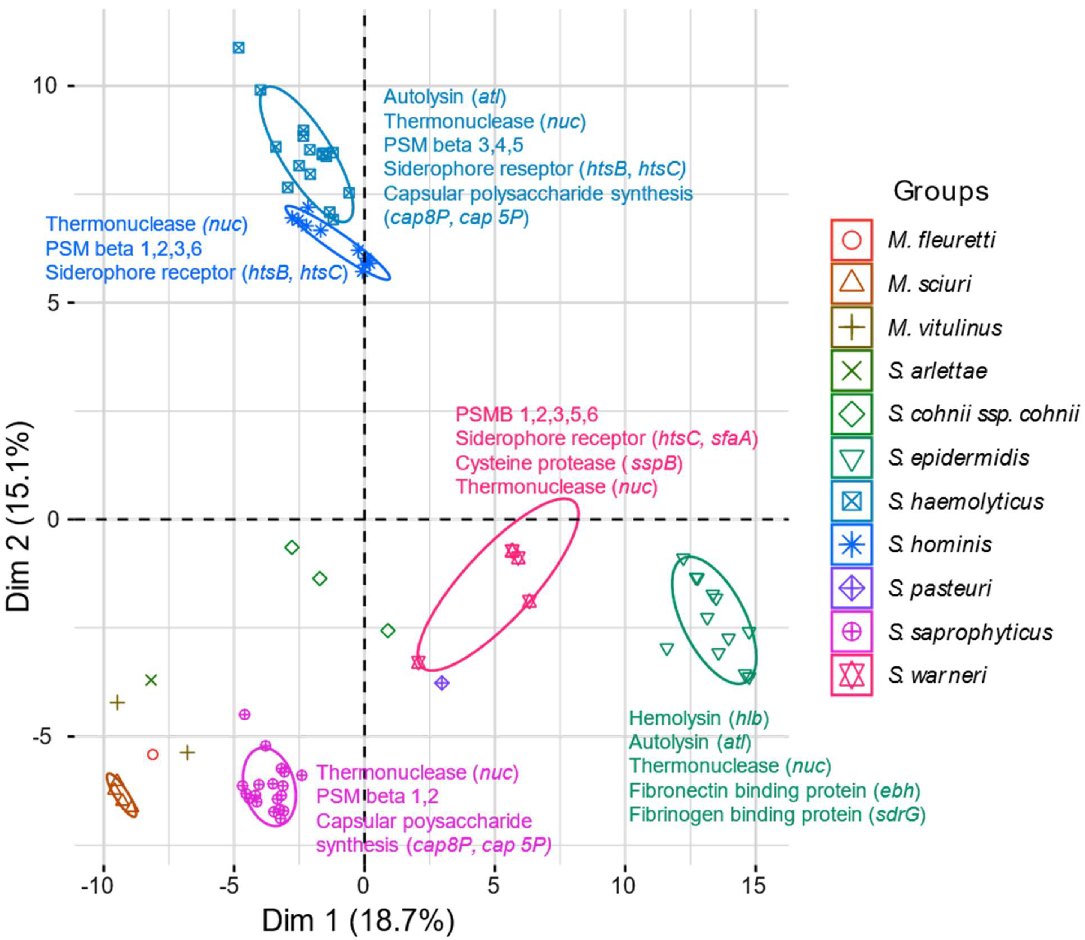
2.6. Virulence Genes

The hits in the virulence gene database are presented in Table S2, and the respective Ha scores for each virulence gene are shown. The exfoliative toxin-encoding gene etc was detected in all sequenced isolates. Furthermore, we observed a high frequency of phenol soluble modulin-encoding genes, thermonuclease-encoding nuc genes and siderophore-encoding genes in most MRCoNS isolates. Overall, the MRCoNS and MRM showed a high degree of species specificity in the virulence gene analysis, apart from S. haemolyticus and S. hominis, which clustered together (Figure 2). We detected a high occurrence of genes involved in adherence in the S. epidermidis isolates (atl, ebh and sdr genes). In addition, we observed two subpopulations of S. epidermidis isolates based on the presence of Type VII secretion-associated genes (Table S2). The tendency of two subpopulations was also evident among the S. haemolyticus isolates. Based on Ha scores of several capsular polysaccharide synthesis enzymes involved in immune evasion (cap genes), the group with high Ha scores consisted of six isolates, of which three were recovered from humans and two were of environmental origin. Isolates with low Ha scores for cap genes were solely detected in the environment.
Subgrouping based on the Ha scores of cap genes was also observed among the S. hominis and S. saprophyticus isolates. The MRM had, in general, few hits with high Ha scores, apart from the etc gene and the capO and capP genes.

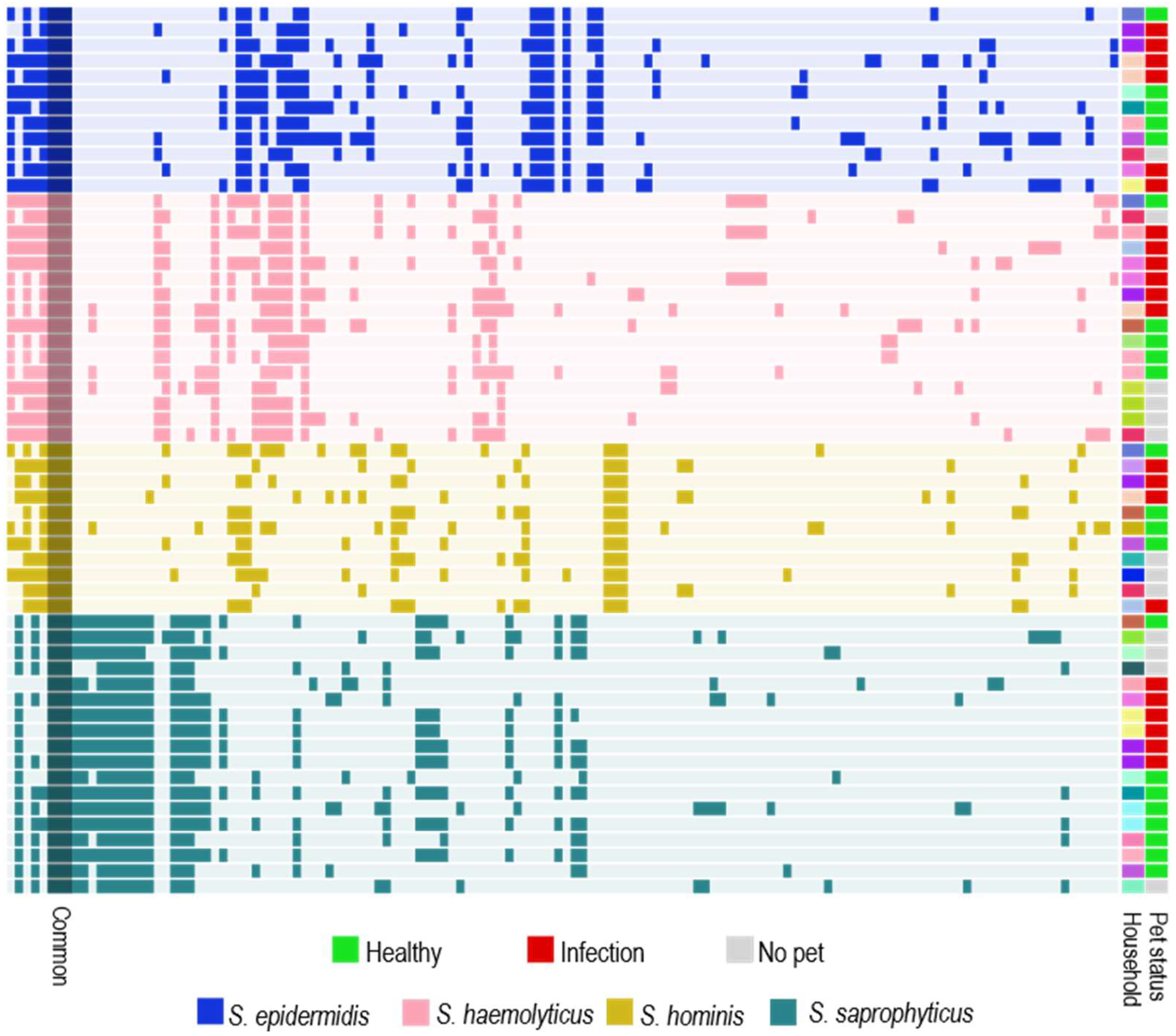
2.7. Mobilome Analysis

The four most prevalent species found in the households, S. saprophyticus, S. epidermidis, S. haemolyticus and S. hominis, were included in the mobilome analysis. Three common gene clusters encoding an IS6 family transposase, the competence protein ComGC and an uncharacterized SPBc2 prophage-derived protein YoqJ (annotated “Common” in Figure 3) were identified in all the isolates. Otherwise, the mobilomes were mainly species-specific (Figure 3). We observed a few examples of similar gene sequences in different species at the household level. For instance, S. haemolyticus and S. saprophyticus isolates from the same household carried phage major capsid protein-encoding genes and phage portal protein-encoding genes with 76.8% and 80% amino acid identities, respectively. In addition, a site-specific tyrosine-type recombinase/integrase was shared by S. epidermidis and S. saprophyticus in two households, and an IS256-like transposase was shared by S. haemolyticus and S. hominis in two other homes.

3. Discussion

MRCoNS are opportunistic pathogens prevalent in hospital environments, often due to their hardy nature, ability to form biofilms and resistance to antimicrobials [13]. The newly described genus Mammaliicoccus shares many properties with staphylococci, like habitat and methicillin resistance [14]. In a former study of the dissemination of clinical MRS in households with infected pets [12], we detected a broad range of MRCoNS and MRM in the home environments and from pets and their owners. To follow up on this observation, we decided to screen different categories of households for the presence of MRCoNS and MRM. To our surprise, MRCoNS and MRM were nearly ubiquitous in the home environments regardless of the presence of pets or health status. The finding of S. epidermidis, S. haemolyticus, S. hominis and S. saprophyticus as the predominant species in the households is reasonable since these species are known as skin commensals in humans. However, they can also cause infections, and the frequent occurrence of methicillin-resistant isolates in home environments is noteworthy. To our knowledge, the home environment has previously not been described as a reservoir for MRCoNS and MRM.
The skin, skin glands and mucous membranes of mammals are considered the main habitats for CoNS [15]. However, in most of the households studied, both the human and the pet tested negative for MRCoNS/MRM while the bacteria were present in the home environment. The absence of MRCoNS/MRM in humans and pets may reflect that the sampling sites in the humans and pets were not optimal for detecting some of the CoNS/mammaliicoccal species. For instance, S. saprophyticus is a frequent colonizer of the perineal region, rectum and urethra in humans, and S. hominis and S. haemolyticus are often isolated from axillae and pubic areas high in apocrine glands [1]. These sites were not included in the sampling procedures. On the other hand, S. epidermidis is a common human, canine and feline nasal mucosa colonizer [16,17]. Therefore, we find it peculiar that we identified relatively few carriers of MRSE, considering that MRSE was present in around one-third of the households. An explanation may be that we only sampled one human member in each household, thus missing possible carriers of the MRCoNS and MRM. Another factor contributing to the high number of MRCoNS/MRS in the households could be that the bacteria had been introduced via visitors, soil or other external sources.
Carriage of MRCoNS in pets was exclusively found in infected dogs. The owners of the three MRSE-positive dogs all tested positive for MRSE. Interestingly, the isolates from the dogs and owners differed in two of the cases, indicating a diversity of MRCoNS not only between households but also within the household. Moreover, we observed that homes with infected pets had a large diversity of MRCoNS species recovered from the home environment. Five of the eight dogs in this group had been treated with beta-lactam antimicrobials within the past three months before sampling, two of which had undergone antimicrobial treatment several times during the past year. The carriership and the diversity may reflect the MRCoNS’s and MRM’s competitive advantage when exposed to beta-lactam antimicrobials. Furthermore, five dogs in this group had been hospitalized within the past twelve months, and two owners were human health care workers. Hence, it is not unlikely that the pets or owners have been exposed to MRCoNS/MRM in these environments and transmitted them further to their home environment. Still, MRCoNS and MRM were present in many households where neither humans nor dogs had been in contact with health care facilities, again emphasizing that MRCoNS and MRM are indeed found outside clinical environments.
The phenotypic resistance analysis revealed that just over 50% of the MRM and MRCoNS were multidrug-resistant. This is consistent with previous reports on CoNS and MRCoNS in non-clinical settings [9,18]. Mobile genetic elements play a central role in spreading ARGs among bacteria [19]. Considering that MRCoNS constitute reservoirs for ARGs, we conducted a mobilome analysis primarily to investigate whether the most prevalent species in the households had mobile genetic elements in common, which could indicate genetic exchange at the household level. Nonetheless, the detected mobile genetic elements displayed mainly a species-specific profile rather than a household-related pattern. This could indicate that mobile genetic elements are not easily transmitted between different staphylococcal species. However, it must be emphasized that this analysis is based on short-read data. To gain further insight in the ARGs’ location relative to the mobile genetic elements, it would be necessary to combine short-read and long-read data.
The inconsistent phenotypic expression of resistance to cefoxitin among the MRCoNS isolates was noteworthy. EUCAST and CLSI operate with different zone diameters when assessing cefoxitin resistance. By following the CLSI breakpoints rather than the EUCAST breakpoints, eight of the ten cefoxitin susceptible CoNS isolates would have been classified as resistant. On the other hand, two of the MRSE isolates would have been reported susceptible to cefoxitin. According to the EUCAST guidelines, cefoxitin should be used when screening for methicillin resistance in CoNS [20]. However, CLSI emphasizes that the cefoxitin disk diffusion test may not perform reliably in detecting methicillin resistance for all CoNS species (e.g., S. haemolyticus) [21]. Although cefoxitin is the recommended agent for most CoNS when screening for methicillin resistance, our results show that oxacillin is more reliable than cefoxitin for the purpose.
MRS are considered resistant to most beta-lactam agents, i.e., penicillins, beta-lactam combination agents and cephems, except for ceftaroline [22,23]. However, we observed a high frequency (20/75) of amoxicillin–clavulanic-acid-susceptible isolates. Sixteen of the susceptible isolates carried blaZ, which encodes a beta-lactamase that inactivates amoxicillin. Admittedly, this could be due to the lack of official breakpoints for amoxicillin–clavulanic acid disk diffusion. Still, six of these isolates were susceptible to cefoxitin, thus demonstrating that even if the mecA gene is present, it is not necessarily expressed towards cefoxitin and amoxicillin–clavulanic acid in vitro.
We could not predict STs or SCCmec cassettes for most MRCoNS/MRM isolates. The pubMLST database only contains data for S. epidermidis, S. haemolyticus, S. hominis and S. chromogenes, and the missing ST identifications may be due to a lack of characterized environmental genomes in the database. The high proportion of non-typeable SCCmec cassettes is consistent with previous reports [24,25]. In many cases, the cassettes shared homology with previously described SCCmec but lacked identifiable ccr genes needed to determine type. This was especially evident for the NT S. haemolyticus cassettes that had the best homology with SCCmec type V. The combination of NT SCCmec elements combined with non-identifiable STs demonstrates the large diversity among the staphylococcal and mammaliicoccal isolates in the home environments and the gaps in knowledge about the epidemiology/ecology of staphylococci from environmental reservoirs.
In general, CoNS and MRM are considered less virulent than S. aureus. Still, CoNS and MRM cause a substantial number of infections, presumably possessing virulence genes enabling them to do so. Virulence genes in CoNS and mammaliicocci are far less studied than the virulence genes of S. aureus. Consequently, we used a database mainly consisting of amino acid sequences from putative and known virulence factors in S. aureus to characterize virulence genes in our MRCoNS and MRM isolates [26]. Admittedly, this is not optimal and will cause uncertainty around the hits with low and medium Ha scores. We focused on the highest scores within each species. However, we cannot be certain that hits with lower scores are of limited importance. Overall, the MRCoNS and MRM displayed species-specific virulence gene patterns, apart from the ubiquitous etc gene. The virulence gene patterns revealed subgroups within the S. epidermidis, S. haemolyticus, S. hominis and S. saprophyticus isolates based on the presence of type VII secretion-associated genes for the former and cap genes for the three latter. The isolation source seemed to matter for multiple cap genes only in the S. haemolyticus isolates, as all the human isolates were in this subgroup.

4. Materials and Methods

4.1. Participants

Participants were recruited through social media and small animal clinics in Oslo and the surrounding areas. All participants signed individual consent forms and completed questionnaires regarding their professions, antimicrobial consumption and hospital admissions within the past 12 months. Thirty-three households participated in the study. Of these, eight were households with dogs diagnosed with an MRS infection; eight were households with clinically healthy dogs; six were households with clinically healthy cats; and eleven were households without pets. The inclusion criteria included cats with MRS infections. However, during the time we recruited participants for the study, no cats with MRS infections were diagnosed in our recruiting clinics. One human and one pet from each home participated in the study. The inclusion criteria for healthy pets were the following: clinically healthy pets without symptoms of infection when examined by a veterinarian. The humans sampled were healthy according to their own statements.

4.2. Sampling

The samples were collected in the period from October 2019 to October 2021. The same veterinarian was responsible for sampling all the household environments and the participating pets. Pets diagnosed with an MRS infection were sampled from the infection site, the perineum and the oral mucosa using nylon flocked swabs (Eswab™ 480C, Copan group, Brescia, Italy). These dogs participated parallelly in another study [12]. Healthy dogs and cats were sampled from the oral mucosa and perineum. Human participants collected swab samples from their nostrils and throats according to the instructions of the veterinarian present at the time of sampling. The home environments were sampled using cloths (Sodibox® Swab cloth, Nevez, France) for swabbing of the most relevant areas such as the pets’ food bowls and sleeping areas, living room floors, bathrooms (sink faucet and hand towel) and kitchens (kitchen counter, dish towel, cloth and sink faucet). In the households without pets, the three latter locations were sampled.

4.3. Culturing and Identification

The samples were cultured as described by Røken et al. [12]. Briefly, all samples were enriched overnight in Müller–Hinton (MH) broth supplemented with 6.5% NaCl. Then, 20 µL of MH broth was inoculated on Oxacillin Resistance Screening Agar Base (ORSAB, Oxoid, Basingstoke, UK) supplemented with 2 µg oxacillin and incubated for 24 h at 35 °C. Blue, blue-white and white colonies growing on the ORSAB agar were sub-cultured on 5% bovine blood agar overnight. The species were identified using Matrix-assisted laser desorption/ionization (MALDI-TOF MS) (VITEK® MS, bioMérieux, Craponne, France). Isolates were tested for the presence of the mecA gene by PCR on a Bio-Rad T100 Thermal cycler (Bio-Rad, Hercules, CA, USA) [27].

4.4. Susceptibility Testing

Isolates were susceptibility tested against 12 antibiotics using the agar disk diffusion method [28]. The test panel included aminoglycoside (gentamicin 10 µg), amphenicol (chloramphenicol 30 µg), beta-lactams (amoxicillin–clavulanic acid 20/10 µg, oxacillin 1 µg, cefoxitin 30 µg, cefalexin 30 µg), fluoroquinolone (enrofloxacin 5 µg), fusidane (fusidic acid 100 µg), folate pathway antagonist (trimethoprim/sulfamethoxazole 1.25/23.75 µg), macrolide (erythromycin 15 µg), lincosamide (clindamycin 2 µg) and tetracycline (tetracycline 30 µg). When testing resistance to oxacillin, we used Müller–Hinton agar supplemented with 4% NaCl. Müller–Hinton plates were incubated at 35 °C for 18–20 h before reading the zone diameters. As there are no official breakpoints for amoxicillin–clavulanic acid and cefalexin, we used the breakpoints ≥25 mm for susceptible and ≤24 mm for resistant. Isolates displaying intermediate resistance to antimicrobial agents were registered as resistant in the phenotypic analysis. Isolates expressing resistance to three or more classes of antimicrobials were defined as multidrug-resistant [29].

4.5. Selection of Isolates

One MRCoNS/MRM from each species was included from each sampling location. Based on the phenotypic resistance profiles, species and households, isolates were selected for whole-genome sequencing (WGS). All WGS isolates went through an additional species identification checks using the Microbial Genomes Atlas (MiGA) webserver against the TypeMat database [30] (Table 5). If the species identities differed between the MALDI-TOF and TypeMat databases, we used the TypeMat output. Furthermore, all sequenced isolates were included in the resistome, virulence gene and SCCmec analyses. However, isolates that turned out to be redundant were excluded when presenting the results (isolates from the same household, identified as the same species with identical resistance genes, virulence genes, SCCmec elements and sequence types (STs)). Non-redundant sequenced S. epidermidis, S. hominis, S. haemolyticus and S. saprophyticus isolates were included in an additional mobilome analysis.

4.6. DNA Extraction, Library Preparation, Sequencing and Assembly

DNA was extracted using a modified version of the Master Pure™ Gram-Positive DNA Purification protocol (Lucigen Corporation, Middleton, WI, USA) [12]. The DNA quality was assessed by NanoDrop® ND-1000 (Thermo Scientific, Wilmington, CA, USA), and the DNA quantity was determined using a Qubit fluorometer with the dsDNA Broad Range Assay kit (Invitrogen, Eugene, OR, USA). Quality-controlled DNA was submitted to the Norwegian Sequencing Centre for library preparation and sequencing. The library prep was performed in two batches using Swift Turbo 2s flex DNA library prep on batch one and Nextera DNA Flex Prep on batch two. The samples were sequenced on the Illumina MiSeq platform v3, resulting in 300 bp paired-end reads. The fastq files were quality checked using FastQC version 0.11.9. Adapters and low-quality sequences were removed using Trim Galore version 0.6.7 [31]. We used SPAdes version 3.15.3 for genome assembly [32].

4.7. Bioinformatics

We used ABRicate version 1.0.1 for the resistome analysis [33]. The assembled sequences were run against the CARD database with cutoff values of 80% nucleotide identity and 80% coverage [34]. We used SCCmecFinder v. 1.2 with default settings (nucleotide identity 90% and minimum sequence length of 60%) to type SCCmec elements [35]. S. epidermidis, S. hominis and S. haemolyticus assemblies were run against a default PubMLST scheme using MLST version 2.19.0 [36].
For the virulence gene analysis, we aligned a custom database containing amino acid sequences of staphylococcal virulence factors [26] against assembled staphylococcal and mammaliicoccal genomes using tblastn (v. 2.5.0) with the default settings, except for high-scoring segment pair (HSP) set to 1 and the culling limit of 1. The resulting sample/VF matrix values were expressed as Ha scores ranging from 0 to 1 [26]. Briefly, the scores were calculated using the following formula:
Ha = (pident × length)/(qlen × 100)
where “pident” represents the proportion of amino acid sequence identities between the VF query and translated proteins from the bacterial genomes in this study, “length” represents the alignment length of a hit and “qlen” is the length of the query sequence drawn for each VF.
The mobilome analysis was conducted using Anvi’o bioinformatics suite version 7.1. [37]. Before the pangenome analysis, we excluded one S. saprophyticus from the dataset due to a high number (>2000) of partial genes. We created the Anvi’o contigs database with the “anvi-gen-contigs-database” program using Prodigal to identify open reading frames [38]. The resulting genes were associated with the functions from the NCBI’s Clusters of Orthologous Groups (COGs) database [39]. The pangenome was computed by the core Anvi’o program “anvi-pan-genome” (default settings), which in turn utilized DIAMOND [40] and MCL [41]. Metadata were integrated into the pangenome results with the “anvi-import-misc-data” program. After the pangenome visualization, we extracted all gene clusters annotated with the “Mobilome” COGs category with the “anvi-split” program for further manual inspection. We used COG annotations or an ad hoc protein web-blast search to characterize the gene clusters of the staphylococcal mobilome.

4.8. Statistical Analysis

We used one-way ANOVA to compare the number of different MRCoNS and MRM species between the households with infected pets, with healthy pets and without pets. Tukey’s honest significant difference (HSD) test was applied to test the pairwise difference between the three household groups. The significance level was set to 0.05.
The virulence factor (VF) matrix was transferred to R version 4.1.0 for further principal component analysis (PCA) using the “prcomp” function of the “stats” package (v. 4.0.1), followed by a visualization using the “fviz_pca” function of the “factoextra” package (v. 1.0.7).

5. Conclusions

In conclusion, we have documented that the home environment is a reservoir for MRCoNS and MRM regardless of the type of household and the carrier status of humans and pets. However, homes with infected pets had a larger diversity in MRCoNS and MRM species than households without pets, which might be due to the recent use of antimicrobials and contact with human and veterinary hospitals. The large diversity in SCCmec elements and sequence types among and within the households indicates no clonal spread of specific strains. The limited common virulomes and mobilomes indicate a high degree of species specificity rather than exchanges of genetic elements between species in the home environment.

Supplementary Materials

The following supporting information can be downloaded at: https://www.mdpi.com/article/10.3390/antibiotics13030279/s1, Table S1: Data sequenced isolates; Table S2: Virulence matrix.

Author Contributions

Conceptualization, M.R., A.M.B., Y.W. and A.H.H.; methodology, M.R.; software, S.I. and M.R.; formal analysis, M.R. and S.I.; investigation, M.R.; data curation, M.R. and S.I.; writing—original draft preparation, M.R.; writing—review and editing, M.R., A.M.B., Y.W., A.H.H. and S.I.; visualization, M.R. and S.I.; supervision, A.M.B., Y.W. and A.H.H.; project administration, M.R. and A.M.B.; funding acquisition, A.M.B., Y.W., A.H.H. and M.R. All authors have read and agreed to the published version of the manuscript.

Funding

This research was funded by the Faculty of Veterinary medicine, Norwegian University of Life Sciences, and by NORM, grant number 19_05.

Institutional Review Board Statement

The study was approved by the Norwegian National Research Ethics Committee (REK Sør-Øst) (Protocol code: 2019/97, 3 April 2019).

Informed Consent Statement

Written informed consent was obtained from all subjects involved in the study.

Data Availability Statement

The sequences included in the analyses are available at https://www.ncbi.nlm.nih.gov/bioproject/?term=PRJNA856113 (accessed on 6 July 2022).

Acknowledgments

We want to thank the NMBU University Animal Hospital Clinic and Kolbotn Animal Clinic for their assistance with recruiting dogs for this study. We would also like to thank the participating persons and pets for their kind contribution to the project.

Conflicts of Interest

Author S.I. was employed by the company Previwo AS. The remaining authors declare that the research was conducted in the absence of any commercial or financial relationships that could be construed as a potential conflict of interest. Authors M.R., A.H.H., A.M.B. and Y.W. declare no conflicts of interest.

References

  1. Becker, K.; Heilmann, C.; Peters, G. Coagulase-Negative Staphylococci. Clin. Microbiol. Rev. 2014, 27, 870. [Google Scholar] [CrossRef]
  2. Piette, A.; Verschraegen, G. Role of coagulase-negative staphylococci in human disease. Vet. Microbiol. 2009, 134, 45–54. [Google Scholar] [CrossRef]
  3. Becker, K.; Both, A.; Weißelberg, S.; Heilmann, C.; Rohde, H. Emergence of coagulase-negative staphylococci. Expert Rev. Anti-Infect. Ther. 2020, 18, 349–366. [Google Scholar] [CrossRef]
  4. Hovelius, B.; Mårdh, P.-A. Staphylococcus saprophyticus as a Common Cause of Urinary Tract Infections. Rev. Infect. Dis. 1984, 6, 328–337. [Google Scholar] [CrossRef]
  5. Kranz, J.; Schmidt, S.; Lebert, C.; Schneidewind, L.; Mandraka, F.; Kunze, M.; Helbig, S.; Vahlensieck, W.; Naber, K.; Schmiemann, G.; et al. The 2017 Update of the German Clinical Guideline on Epidemiology, Diagnostics, Therapy, Prevention, and Management of Uncomplicated Urinary Tract Infections in Adult Patients. Part II: Therapy and Prevention. Urol. Int. 2018, 100, 271–278. [Google Scholar] [CrossRef] [PubMed]
  6. Klingenberg, C.; Aarag, E.; Rønnestad, A.; Sollid, J.E.; Abrahamsen, T.G.; Kjeldsen, G.; Flægstad, T. Coagulase-Negative Staphylococcal Sepsis in Neonates: Association Between Antibiotic Resistance, Biofilm Formation and the Host Inflammatory Response. Pediatr. Infect. Dis. J. 2005, 24, 817–822. [Google Scholar] [CrossRef] [PubMed]
  7. Rolo, J.; Worning, P.; Nielsen Jesper, B.; Bowden, R.; Bouchami, O.; Damborg, P.; Guardabassi, L.; Perreten, V.; Tomasz, A.; Westh, H.; et al. Evolutionary Origin of the Staphylococcal Cassette Chromosome mec (SCCmec). Antimicrob. Agents Chemother. 2017, 61, e02302-16. [Google Scholar] [CrossRef] [PubMed]
  8. Klingenberg, C.; Glad, T.; Olsvik, Ø.; Flægstad, T. Rapid PCR Detection of the Methicillin Resistance Gene, mecA, on the Hands of Medical and Non-medical Personnel and Healthy Children and on Surfaces in a Neonatal Intensive Care Unit. Scand. J. Infect. Dis. 2001, 33, 494–497. [Google Scholar] [CrossRef] [PubMed]
  9. Xu, Z.; Mkrtchyan, H.; Cutler, R. Antibiotic resistance and mecA characterization of coagulase-negative staphylococci isolated from three hotels in London, UK. Front. Microbiol. 2015, 6, 1–10. [Google Scholar] [CrossRef] [PubMed]
  10. Stepanović, S.; Ćirković, I.; Djukić, S.; Vuković, D.; Švabić-Vlahović, M. Public transport as a reservoir of methicillin-resistant staphylococci. Lett. Appl. Microbiol. 2008, 47, 339–341. [Google Scholar] [CrossRef] [PubMed]
  11. Barbier, F.; Ruppé, E.; Hernandez, D.; Lebeaux, D.; Francois, P.; Felix, B.; Desprez, A.; Maiga, A.; Woerther, P.-L.; Gaillard, K.; et al. Methicillin-Resistant Coagulase-Negative Staphylococci in the Community: High Homology of SCCmec IVa between Staphylococcus epidermidis and Major Clones of Methicillin-Resistant Staphylococcus aureus. J. Infect. Dis. 2010, 202, 270–281. [Google Scholar] [CrossRef]
  12. Røken, M.; Iakhno, S.; Haaland, A.H.; Wasteson, Y.; Bjelland, A.M. Transmission of Methicillin-Resistant Staphylococcus spp. from Infected Dogs to the Home Environment and Owners. Antibiotics 2022, 11, 637. [Google Scholar] [CrossRef]
  13. Weiß, S.; Kadlec, K.; Feßler, A.T.; Schwarz, S. Identification and characterization of methicillin-resistant Staphylococcus aureus, Staphylococcus epidermidis, Staphylococcus haemolyticus and Staphylococcus pettenkoferi from a small animal clinic. Vet. Microbiol. 2013, 167, 680–685. [Google Scholar] [CrossRef]
  14. Nemeghaire, S.; Argudín, M.A.; Feßler, A.T.; Hauschild, T.; Schwarz, S.; Butaye, P. The ecological importance of the Staphylococcus sciuri species group as a reservoir for resistance and virulence genes. Vet. Microbiol. 2014, 171, 342–356. [Google Scholar] [CrossRef]
  15. De Vos, P.; Garrity, G.M.; Bergey, D.H. Bergey’s Manual of Systematic Bacteriology: Volume 3: The Firmicutes, 2nd ed.; Springer: New York, NY, USA, 2009; Volume 3. [Google Scholar]
  16. Han, J.-I.; Yang, C.-H.; Park, H.-M. Prevalence and risk factors of Staphylococcus spp. carriage among dogs and their owners: A cross-sectional study. Vet. J. 2016, 212, 15–21. [Google Scholar] [CrossRef] [PubMed]
  17. Bierowiec, K.; Korzeniowska-Kowal, A.; Wzorek, A.; Rypuła, K.; Gamian, A. Prevalence of Staphylococcus Species Colonization in Healthy and Sick Cats. Biomed. Res. Int. 2019, 2019, 4360525. [Google Scholar] [CrossRef] [PubMed]
  18. Xu, Z.; Shah, H.N.; Misra, R.; Chen, J.; Zhang, W.; Liu, Y.; Cutler, R.R.; Mkrtchyan, H.V. The prevalence, antibiotic resistance and mecA characterization of coagulase negative staphylococci recovered from non-healthcare settings in London, UK. Antimicrob. Resist. Infect. Control 2018, 7, 73. [Google Scholar] [CrossRef]
  19. Malachowa, N.; DeLeo, F.R. Mobile genetic elements of Staphylococcus aureus. Cell. Mol. Life Sci. 2010, 67, 3057–3071. [Google Scholar] [CrossRef] [PubMed]
  20. EUCAST. Breakpoint Tables for Interpretation of MICs and Zone Diameters Version 12.0. Available online: https://www.eucast.org/clinical_breakpoints/ (accessed on 10 May 2022).
  21. CLSI. Performance Standards for Antimicrobial Susceptibility Testing, 32nd ed.; CLSI supplement M100; Clinical and Laboratory Standards Institute: Wayne, PA, USA, 2022; pp. 66–77. [Google Scholar]
  22. Cosimi, R.A.; Beik, N.; Kubiak, D.W.; Johnson, J.A. Ceftaroline for Severe Methicillin-Resistant Staphylococcus aureus Infections: A Systematic Review. Open Forum Infect. Dis. 2017, 4, ofx084. [Google Scholar] [CrossRef]
  23. Stein, G.E.; Johnson, L.B. Ceftaroline: A Novel Cephalosporin with Activity against Methicillin-resistant Staphylococcus aureus. Clin. Infect. Dis. 2011, 52, 1156–1163. [Google Scholar] [CrossRef]
  24. Chen, X.-P.; Li, W.-G.; Zheng, H.; Du, H.-Y.; Zhang, L.; Zhang, L.; Che, J.; Wu, Y.; Liu, S.-M.; Lu, J.-X. Extreme diversity and multiple SCCmec elements in coagulase-negative Staphylococcus found in the Clinic and Community in Beijing, China. Ann. Clin. Microbiol. Antimicrob. 2017, 16, 57. [Google Scholar] [CrossRef]
  25. Gómez-Sanz, E.; Ceballos, S.; Ruiz-Ripa, L.; Zarazaga, M.; Torres, C. Clonally Diverse Methicillin and Multidrug Resistant Coagulase Negative Staphylococci Are Ubiquitous and Pose Transfer Ability between Pets and Their Owners. Front. Microbiol. 2019, 10. [Google Scholar] [CrossRef] [PubMed]
  26. Naushad, S.; Naqvi, S.A.; Nobrega, D.; Luby, C.; Kastelic John, P.; Barkema Herman, W.; De Buck, J.; Kent Angela, D. Comprehensive Virulence Gene Profiling of Bovine Non-aureus Staphylococci Based on Whole-Genome Sequencing Data. mSystems 2019, 4, e00098-18. [Google Scholar] [CrossRef]
  27. Stegger, M.; Andersen, P.S.; Kearns, A.; Pichon, B.; Holmes, M.A.; Edwards, G.; Laurent, F.; Teale, C.; Skov, R.; Larsen, A.R. Rapid detection, differentiation and typing of methicillin-resistant Staphylococcus aureus harbouring either mecA or the new mecA homologue mecALGA251. Clin. Microbiol. Infect. 2012, 18, 395–400. [Google Scholar] [CrossRef] [PubMed]
  28. EUCAST. EUCAST Disk Diffusion Method for Antimicrobial Susceptibility Testing; EUCAST: Basel, Switzerland, 2019. [Google Scholar]
  29. Magiorakos, A.P.; Srinivasan, A.; Carey, R.B.; Carmeli, Y.; Falagas, M.E.; Giske, C.G.; Harbarth, S.; Hindler, J.F.; Kahlmeter, G.; Olsson-Liljequist, B.; et al. Multidrug-resistant, extensively drug-resistant and pandrug-resistant bacteria: An international expert proposal for interim standard definitions for acquired resistance. Clin. Microbiol. Infect. 2012, 18, 268–281. [Google Scholar] [CrossRef]
  30. Rodriguez-R, L.M.; Harvey, W.T.; Rosselló-Mora, R.; Tiedje, J.M.; Cole, J.R.; Konstantinidis, K.T. Classifying prokaryotic genomes using the Microbial Genomes Atlas (MiGA) webserver. In Bergey’s Manual of Systematics of Archaea and Bacteria; John Wiley and Sons: Hoboken, NJ, USA, 2018; pp. 1–11. [Google Scholar]
  31. Krueger, F.; Galore, T. A Wrapper Tool around Cutadapt and FastQC to Consistently Apply Quality and Adapter Trimming to FastQ Filesl; Babraham Institute: Cambridge, UK, 2015; Volume 516. [Google Scholar]
  32. Bankevich, A.; Nurk, S.; Antipov, D.; Gurevich, A.A.; Dvorkin, M.; Kulikov, A.S.; Lesin, V.M.; Nikolenko, S.I.; Pham, S.; Prjibelski, A.D.; et al. SPAdes: A new genome assembly algorithm and its applications to single-cell sequencing. J. Comput. Biol. 2012, 19, 455–477. [Google Scholar] [CrossRef] [PubMed]
  33. Seemann, T. Abricate: Mass Screening of Contigs for Antimicrobial and Virulence Genes; Department of Microbiology and Immunology, The University of Melbourne: Melbourne, Australia, 2018; Available online: https://github.com/tseemann/abricate (accessed on 28 February 2019).
  34. Alcock, B.P.; Raphenya, A.R.; Lau, T.T.Y.; Tsang, K.K.; Bouchard, M.; Edalatmand, A.; Huynh, W.; Nguyen, A.-L.V.; Cheng, A.A.; Liu, S.; et al. CARD 2020: Antibiotic resistome surveillance with the comprehensive antibiotic resistance database. Nucleic Acids Res. 2020, 48, D517–D525. [Google Scholar] [CrossRef] [PubMed]
  35. Kaya, H.; Hasman, H.; Larsen, J.; Stegger, M.; Johannesen, T.B.; Allesøe, R.L.; Lemvigh, C.K.; Aarestrup, F.M.; Lund, O.; Larsen, A.R. SCCmecFinder, a Web-Based Tool for Typing of Staphylococcal Cassette Chromosome mec in Staphylococcus aureus Using Whole-Genome Sequence Data. mSphere 2018, 3, e00612-17. [Google Scholar] [CrossRef] [PubMed]
  36. Seemann, T. MLST. Available online: https://github.com/tseemann/mlst (accessed on 9 February 2022).
  37. Eren, A.M.; Esen, Ö.C.; Quince, C.; Vineis, J.H.; Morrison, H.G.; Sogin, M.L.; Delmont, T.O. Anvi’o: An advanced analysis and visualization platform for ‘omics data. PeerJ 2015, 3, e1319. [Google Scholar] [CrossRef]
  38. Hyatt, D.; Chen, G.-L.; LoCascio, P.F.; Land, M.L.; Larimer, F.W.; Hauser, L.J. Prodigal: Prokaryotic gene recognition and translation initiation site identification. BMC Bioinform. 2010, 11, 119. [Google Scholar] [CrossRef]
  39. Tatusov, R.L.; Galperin, M.Y.; Natale, D.A.; Koonin, E.V. The COG database: A tool for genome-scale analysis of protein functions and evolution. Nucleic Acids Res. 2000, 28, 33–36. [Google Scholar] [CrossRef] [PubMed]
  40. Buchfink, B.; Xie, C.; Huson, D.H. Fast and sensitive protein alignment using DIAMOND. Nat. Methods 2015, 12, 59–60. [Google Scholar] [CrossRef] [PubMed]
  41. van Dongen, S.; Abreu-Goodger, C. Using MCL to extract clusters from networks. Methods Mol. Biol. 2012, 804, 281–295. [Google Scholar] [CrossRef] [PubMed]
Figure 1. Number of phenotypic resistance classes in the MRCoNS and MRM isolates, n = 75. The crosses represent the mean number of antimicrobial resistance (AMR) classes, the horizontal lines represent the median number, the boxes represent the quartiles, while the whiskers represent minimum and maximum number or resistance classes.
Antibiotics 13 00279 g001
Figure 2. Principal component analysis plot of virulence gene similarity in MRCoNS and MRM isolates. The exfoliative toxin gene etc was present in all isolates. The annotations refer to the genes with the highest Ha scores among the isolates.
Antibiotics 13 00279 g002
Figure 3. Mobilome of S. epidermidis, S. haemolyticus, S. hominis and S. saprophyticus, sorted by species. The colors in the “Household” column represent the different households the isolates were isolated from. Gene clusters identified in all isolates are annotated as “Common” and include genes encoding an IS6 family transposase, the competence protein ComGC and an uncharacterized SPBc2 prophage-derived protein YoqJ.
Antibiotics 13 00279 g003
Table 1. Overview of antimicrobial use (AM), hospital admission and location of methicillin-resistant coagulase-negative Staphylococcus spp. (MRCoNS) and methicillin-resistant Mammaliicoccus spp. (MRM) in the 33 households. B = Bathroom; FB = Food bowl; FL = Floor; K = Kitchen; S = Sleeping place.
StatusHouseholdPet AM Treatment (within Months)Human AM Treatment (within 12 Months)Hospital Admission (within 12 Months)Work in Health CareHome EnvironmentHuman
Carriage
Pet
Carriage
S. arlettaeS. cohnii ssp. cohniiS. epidermidisM. fleurettiiS. haemolyticusS. hominisS. pasteuriS. saprophyticusM. sciuriM. vitulinusS. warneri
Household with
dogs with infection
A Yes B, FB, KFB, K B, FL, K
B Pet B, FL K
CCefalexin (0–3)
Polymyxin B (3–6)
Unknown agent B, K FB, FLB FB, SS. epidermidisS. epidermidis
DCefalexin (0–3) Pet S B, FL, K, S BFL, S S. haemolyticus
EAmoxicillin (0–3)
Trim/sulfa (0–3)
Pet B K FBFL
FAmoxicillin 0–3)
Cefalexin (3–6)
Enrofloxacin (3–6)
Pet.
Human
Yes K, SBBFB FL, K FL, SS. epidermidisS. epidermidis
G Pet FL
HAmoxicillin (0–3)
Fusidic acid (6–12)
K FB, K S. epidermidisS. epidermidis
Household with
healthy dogs
1 FL, SFB FL, K
2
3 ClindamycinHuman S KS. warneri
4Amoxicillin–clav (0–3)Penicillin S
5Fusidic acid (0–3)ChloramphenicolPetYes B, FL, S
6
7 Human B FB, FL
8 ErythromycinHuman B, FB, FL, S, K BFLFB
Household with
heathy cats
9 Pivemecillinam B, FL KB, FB, S
10Amoxicillin (0–3) FB FL
11 FB FB, S
12 FL
13 FL, S
14 B FL FL S. haemolyticus
Household without petsI Yes B
II Yes FL, K
III BK
IV Penicillin FL B, K
V FL
VI Yes B
VII Penicillin
VIII Human B, FL FL
IX Unknown agentHuman K
X FL, K
XI Tetracycline K FL, KFL K B
Table 2. Percentage of phenotypically expressed resistance in the 75 sequenced MRCoNS and MRM isolates. A darker shade represents a higher percentage. Gen: Gentamicin; Chl: Chloramphenicol; Oxa: Oxacillin; Cfox: Cefoxitin; AmCl: Amoxicillin–clavulanic acid; Clex: Cefalexin; Enr: Enrofloxacin; T/S: Trimethoprim sulfamethoxazole; Fus: Fusidic acid; Cli: Clindamycin; Ery: Erythromycin; Tet: Tetracycline.
Antimicrobial Agent
SpeciesnGenChlOxaCfoxAmClClexEnrT/SFusCliEryTet
All isolates7520599887395122151215120
S. arlettae1 100100100100 100100
S. cohnii ssp. cohnii3 3310010033100 33336710067
S. epidermidis128 100100338381750175017
M. fleuretti1 100100100100 100100
S. haemolyticus165061008888100382525315031
S. hominis1146 91554682184655276427
S. pasteuri1 100100100100 100
S. saprophyticus19 1110095100100 165355811
M. sciuri4 100100100100 10025
M. vitulinus2 1005050100 100
S. warneri520 1001008080 20802020
Table 3. Percentage of the 75 isolates testing positive for antimicrobial resistance genes (ARGs). All numbers are percentages of the number of isolates shown in column 2. A darker shade represents a higher percentage.
Antimicrobial Class ID S. arlettaeS. cohnii ssp. cohniiS. epidermidisM. fleurettiiS. haemolyticusS. hominisS. pasteuriS. saprophyticusM. sciuriM. vitulinusS. warneri
ARGAll Isolates
n = 75
131211611119425
Aminoglycosideaac(6′)-le/aph(2″)-la20 8 5646
ant(4′)-lb13 6725 31
aph(3′)-IIIa4 19
sat45 8 19
Amphenicolcat4 33 11
catA1 6
Beta-lactamarl1100
blaZ63 33100 9482 32 80
mecA100100100100100100100100100100100100
Folate pathway antagonistdfrC27 92 9 21 80
dfrG8 8 31
FosfomycinfosB64 27
fosD1 25
FusidanefusB29 50 2536 21 80
fusC7 8 627
fusD25 100
fusF4 100
Macrolide, lincosamide, streptograminermC13 8 3118 5 20
lnuA15 67 27 32
mphC29 10017 38910047
msrA4310010042 384210046 20
salA5 100
vgaA4 8 9
vgaALC5 19
MultidrugmgrA61 3317 10092100 100
norA24 100 8100 80
Pseudomonic acidmupA5 1317
Quaternary ammonium compoundsqacA41 3342 6991 11 40
qacB3 33 100
TetracyclinetetK201006717 3818 11
tetL3 13
Table 4. Predicted SCCmec elements based on detected genes in the sequenced MRCoNS and MRM isolates.
Species IDII (2A)III (3A)IV (2B)IVa (2B)IVd (2B)IVc (2B)V (5C2 and 5)VIII (4A)Non-Typeable
S. arlettae 1
S. cohnii ssp. cohnii 3
S. epidermidis11261 1
M. fleurettii 1
S. haemolyticus 1114
S. hominis 47
S. pasteuri 1
S. saprophyticus 10 9
M. sciuri 4
M. vitulinus 2
S. warneri 31 1
Table 5. Criteria for including isolates in the different analyses.
AnalysisIsolates Included in the AnalysisIsolates Presented in the Results
Section
mecA PCRAll cultured isolates
Species identification
(MALDI-TOF MS)
All cultured isolates
Susceptibility testingAll cultured isolatesNon-redundant WGS isolates
Whole-genome sequencing (WGS)Non-redundant isolates based on phenotypical resistance profiles, species and household
Additional species identification
(in silico, TypeMat)
All WGS isolates
Resistome analysisAll WGS isolatesNon-redundant WGS isolates
Virulence gene analysisAll WGS isolatesNon-redundant WGS isolates
SCCmec typingAll WGS isolatesNon-redundant WGS isolates
Sequence typing
(in silico MLST)
All WGS S. epidermidis, S. hominis and S. haemolyticus isolatesNon-redundant WGS S. epidermidis, S. hominis and S. haemolyticus isolates
Mobilome analysisAll whole genome-sequenced S. epidermidis, S. haemolyticus, S. hominis and S. saprophyticus isolatesNon-redundant S. epidermidis, S. haemolyticus, S. hominis and S. saprophyticus isolates
Disclaimer/Publisher’s Note: The statements, opinions and data contained in all publications are solely those of the individual author(s) and contributor(s) and not of MDPI and/or the editor(s). MDPI and/or the editor(s) disclaim responsibility for any injury to people or property resulting from any ideas, methods, instructions or products referred to in the content.

