Article

A Little Helper: Beneficial Bacteria with Growth-Promoting Mechanisms Can Reduce Asian Soybean Rust Severity in a Cell-Free Formulation

by
Victor Hugo Buttrós
1,*,
Neílton Antônio Fiusa Araújo
2,
Vinícius de Abreu D’Ávila
3,
Maysa Mathias Alves Pereira
2,
Dirceu de Sousa Melo
1,
Moacir Pasqual
2 and
Joyce Dória
2,*
1
Department of Biology, Federal University of Lavras, Lavras 37200-900, Minas Gerais, Brazil
2
Department of Agriculture, Federal University of Lavras, Lavras 37200-900, Minas Gerais, Brazil
3
Department of Agronomy, State University of Minas Gerais, Belo Horizonte 37900-106, Minas Gerais, Brazil
*
Authors to whom correspondence should be addressed.
Agronomy 2022, 12(11), 2635; https://doi.org/10.3390/agronomy12112635
Submission received: 31 August 2022 / Revised: 22 October 2022 / Accepted: 24 October 2022 / Published: 26 October 2022

Abstract

:
Growth-promoting bacteria are already used in sustainable agricultural systems in Brazil. The market is dominated by inoculants and biological pesticides, which do not reach the full potential of this tool in the agricultural sector. This study aimed to evaluate four bacterial strains for the presence of growth promotion mechanisms, as well as the reduction of Asian rust severity in soybean plants and its effects on three antioxidant enzymes during pathogenesis. The plants were treated using the bacterial cells and/or their biosurfactants before inoculation of the pathogen (IOP). Severity was measured based on a diagrammatic scale at 14, 18 and 21 days after IOP, and the activities of the enzymes SOD, CAT, and APX were evaluated 21 days after IOP. Treatments containing only bacterial cells were not efficient in reducing the severity, with losses of leaf area reaching 15%, while the addition of biosurfactants led to a result that is similar to the biofungicide, based on Bacillus subtilis (Serenade®). The presence of direct growth promotion mechanisms can be observed in all isolates, as well as the role of bacterial metabolites, especially lipopeptides, in the biological control of diseases and the modulation of the plant’s immune response.

1. Introduction

Several microorganisms are known for their close association with plants, exerting a beneficial, neutral, variable, or deleterious effect [1]. Plant growth-promoting bacteria (PGPB) are among the microorganisms capable of exerting beneficial effects through different mechanisms, which can be direct: (1) Soil phosphate solubilization; (2) nitrogen fixation; (3) production of siderophores; (4) production of hormones; and (5) production of enzymes (ACC deaminase). The indirect mechanisms are: (1) Biological control of pathogens and pests, and (2) relief from abiotic stress [2,3].
The promotion of plant growth mediated by PGPB cannot be attributed to just one of the mechanisms described, as they can occur simultaneously or sequentially, where the increase observed in the evaluated agronomic characteristics is due to the sum of the effects of each mechanism present. Several genera of bacteria, such as Pseudomonas, Azospirillum, Bacillus, Pantoea, Burkholderia, Rhizobium, and Enterobacter, have already been attributed to one or more mechanisms of growth promotion in plants [4,5].
With the growing demand for agricultural inputs with less environmental impact, the use of PGPB associated with other integrated crop management tools follows this demand, making its use promising, especially in crops of high economic value. Focusing on the promising role of the use of PGPB in high-value crops, soybean (Glycine max L.) stands out as one of the strongest elements of the Brazilian economy, currently the largest producer of this grain, with a yield of 122,647 million tons and a planted area of 36.950 million hectares [6].
Among the main factors that limit the maximum exploitation of soybeans’ productive potential, diseases, whether caused by viruses, bacteria, fungi, or nematodes, are responsible for most of the economic damage. Asian soybean rust, caused by the fungus Phakopsora pachyrhizi (Sydow), is the main disease of the crop, and the uninterrupted cultivation, associated with the continuous presence of host plants, including soybeans and other legumes in the off-season, makes this disease a constant threat since its arrival in Brazil [7].
The first sign of rust is the appearance of small spots of that are darker in color in comparison to the healthy tissue and approximately 1 mm in diameter, and is seen on the adaxial face of the leaf [8]. After a latent period, under such points, the asexual reproduction structures, the uredia, are formed, which will give rise to the uredospores, initially hyaline in color, but which become brownish and accumulate in the opening of the pore to be carried by the wind. As sporulation progresses, the leaf tissue around the first uredia acquires a light brown color in the so-called “tan” type lesions due to its brownish appearance. The loss of photosynthetic area due to the rapid yellowing and/or premature leaf fall impairs the full formation of the grains, which direct affects productivity [9].
During pathogenesis, tissue colonization and toxins released by the pathogen can induce an increase in the release of reactive oxygen species (ROS), which, due to its antimicrobial action and effect on tissue lignification, constitutes an important defense strategy for plants. However, this increases in ROS, which has highly oxidizing molecules, can cause structural and molecular damage to the plant. To counterbalance this increase, a series of enzymes (Superoxide dismutase = SOD; Catalase = CAT, and Ascorbate-peroxidase = APX) capable of removing ROS are released; their increase is commonly related to the onset and progress of the disease [10].
Soybean rust control takes place through a series of procedures adopted together when signs of the pathogen are already noticed in the area. Chemical control is the most widespread method, but other measures to reduce the damage caused by the disease can be allied. These consist of using earlier cultivars, sown at the beginning of the recommended season for each region, to avoid a prolonged sowing period, to constantly inspect the crops and crop rotation with non-host species of the pathogen, in addition to the sanitary void [7].
There is a growing trend in the search for new methods to control diseases in plants, seeking either to reduce the environmental damage caused by traditional methods or to delay the emergence of resistant populations of the pathogen caused by the indiscriminate use of synthetic pesticides. There is also a concern about organic crops, with Brazil being a standout example. Alternative products that serve as defensives and cause less damage to the environment, whether synthetic or natural, have been following this trend, including the various biofertilizers, sprays, essential oils, and biocontrol agents currently available on the market [11].
Disease control in plants using microbial agents, such as bacteria, is already widely used and is considered to be a promising alternative to traditional pesticides [12]. The genus Bacillus is one of the most common genera of gram-positive bacteria that is capable of using different carbon sources for their growth [12,13]; it is strictly aerobic and forms endospores that are resistant to elevated temperature, pressure, and radiation, thus serving as resistance spores of bacteria in inhospitable environments and situations [14].
Several species of Bacillus are known for their efficiency in the biological control of plant diseases and compose commercial formulations of fungicides of biological origin, such as Serenade®, containing B. subtilis, and Sonata®, with B. pumilus. In addition to these, B. thuringiensis, B. formosus, and B. amyloliquefaciens stand out as important species due to the applicability of their antifungal and antibacterial effects in the biological control of plant pathogens [15,16]. Bacteria of the genus Bacillus, and phylogenetically-close groups, are capable of producing complex mixtures of amphipathic substances known as biosurfactants, composed of nonpolar fatty acids linked to a polar radical at the end. Biosurfactants affect the integrity of the plasma membranes of different microorganisms, including fungi, which is linked to the ability of bacteria to control several diseases, such as gray mold (Botrytis cinerea), anthracnose (Colletotrichum gloeosporioides), and early-blight (Alternaria solani) [17].
The principle explored in fungicides of microbiological origin, such as Serenade®, is the interaction of lipopeptides, as amphipathic molecules, with the cell membrane of the pathogens’ reproductive and resistance structures, causing a loss in conformation, functionality, and ruptures. In addition, the presence of the antagonist establishes direct competition with the pathogen for space and nutrients on the plant surface and in the rhizosphere [18]. In addition to the structural damage caused to the pathogen, the recognition of cyclic lipopeptides produced by Bacillus sp. can modulate the jasmonic acid (JA) and ethylene (ET) pathways, in a mechanism known as induced systemic resistance (ISR) [19]. The ISR occurs without interaction with the pathogen, but the so-called primed state that is acquired enables faster and more intense responses against future pathogen attacks [20].
Bio-formulations containing cells have limitations, mainly a short shelf life and sub-optimal conditions encountered by microorganisms after application to soil and leaf surfaces [21]. After application, the active ingredient encounters adverse conditions, both in the soil and on the leaf surface, and has to compete with the autochthonous microbiota for carbon and other nutrients [22]. Prolonged storage of biological products with cells and in liquid formulation suffers a significant decrease in cell viability, in addition to the possible accumulation of inhibitors in the medium, which contributes to a reduction in the efficiency of the applied product [23].
By attributing the desired effect of the application of a microorganism to one or more of its metabolites, the feasibility of formulating cell-free products is evidenced. The use of one or more processes for cell removal and concentration of active metabolites makes it possible to not only circumvent the limitation of cell viability in storage conditions, but also to guarantee an immediate effect of the product and a lower volume of application [24].
Therefore, the objective of this work was to confirm the presence of the main direct mechanisms of plant growth promotion, as well as to evaluate the effect of treating soybean plants with microorganisms of the genus Bacillus and their lipopeptides, in formulations with and without the presence of bacterial cells, on the severity of Asian soybean rust and the activity of three enzymes in the antioxidant system (SOD, CAT, and APX) during pathogenesis.

2. Materials and Methods

The experiments were conducted on the university campus of the Federal University of Lavras, Department of Agriculture, Laboratory for Cultivation of Microorganisms, Laboratory of Plant Tissue Culture and attached greenhouses, and in the Department of Biology, Microbiology sector, Enzyme Laboratory.

2.1. Obtaining and Cultivation of Microorganisms and Biosurfactants

The microorganisms (Table 1) were provided by the Agricultural Microbiology Culture Collection (CCMA) of the Federal University of Lavras and by the Laboratory of Biotechnology and Biodiversity for the Environment of the Federal University of Viçosa, as follows:
To conduct the experiments, the nutrient broth culture medium was prepared and poured into 810 mL glass flasks with a lid adapted for the insertion of 8 mm metal connections allowing for the placement of 0.22 µm syringe filters in the inlet cannula. The outlet cannula was partially blocked with cotton, allowing pressure to release. After inoculation, the flasks were connected to an air pump with a capacity of 30 L/min, divided into twelve outlets of 2.5 L/min each. The flasks were kept under constant aeration at 27 °C ± 2 for 72 h.
For the separation of bacterial cells, the fermented broth was centrifuged for 10 min at 5000 rpm, the supernatant was separated, and the bacteria were suspended in a phosphate-buffered saline (PBS). As for the separation of the biosurfactants, acid precipitation of the supernatant previously obtained was conducted until reaching values of pH 2.0 ± 1. After decanting in a refrigerator, the excess culture medium was removed; the precipitate was washed with distilled water (pH 2.0 adjusted with HCl 1M). The second centrifugation followed by discarding the supernatant was performed to concentrate the biosurfactants, which were separated into 10 mL samples in falcon tubes with a capacity of 50 mL, which were then stored in a freezer at −20 °C for later lyophilization [16,25]. After lyophilization and comparison of the masses, an average content of 40% of dry matter was observed in the form of precipitable compounds in this pH range.

2.2. Confirming Direct Growth Promotion Mechanisms

Specific tests for each mechanism were performed, as follows:

2.2.1. Production of Siderophores

To evaluate the microorganisms for the production of siderophores, a qualitative colorimetric test was conducted to detect the iron ion dispersed in the solution. To perform the test, four solutions were first prepared:
  • 0.06 g of Chromium azurol S (CAS) in sterile distilled water (QSP 50 mL).
  • 0.0027 g of FeCl3.H2O in 10 mM HCl solution (QSP 10 mL).
  • 0.073 g of Cetrimonium bromide in sterile distilled water (QSP 50 mL).
  • Casamino acid solution was obtained by dissolving 3 g casamino acid in 27 mL of distilled water and 3 mL of a 3% v/v hydroxyquinolone-chloroform solution.
All of them were filtered through a 0.22 µm membrane filter. Solutions 1, 2, and 3 were then mixed in a flask and autoclaved at 121 °C for 15 min.
Separately, 100 mL of M9 (5×) culture medium was prepared, which was then added to 150 mL of distilled water and 32.24 g of PIPES buffer. The pH was adjusted to 6.8 and the medium was sterilized at 121 °C for 15 min. After cooling to temperatures below 50 °C, 10 mL of 20% glucose solution, 30 mL of filtered casamino acid solution, and 100 mL of dye solution (solutions 1, 2, and 3) were added. The medium was poured into Petri dishes and after inoculation of the bacteria and incubation at 28 °C for 24 h, the plates with the presence of a yellow halo around the colony were considered positive for siderophore production [26,27].

2.2.2. Auxin Production

For the detection of indol-acetic acid from microorganisms, a quantitative test was performed, which consisted of the inoculation of microorganisms in nutrient broth (NB) medium followed by incubation at 28 °C for 48 h. After bacterial growth, the optical density (OD600) was then adjusted to 0.5 (107–108 CFU mL−1). An aliquot of this adjusted solution was then added to a fresh nutrient broth medium supplemented with tryptophan (100 µg·mL−1), in a proportion of 5% adjusted solution and 95% sterile nutrient broth, which was followed by further incubation at 30 °C for 72 h in the dark. After microbial growth, centrifugation was performed at 12,000 rpm for 5 min and the supernatant was recovered.
Auxin production was then measured from the combination of the supernatant obtained above and the Salkowski reagent (1875 g FeCl3.6H2O, 100 mL of distilled water, and 150 mL of 35% H2SO4 solution) in a 1:1 ratio. The solution was incubated in the dark at 30 °C for 15 min and the absorbance of each sample was measured at a wavelength of 530 nm. Auxin quantification followed values available in the standard curve prepared for Azospirillum brasilense Ab-V5, which was also the positive control for this test [28,29]. Statistical analyzes were performed using the R Studio software [30] to compare the means using the Tukey test at a 5% probability.

2.2.3. Phosphate Solubilization

To assess the competence of the test microorganisms for phosphate solubilization, they were inoculated in an NB culture medium and incubated at 30 °C for 48 h. The optical density was then adjusted (OD600) to 0.5 (107–108 CFU mL−1) and the suspension was used for inoculation in NBRIP culture medium [31] poured into Petri dishes, where 3 drops of 10 µL of bacterial suspension were deposited at equidistant points.
The evaluation was performed every 3 days for 12 days, and the test was considered positive in colonies surrounded by a characteristic translucent halo. The diameter of the halo and the colony was used to calculate the solubilization index (SI), expressed by the ratio between the diameter of the halo (mm) and the diameter of the colony (mm) [32]. The SI of each bacterium can be classified between: Low (SI < 2.0); Medium (2.0 < SI < 4.0) and High (SI > 4.0), classifications linked to a greater or lesser capacity and efficiency of solubilization [33]. Statistical analyzes were performed using the R Studio software [30] to compare the means using the Tukey test at a 5% probability.

2.3. Inhibition of Uredospore Germination

To evaluate the ability of cells and/or biosurfactants to inhibit P. pachyrhizi uredospores germination, fourteen treatments (Table 2) were applied, as follows:
Treatments containing only bacterial cells were obtained from the centrifugation of fermented nutrient broth medium, as described in Item 2.1, followed by suspension of cells in PBS buffer until optical density (OD600) was equal to 0.2. In treatments composed of filtered nutrient broth, the supernatant from the previous centrifugation was passed through a 0.22 µm membrane filter, resulting in a transparent filtrate containing only the dissolved compounds. In the treatments that correspond to cells and filtrate, the cells were suspended in the filtrate until the optical density (OD600) was equal to 0.2. The suspension of P. pachyrhizi uredospores were prepared in distilled water with sucrose (0.05%) and the concentration was adjusted to 3.5 × 10⁴ uredospores.mL−1 with the aid of a Neubauer chamber.
In 96-well ELISA plates, 125 µL of the uredospore suspension was deposited with the aid of a micropipette and then 125 µL of the solution corresponding to each treatment was added in a quadruplicate scheme (4 wells per treatment). After 24 h, each treatment was observed under a microscope to verify the occurrence or absence of germination. In cases where germination occurred, it was classified as total (1.0) or partial (0.5). A uredospore whose germ tube was at least twice the length of the uredospore itself was considered germinated.

2.4. Assessment of the Severity of Asian Rust

To evaluate the control of Asian soybean rust (Phakopsora pachyrhizi) by the selected microorganisms, an experiment was conducted in a greenhouse simulating ideal temperature and humidity conditions for the pathogenesis. The soybean plants (Glycine max L. var. BRS 519) were obtained after sowing five seeds in 1 L pots containing Tropstrato® substrate. After planting, the pots were kept in a greenhouse (25–28 °C) until seedling emergence. A thinning was performed, leaving two plants per pot, each treatment consisted of six plants, for a total of eighty-four plants. Treatments with microorganisms and/or biosurfactants were started 40 days after planting the seeds. Seeking to individualize the effects of biosurfactants, and cells and also to evaluate the effects of the combination of both on the severity of Asian soybean rust, fourteen treatments (Table 2) were performed. The treatments were composed of two controls, a negative consisting only of distilled water, and a positive consisting of the application of a commercial biological fungicide based on B. subtilis, Serenade®. In addition to the controls, each of the four microorganisms was applied in three ways: (1) Only bacterial cells in suspension; (2) lipopeptides only, and (3) a combination of both.
Treatments containing only bacterial cells were obtained by centrifuging the nutrient broth in which bacteria were grown, as described in item 2.1, followed by suspension of cells in sucrose solution (3 g·L−1) until an optical density (OD600) of 1.0 (108–109 CFU.mL−1) was obtained. The addition of sucrose in all the treatments aimed to break the surface tension and promote spreading and adherence in the application, in addition to maintaining the viability of the inoculum.
The treatments only composed of biosurfactants were obtained from the addition of the biosurfactant concentrate previously obtained in item 2.1 to a sucrose solution at 3 g·L−1 in the proportion of 1% (v/v). The treatments composed of cells and biosurfactants simultaneously present the same concentration of cells (1 × 10⁸–109 CFU.mL−1) and of biosurfactants (1% v/v of concentrated or approximately 4 g·L−1 of dry precipitate) of the isolated treatments. Treatment containing Serenade® was conducted following the dilution instructions on the package insert. With the aid of a garden sprayer, two sprays were made with microorganisms and/or lipopeptides, with a five-day interval between applications. Applications were always made in the late afternoon and until the point of run-off of the active ingredient.
After 48 h of the second spraying with the active ingredient, the plants then inoculated the pathogen. A suspension of uredospores at 2.9 × 10⁴ uredospores.mL−1 was prepared using a Neubauer chamber, with distilled water and sucrose (0.05%) as a spreader and adhesive. The uredospore suspension was applied with the aid of a garden sprayer, to the point of runoff. After inoculation of the pathogen, the plants were wrapped in opaque black bags to simulate a dark humid chamber to optimize pathogen germination, where they remained for 48 h. After this period, the plants were uncovered and conducted in a greenhouse, with daily localized irrigation, without leaf wetness. As a parameter for soybean rust severity, the diagrammatic scale (Figure 1) proposed by Godoy et al. in 2006 was used, based on the leaf area lost by lesions and yellowing [34].
Three evaluations were performed, the first one at 14 days after inoculation of the pathogen (DAI), followed by one at 18 days after inoculation, and ending with the last one at 21 days after inoculation. All evaluations were made by always observing one of the leaves of the first pair just above the cotyledonary leaves, in which one of them was randomly detached from the plant in the first evaluation.
Statistical analyzes were performed with the aid of the R Studio software [30], through which the Tukey test was performed at a 5% probability to compare the means. The software was also used to set up graphs to observe the distribution of values.

2.5. Activity of Antioxidant Enzymes during Pathogenesis

Leaves of two random plants were collected, stored in aluminum foil envelopes, and placed in Styrofoam boxes containing liquid nitrogen. The samples were macerated in porcelain mortar on ice, in the presence of liquid nitrogen and polyvinylpyrrolidone (PVP) and stored in an ultra-freezer at −86 °C.
For the extraction of catalase (CAT; EC 1.11.1.6), ascorbate peroxidase (APX; EC 1.11.1.1), and superoxide dismutase (SOD; EC 1.15.1.1) from the plant material, 100 mg of each treatment was used and was homogenized in a 400 mM potassium phosphate buffer at pH 7.8 (375 µL), 10 mM EDTA (15 µL), 200 mM ascorbic acid (15 µL) and water (1035 µL). Subsequently, the extract was centrifuged at 14,000 revolutions per minute (RPM) at 4 °C. Enzyme activity was determined by the Enzyme Linked Immuno Sorbent Assay (ELISA) technique, with a 96-well polystyrene microplate.
For SOD evaluation, 10 µL of the crude enzyme extract and 190 µL of the reaction medium were used, consisting of 100 mM sodium phosphate buffer pH 7.8 (100 µL), 70 mM methionine (40 µL), EDTA 10 mM (3 µL), water (30 µL), 1 mM p-nitro blue tetrazolium (NBT) (15 µL) and 2 µM riboflavin (2 µL). The reaction was conducted at 25 °C in a reaction chamber illuminated with a 15 W fluorescent lamp. After 7 min of light exposure, absorbance was read at 560 nm. The results of SOD activity was expressed in U SOD·min−1·g·FM−1 [35].
For CAT, 10 µL of the crude enzyme extract and 190 µL of the reaction medium were used, consisting of 200 mM potassium phosphate buffer at pH 7.0 (100 µL), water (80 µL), and 250 mM hydrogen peroxide (10 µL). The decrease in absorbance at 240 nm at 28 °C was measured during three minutes of reaction. The CAT result was expressed in mmol H2O2.min−1.gFM−1 (in the graph, the value is multiplied by 108 to facilitate graphical representation) [36].
For APX, 10 μL of the crude enzymatic extract and 190 µL of the reaction medium were used, consisting of 200 mM potassium phosphate buffer, pH 7.8 (100 µL), 10 mM ascorbic acid (10 µL), water (70 µL), and 2 mM hydrogen peroxide (10 µL). The decrease in absorbance was observed at 290 nm for three minutes at 28 °C. The APX result was expressed in mmol.ASA.gFM−1.min−1 (in the graph, the value is multiplied by 107 to facilitate graphical representation) [37].

3. Results

3.1. Assessment of Direct Growth-Promotion Mechanisms

The results related to the direct mechanisms of plant growth promotion are shown in Table 3. For phosphate solubilization, they were expressed by measuring the diameter of the colony and the indicative halo around the colony, and the solubility index (SI) was then calculated with the following equation:
SI = Halo   diameter   ( mm ) Colony   diameter   ( mm )
The SI value was then classified as high (SI > 4.0), medium (2.0 < SI < 4.0) and low (SI < 2.0).
The four isolates demonstrated the ability to solubilize phosphate, however the UFV LPP155 isolate stood out (SI > 5.0 at 12 days) with a statistically superior mean even when compared to another isolate of the same species (CCMA1347). There was no statistical difference between the solubilization rates of the other strains.
All isolates were able to synthesize indol-acetic acid at various levels, where the P. aeruginosa isolate (CCMA-0106) presented the highest mean (9.76 µg/mL) among all treatments. All tested strains were able to produce siderophores.
The tests of direct mechanisms of growth promotion aimed to confirm their presence in the studied strains. In future quantitative tests, it will be possible to rank the most efficient microorganisms for each mechanism and then to verify the existence of a synergistic relationship in the combination of more than one microorganism.

3.2. Inhibition of Uredospore Germination

From the germination test (Table 4), it is possible to observe which strains are capable of inhibiting the pathogen’s germination, as well as to identify which fraction of the formulation (biosurfactants, bacterial cells, or a combination of both) is responsible for the effect.
The results point to the importance of metabolites, known as biosurfactants, in the potential for biological control of the disease since the application of only isolated cells (T1, T4, T7 and T10) was not able to prevent the germination of the pathogen.
In the treatment composed of isolated cells of the strain CCMA1347 of B. subtilis, there was partial inhibition of germination, in which the spores considered germinated showed morphological alterations when compared to the spores in the treatment composed of distilled water. In the latter, the germ tubes were thinner and did not present appressorium formation.
In light microscopy (Figure 2), it was possible to see the differences in uredospores under optimal conditions for germination and their appearance when in contact with inhibitors. In the observation of the experiment, higher turbidity of the treatments containing bacterial cells after 24 h can be observed, indicating bacterial growth even after the assembly of the assay.

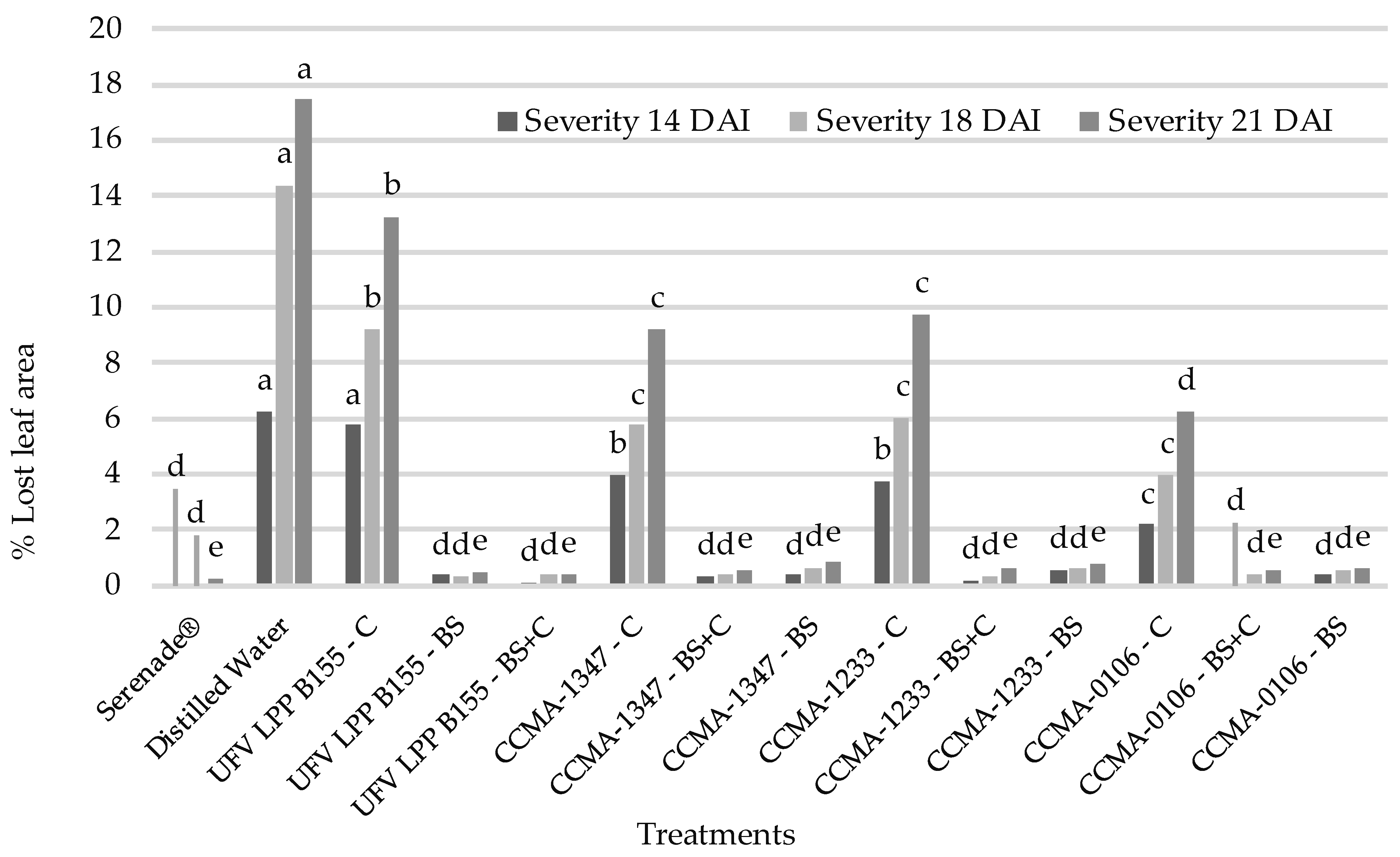
3.3. Assessment of the Severity of Asian Rust and Antioxidant Pattern during Pathogenesis

In the first evaluation, 14 days after inoculation (DAI), it was already possible to observe (Figure 3) a slight progression of the disease in all treatments, except the one where Serenade® was applied. However, the severity of the disease was significantly lower in the treatments composed of biosurfactants and by biosurfactants and bacterial cells, which did not differ statistically from each other. The observed severity is quantified based on a diagrammatic scale that considers the leaf area lost to the lesions.
At 18 days after the introduction of the pathogen, a slower progression of the disease can be observed in treatments containing biosurfactants, regardless of the presence of bacterial cells. It is possible to observe that T3 did not differ from the negative control (distilled water) at first (14DAI), but after 4 days (18DAI), it was possible to observe a slight inhibition in severity. However, this inhibition was lower than that observed for the treatments T6, T9, and T12, which, even if composed only of cells, showed a greater reduction in the severity of the disease. The treatments that contained biosurfactants (T4, T5, T7, T8, T10, T11, T13, and T14) continued not to differ from the positive control, namely the Serenade® fungicide.
At 21 days after the introduction of the pathogen, in all treatments, there was a sign of the pathogen, but the severity of the disease was lower in treatments containing biosurfactants and/or bacterial cells, which, in this case, followed the behavior of the control treatment with fungicide.
There was no statistical difference between the strains evaluated when the treatment included biosurfactants from the same bacteria, which indicates that all of them have the potential for application in the biological control of the disease. The use of bacterial cells in the tested formulations did not increase the efficiency, indicating the fundamental role of biosurfactants in the anti-pathogenic action, regardless of the presence of bacterial cells.
The loss of chlorophyll area can be observed in the progression of the disease (Figure 4b), which would translate into lower production, and consequently, economic loss. With applications of bioformulation-containing biosurfactants, it was possible to reduce the severity of the disease (Figure 4a) in levels compared to commercial fungicides, even in the presence of the pathogen and all conditions ideal for its development.
SOD activity (Figure 5) remained almost constant in all treatments. Treatments with the B. subtilis strain LPP B155, where only cells (C) or only biosurfactant (BS) were applied, showed differences in enzyme activity, where BS led to higher SOD activity when compared to C and the combination BS + C led to intermediate values of activity of this enzyme. In BS and BS + C for LPP B155, there was no statistical difference in SOD activity or disease severity, which increased once the biosurfactants were removed. For CCMA-1233 (B. licheniformis), the lack of bacterial cells (BS) led to an evident reduction in SOD activity, the opposite of severity, which increases when biosurfactants are removed from the formulation. Treatments based on the P. aeruginosa strain CCMA0106 showed higher values of enzymatic activity even in the absence of cells (BS), but the absence of biosurfactants leads to an increase in rust severity.
The APX enzyme (Figure 6) showed greater variation between treatments, where a significant increase can be observed for treatments containing cells (C and BS + C) of P. aeruginosa strain CCMA0106, where their presence led to the highest values for the activity of this enzyme. For the B. subtilis strain LPP B155, the presence of biosurfactants led to lower enzyme activity, as well as lower disease severity, which also increased in the absence of biosurfactants. For the treatments containing the strains CCMA1347 and CCMA1233, the combination of cells and biosurfactants (BS + C) led to a higher value in enzyme activity when compared to the isolated treatments (BS and C).
For CAT (Figure 7), there was also a high variation between treatments. For strain CCMA0106 (P. aeruginosa), the presence of isolated cells (C) led to a significant increase in catalase activity when compared to the treatment in the presence of biosurfactants (BS and BS + C), where the severity was significantly lower. The absence of cells in the treatment based on B. licheniformis (CCMA1233) led to the lowest values observed for the activity of this enzyme, with similar behavior in LPP B155 (B. subtilis).

4. Discussion

4.1. Direct Growth Promotion Mechanisms

The four bacterial strains were able to solubilize phosphate, however the isolate UFV LPP155 of B. subtilis stood out with a statistically superior mean, even when compared to another isolate of the same species (CCMA 1347), an event that is well described in the literature, where different strains of the same species tend to show different degrees of phosphate solubilization [38,39].
All bacterial strains were able to synthesize indol-acetic acid at distinct levels, where the P. aeruginosa isolate CCMA-0106 presented the highest average among all treatments. In growth promotion studies, Pseudomonas sp. stands out for its ability to produce high amounts of auxin analogs, with values even higher than was observed in this work [40,41].
All the strains were able to produce siderophores, whereas all species have already been described in the literature as having the ability to produce chelating compounds [42]. Quantitative analytical tests are necessary to determine the amount of the iron–siderophore complex formed in the solution.
The presence of direct and indirect mechanisms simultaneously guarantees not only the control of the disease, but also allows for improvements in plant growth and development, combined with the reduction of the use of inputs of chemical origin, reducing the cost of production and preserving finite reserves, such as in the case of phosphate [43].

4.2. Inhibition of Uredospore Germination

In this study, the compounds produced were effective in reducing the severity of Asian soybean rust. Bacillus sp. isolates are already known for their ability to inhibit the germination of uredospores of coffee rust (Hemileia vastatrix) [44]. The inhibition in germination may be a consequence of antifungal metabolites secreted by the bacteria during growth in a liquid medium and/or by the action of surface-active compounds (biosurfactants) that interact with the uredospore membrane, compromising its integrity and functionality [45,46].
The amphipathic compounds called biosurfactants are already widely known for their physicochemical characteristics and physiological functions for microorganisms, key metabolites for the interaction of these microorganisms with the environment, and for accessing substrates that are often insoluble. By amphipathic, it is understood that they have a hydrophilic and a hydrophobic portion, capable of interacting intimately with the membranes of other cells, including pathogens [47,48,49]. Such compounds can be used to combat the disease and prevent the development of resistance to chemical fungicides, as already observed in the soybean/Asian rust pathosystem in Brazil [50].

4.3. Assessment of the Severity of Asian Rust and Antioxidant Pattern during Pathogenesis

Several studies point to the ability of bacteria of the genus Bacillus and Pseudomonas, especially concerning their metabolites, including biosurfactants, in controlling plant diseases. All tested strains are capable of producing amphipathic compounds, such as surfactin, which is considered the most potent biosurfactant ever discovered. Other lipopeptides, such as iturin and fengicin, in the case of bacteria of the genus Bacillus and rhamnolipids in the case of P. aeruginosa can also be present [51,52]. In addition to biosurfactants, other compounds, such as bacillomycin [53] and zwittermycin [54], are also described as strong antifungal agents and may also be linked to a reduction in the severity of Asian soybean rust.
In vitro assays performed by Khedher et al. [47] showed a minimum inhibitory concentration of 2 mg.mL−1 of bioformulation, which is a concentration that was successful in controlling gray mold (Botrytis cinerea) in post-harvest grapes, and half the concentration was used in this work. Microscopic observation of hyphae subjected to contact with B. subtilis biosurfactants shows a series of morphological alterations that lead to a reduction in the severity of the disease [47]. In the biological control experiments of Moniliophthora sp. in cocoa plants, the raw extract of biosurfactants was used in concentrations from 0.07 to 0.035 mg.mL−1. Such concentrations were able to cause morphological changes in the vegetative structures of the fungus [55]. It was possible to observe the inhibition of pathogen spores’ germination and its deformations under the microscope, where the presence of biosurfactants completely inhibited the process.
In addition to physical barriers against pathogen attack, plants have developed a series of internal defenses that are activated upon recognition of the pathogen [56]. Such metabolic pathways can be activated without recognition of the pathogen, in a process called “priming”, which can be applied to induce plant resistance to diseases, in the so-called induction of systemic resistance (ISR) [57]. After penetration of the pathogen, an explosion of reactive oxygen species is released inside the plant, and the oxidative burst is essential for the signaling and defense of the plant. The initial oxidative burst, in addition to acting on the pathogen, signals the strengthening of the cell walls surrounding the infection [58].
As all the plants in this study came into contact with the pathogen, the elevated levels for SOD activity are plausible, since, at the beginning of the pathogenesis, there is an oxidative burst of superoxide radicals (O2) [58,59], which in turn lead to an increase in SOD activity, which is responsible for the dismutation of free radical O2 into H2O2. The increase in the hydrogen peroxide level leads to a further increase in the activity of CAT and APX, which is specifically responsible for reducing hydrogen peroxide levels. The balance between SOD and APX/CAT is essential for the control of O2 and H2O2 species in the plant and is linked to the management of biotic and abiotic stress [20,60].
After recognition of the pathogen, SOD levels can remain elevated, even without disease progression, increasing the efficiency of the response in the course of the next infection, which may explain the almost constant level of SOD activity between treatments, since all had contact with the pathogen [61,62,63]. The action of ROS during pathogenesis is variable and is able to act directly by killing the pathogen from H2O2 reactions with transition metals, and releasing hydroxyl radicals (OH) in the medium, which are lethal to biomolecules. The presence of H2O2 has already been shown to be capable of signaling a stiffening of the tissue walls, making it difficult for the infection to spread [56,64].
The treatments based on the CCMA0106 strain of P. aeruginosa differ significantly from the treatments containing species of the genus Bacillus, in the activity of CAT and APX. This may be due to the different nature of the biosurfactant compounds produced, since in the genus Pseudomonas, in which the most common type of these amphipathic compounds are rhamnolipids [60,65] (a type of glycolipid) rather than lipopeptides. Studies show that rhamnolipids interact and modulate the antioxidant system of plants at different intensities than lipopeptides depending on the plant species [65,66].
The low values of enzymatic activity observed in the absence of bacterial cells of the CCMA1203 strain (B. licheniformis) can be explained by the absence of plant priming by the bacteria [20,60]. During priming, PR (pathogen-related) proteins accumulate even without the presence of the pathogenic organism. This accumulation of PR proteins causes an increase in the concentration of ROS and consequently the ROS-scavenging system [58]. In experiments to quantify APX activity during pathogenesis by Cicer arietinum L. in chickpea plants by Kandoliya et al., it was observed that naturally resistant plants present this enzyme activity up to three times higher than susceptible plants. In this work, it was possible to observe that the increase in APX activity coincided with a reduction in the severity of the disease [67].
CAT and APX are produced to counterbalance the hydrogen peroxide produced in the event of biotic and abiotic stress. In the case of pathogenesis, its production is essential for plant survival under stress conditions. Studies conducted by Scandalios et al. point out that compounds produced by fungi are responsible for the increase in the activity of the enzyme [68], which may explain the behavior of the enzyme in this work. Except for the CCMA0106 strain of P. aeruginosa, where, even without disease progression, enzymatic activity remained elevated. This behavior in P. aeruginosa may be due to the pathogenic potential that other closely related Pseudomonas species have for some plants. In a study conducted by Fones and Presto, it was demonstrated that Pseudomonas strains can induce anti-ROS production, including CAT, for infection purposes [69].
The maximum enzymatic activity of the antioxidant system, as a result of priming, occurs in the first 24 to 48 h of the interaction [70]. A quantification 24 h after spraying would provide complementary data on the behavior of these enzymes, but greater space between applications is desirable when considering a biofungicide [16,71]. In this work, the introduction of the pathogen took place 48 h after the second spraying of the active ingredient, to synchronize the germination of the pathogen with the peak of enzymes related to the plant’s defense system.
Judging by the clear relationship between the activity of ROS-scavenging enzymes and the severity of Asian soybean rust, as well as the role of bacterial biosurfactants in reducing this severity, it is possible to assume the presence of systemic resistance induction (SRI) by lipopeptides and glycolipids, produced by the bacteria assessed, where there is recognition of the colonization and activation of the plant’s defense system without damage caused by the pathogenesis [19,72].
When it comes to bacterial biosurfactants in the control of diseases in plants, it is unlikely that the observed severity reduction will be the product of a single isolated mechanism, but of a combination of the methods of action of these molecules. It is plausible to assume that the treatment of the leaf surface with biosurfactants interrupts the germination of part of the spores of the pathogen and that the rest, after germination and penetration into the tissues, find less favorable conditions for their establishment due to the high concentration of reactive oxygen species linked to the plant defense system [16,47,73].

5. Conclusions

All the bacterial strains evaluated showed one or more direct mechanisms of plant growth promotion and were efficient in controlling the disease and reducing its severity, so long as the formulation contains the bacteria’s biosurfactants. Based on this study, it is possible to conclude that the biological control of Asian soybean rust is strongly dependent on the presence of biosurfactants in the formulation, regardless of the presence of bacterial cells, which may facilitate the storage of products derived from these strains in the future. Additionally, the maintenance of the viability of live cells in the formulation and the prolonged time until the beginning of the action of the active ingredient still limits the potential of using these products on a larger scale. Studies seeking to prolong the survival of the inoculum after application, consequently increasing the interval between sprays, are needed in the future to make biological products, even with more competitive markets.

Author Contributions

Conceptualization, V.H.B., V.d.A.D., M.M.A.P. and D.d.S.M.; methodology, V.H.B. and V.d.A.D.; software, N.A.F.A.; validation, V.H.B., N.A.F.A. and D.d.S.M.; formal analysis, V.H.B.; investigation, V.H.B.; resources, M.P. and J.D.; data curation, N.A.F.A.; writing—original draft preparation, V.H.B.; writing—review and editing, V.H.B., V.d.A.D. and J.D.; visualization, V.H.B.; supervision, J.D.; project administration, J.D.; funding acquisition, J.D. All authors have read and agreed to the published version of the manuscript.

Funding

This research received no external funding.

Acknowledgments

The authors would like to thank CAPES, CNPq, and FAPEMIG for the financial resources and scholarship payments to the team members.

Conflicts of Interest

This work was conducted without external resources that could be configured as a conflict of interest.

References

  1. Barea, J.M. Future challenges and perspectives for applying microbial biotechnology in sustainable agriculture based on a better understanding of plant-microbiome interactions. J. Soil Sci. Plant Nutr. 2015, 15, 261–282. [Google Scholar] [CrossRef]
  2. Bharti, N.; Barnawal, D. Chapter Five-Amelioration of Salinity Stress by PGPR: ACC Deaminase and ROS Scavenging Enzymes Activity. In PGPR Amelioration in Sustainable Agriculture; Singh, A.K., Kumar, A., Singh, P.K., Eds.; Woodhead Publishing: Thorston, UK, 2019; pp. 85–106. [Google Scholar] [CrossRef]
  3. Lobo, C.B.; Juárez Tomás, M.S.; Viruel, E.; Ferrero, M.A.; Lucca, M.E. Development of low-cost formulations of plant growth-promoting bacteria to be used as inoculants in beneficial agricultural technologies. Microbiol. Res. 2019, 219, 12–25. [Google Scholar] [CrossRef]
  4. Ahmad, M.; Ahmad, I.; Hilger, T.H.; Nadeem, S.M.; Akhtar, M.F.; Jamil, M.; Hussain, A.; Zahir, Z.A. Preliminary study on phosphate solubilizing Bacillus subtilis strain Q3 and Paenibacillus sp. strain Q6 for improving cotton growth under alkaline conditions. PeerJ 2018, 6, e5122. [Google Scholar] [CrossRef] [PubMed] [Green Version]
  5. Martínez-Viveros, O.; Jorquera, M.A.; Crowley, D.E.; Gajardo, G.; Mora, M.L. Mechanisms And Practical Considerations Involved in Plant Growth Promotion By Rhizobacteria. J. Soil Sci. Plant Nutr. 2010, 10, 293–319. [Google Scholar] [CrossRef] [Green Version]
  6. Embrapa. Soja em Números. Available online: https://www.embrapa.br/soja/cultivos/soja1/dados-economicos (accessed on 12 March 2022).
  7. Yorinori, J.T.; Paiva, W.M. Ferrugem da soja: Phakopsora pachyrhizi Sydow. 2002. Available online: http://www.infoteca.cnptia.embrapa.br/infoteca/handle/doc/463002 (accessed on 21 April 2022).
  8. Embrapa. Tecnologias de Produção de Soja-Região Central do Brasil. 2013, Volume 14, p. 265. Available online: https://ainfo.cnptia.embrapa.br/digital/bitstream/item/223209/1/SP-17-2020-online-1.pdf (accessed on 21 April 2022).
  9. Bromfield, K.R. Soybean rust. Am. Phytopathol. Soc. 1984, 11, 65. [Google Scholar]
  10. Fortunato, A.; Debona, D.; Bernardeli, A.; Rodrigues, F. Changes in the Antioxidant System in Soybean Leaves Infected by Corynespora cassiicola. Phytopathology 2015, 105, 8. [Google Scholar] [CrossRef] [PubMed] [Green Version]
  11. Fernandes, M.C.A. Emprego de métodos alternativos de controle de pragas e doenças na olericultura. In Horticultura Brasileira; EPAMIG: São Paulo, Brazil, 2000; pp. 110–112. [Google Scholar]
  12. Zouari, I.; Masmoudi, F.; Medhioub, K.; Tounsi, S.; Trigui, M. Biocontrol and plant growth-promoting potentiality of bacteria isolated from compost extract. Antonie Van Leeuwenhoek 2020, 113, 2107–2122. [Google Scholar] [CrossRef]
  13. Panda, A.; Bisht, S.; De Mandal, S.; Kumar, N.S.; Gurusubramanian, G.; Panigrahi, A. Brevibacillus as a biological tool: A short review. Antonie Van Leeuwenhoek 2014, 105, 623–639. [Google Scholar] [CrossRef] [PubMed]
  14. Izumi, M.; Fujifuru, M.; Okada, A.; Takai, K.; Takahashi, K.; Udagawa, T.; Miyake, M.; Naruyama, S.; Tokuda, H.; Nishioka, G.; et al. Evaluation of Bacillus oleronius as a Biological Indicator for Terminal Sterilization of Large-Volume Parenterals. PDA J. Pharm. Sci. Technol. 2016, 70, 30. [Google Scholar] [CrossRef] [PubMed]
  15. Edwards, S.G.; Seddon, B. Mode of antagonism of Brevibacillus brevis against Botrytis cinerea in vitro. Appl. Microbiol. 2001, 91, 652–659. [Google Scholar] [CrossRef]
  16. Albayrak, Ç.B. Bacillus Species as Biocontrol Agents for Fungal Plant Pathogens. In Bacilli and Agrobiotechnology: Phytostimulation and Biocontrol; Islam, M.T., Rahman, M.M., Pandey, P., Boehme, M.H., Haesaert, G., Eds.; Springer International Publishing: Cham, Switzerland, 2019; Volume 2, pp. 239–265. [Google Scholar] [CrossRef]
  17. Agrofit. Sistema de Agrotóxicos Fitossanitários. 2022. Available online: https://agrofit.agricultura.gov.br/-agrofit_cons/principal_agrofit_cons (accessed on 20 January 2022).
  18. Agrofit. Consulta de Produtos Formulados. Serenade. 2022. Available online: http://agrofit.agricultura.gov.br/-agrofit_cons/agrofit.ap_download_blob_agrofit?p_id_file=259647&p_nm_file=F411163021/Serenade_BULA_AGROFIT_altera%E7%E3o%20belford%20roxo.pdf (accessed on 28 November 2021).
  19. Chandler, S.; Van Hese, N.; Coutte, F.; Jacques, P.; Höfte, M.; De Vleesschauwer, D. Role of cyclic lipopeptides produced by Bacillus subtilis in mounting induced immunity in rice (Oryza sativa L.). Physiol. Mol. Plant Pathol. 2015, 91, 20–30. [Google Scholar] [CrossRef]
  20. Aranega-Bou, P.; de la O Leyva, M.; Finiti, I.; García-Agustín, P.; González-Bosch, C. Priming of plant resistance by natural compounds. Hexanoic Acid A Model 2014, 5, 488. [Google Scholar] [CrossRef] [Green Version]
  21. Bashan, Y.; de-Bashan, L.E.; Prabhu, S.R.; Hernandez, J.-P. Advances in plant growth-promoting bacterial inoculant technology: Formulations and practical perspectives (1998–2013). Plant Soil 2014, 378, 1–33. [Google Scholar] [CrossRef] [Green Version]
  22. Arora, N.K.; Verma, M.; Mishra, J. Rhizobial Bioformulations: Past, Present and Future. In Rhizotrophs: Plant Growth Promotion to Bioremediation; Mehnaz, S., Ed.; Springer: Singapore, 2017; pp. 69–99. [Google Scholar] [CrossRef]
  23. Yadav, A.; Dhull, S.; Sehrawat, A.; Suneja, S. Growth, survival and shelf life enhancement of phosphate solubilizing bacterial liquid inoculants formulations with polymeric additives. Bioscan 2017, 12, 113–116. Available online: http://thebioscan.com/journals/23_7795_AARTI%20YADAV_R.pdf (accessed on 21 April 2022).
  24. Pellegrini, M.; Pagnani, G.; Bernardi, M.; Mattedi, A.; Spera, D.M.; Gallo, M.D. Cell-Free Supernatants of Plant Growth-Promoting Bacteria: A Review of Their Use as Biostimulant and Microbial Biocontrol Agents in Sustainable Agriculture. Sustainability 2020, 12, 9917. [Google Scholar] [CrossRef]
  25. Shah, M.U.H.; Sivapragasam, M.; Moniruzzaman, M.; Yusup, S.B. A comparison of Recovery Methods of Rhamnolipids Produced by Pseudomonas aeruginosa. Procedia Eng. 2016, 148, 494–500. [Google Scholar] [CrossRef] [Green Version]
  26. Loaces, I.; Fau-Scavino, A.F.F.L.; Scavino, A.F. Dynamics, diversity and function of endophytic siderophore-producing bacteria in rice. Microb. Ecol. 2011, 61, 606–618. [Google Scholar] [CrossRef] [PubMed]
  27. Schwyn, B.; Neilands, J.B. Universal chemical assay for the detection and determination of siderophores. Anal. Biochem. 1987, 160, 47–56. [Google Scholar] [CrossRef]
  28. Gordon, S.A.; Weber, R.P. Colorimetric Estimation of Indoleacetic Acid. Plant Physiol. 1951, 26, 192–195. [Google Scholar] [CrossRef] [PubMed]
  29. Szilagyi-Zecchin, V.J.; Ikeda, A.C.; Hungria, M.; Adamoski, D.; Kava-Cordeiro, V.; Glienke, C.; Galli-Terasawa, L.V. Identification and characterization of endophytic bacteria from corn (Zea mays L.) roots with biotechnological potential in agriculture. AMB Express 2014, 4, 26. [Google Scholar] [CrossRef]
  30. R-DCT R: A Language and Environment for Statistical Computing; R Foundation for Statistical Computing: Vienna, Austria, 2020.
  31. Nautiyal, C.S. An efficient microbiological growth medium for screening phosphate solubilizing microorganisms. FEMS Microbiol. Lett. 1999, 170, 265–270. [Google Scholar] [CrossRef] [PubMed]
  32. Akintokun, A.K.; Akande, G.A.; Akintokun, P.O.; Popoola, T.; Babalola, A.O. Solubilization of Insoluble Phosphate by Organic Acid-Producing Fungi Isolated from Nigerian Soil. Int. J. Soil Sci. 2007, 2, 301–307. [Google Scholar] [CrossRef] [Green Version]
  33. Marra, L.; Soares, C.; Oliveira-Longatti, S.; Ferreira, P.; Soares, B.; Carvalho, R.; de Lima, J.; Moreira, F. Biological nitrogen fixation and phosphate solubilization by bacteria isolated from tropical soils. Plant Soil 2012, 357, 289–307. [Google Scholar] [CrossRef]
  34. Godoy, C.V.; Koga, L.J.; Canteri, M.G. Diagrammatic scale for assessment of soybean rust severity. Fitopatol. Bras. 2006, 31, 63–68. [Google Scholar] [CrossRef]
  35. Del Longo, O.T.; González, C.A.; Pastori, G.M.; Trippi, V.S. Antioxidant Defences under Hyperoxygenic and Hyperosmotic Conditions in Leaves of Two Lines of Maize with Differential Sensitivity to Drought. Plant Cell Physiol. 1993, 34, 1023–1028. [Google Scholar] [CrossRef]
  36. Havir, E.A.; McHale, N.A. Biochemical and developmental characterization of multiple forms of catalase in tobacco leaves. Plant Physiol. 1987, 84, 450–455. [Google Scholar] [CrossRef] [PubMed] [Green Version]
  37. Anjum, N.A.; Sharma, P.; Gill, S.S.; Hasanuzzaman, M.; Khan, E.A.; Kachhap, K.; Mohamed, A.A.; Thangavel, P.; Devi, G.D.; Vasudhevan, P.; et al. Catalase and ascorbate peroxidase—Representative H2O2-detoxifying heme enzymes in plants. Environ. Sci. Pollut. Res. 2016, 23, 19002–19029. [Google Scholar] [CrossRef]
  38. Ahmad, M.; Nadeem, S.M.; Naveed, M.; Zahir, Z.A. Potassium-Solubilizing Bacteria and Their Application in Agriculture. In Potassium Solubilizing Microorganisms for Sustainable Agriculture; Meena, V.S., Maurya, B.R., Verma, J.P., Meena, R.S., Eds.; Springer: New Delhi, India, 2016; pp. 293–313. [Google Scholar] [CrossRef]
  39. Saeid, A.; Prochownik, E.; Dobrowolska-Iwanek, J. Phosphorus Solubilization by Bacillus Species. Molecules 2018, 23, 2897. [Google Scholar] [CrossRef]
  40. Júnior, R.F.G.; Pedrinho, E.A.N.; Castellane, T.C.L.; Lemos, E.G.d.M. Auxin-producing bacteria isolated from the roots of Cattleya walkeriana, an endangered Brazilian orchid, and their role in acclimatization. Rev. Bras. Ciência Do Solo 2011, 35, 729–737. Available online: https://www.redalyc.org-/articulo.oa?id=180219357008 (accessed on 21 April 2022).
  41. Susilowati, D.N.; Riyanti, E.I.; Setyowati, M.; Mulya, K. Indole-3-acetic acid producing bacteria and its application on the growth of rice. AIP Conf. Proc. 2018, 2002, 020016. [Google Scholar] [CrossRef]
  42. Ghazy, N.; El-Nahrawy, S. Siderophore production by Bacillus subtilis MF497446 and Pseudomonas koreensis MG209738 and their efficacy in controlling Cephalosporium maydis in maize plant. Arch. Microbiol. 2021, 203, 1195–1209. [Google Scholar] [CrossRef] [PubMed]
  43. Oleńska, E.; Małek, W.; Wójcik, M.; Swiecicka, I.; Thijs, S.; Vangronsveld, J. Beneficial features of plant growth-promoting rhizobacteria for improving plant growth and health in challenging conditions: A methodical review. Sci. Total Environ. 2020, 743, 140682. [Google Scholar] [CrossRef] [PubMed]
  44. Haddad, F.; Saraiva, R.; Mizubuti, E.; Romeiro, R.; Maffia, L. Antifungal compounds as a mechanism to control Hemileia vastatrix by antagonistic bacteria. Trop. Plant Pathol. 2013, 38, 398–405. [Google Scholar] [CrossRef] [Green Version]
  45. Verma, C.; Jandaik, S.; Kashyap, N.; Gupta, B.; Kashyap, S.; Kerketta, A. Microbial metabolites in plant disease management: Review on biological approach. Int. J. Chem. Stud. 2020, 8, 2570–2581. [Google Scholar] [CrossRef]
  46. Chitarra, G. Germination Inhibitors of Fungal Spores: Identification and Mode of Action. 2003. Available online: https://edepot.wur.nl/121471 (accessed on 21 April 2022).
  47. Khedher, S.b.; Boukedi, H.; Laarif, A.; Tounsi, S. Biosurfactant produced by Bacillus subtilis V26: A potential biological control approach for sustainable agriculture development. Org. Agric. 2020, 10, 117–124. [Google Scholar] [CrossRef]
  48. Karanth, N.G.K.; Deo, P.G.; Veenanadig, N.K. Microbial production of biosurfactants and their importance. Curr. Sci. 1999, 77, 116–126. Available online: https://www.jstor.org/stable/24102919 (accessed on 21 April 2022).
  49. Pradhan, A.K. A bioactive microbial compound having various biotechnological activities: Biosurfactant. J. Bacteriol. Infect. Dis. 2017, 1, 1–2. Available online: https://www.alliedacademies.org/download.php?download=articles/a-bioactive-microbial-compound-having-various-biotechnological-activities-biosurfactant.pdf (accessed on 21 April 2022).
  50. Godoy, C. Risk and management of fungicide resistance in the Asian soybean rust fungus Phakopsora pachyrhizi. Fungic. Resist. Crop Prot. Risk Manag. 2011, 7, 87–95. [Google Scholar] [CrossRef]
  51. Desai, J.D.; Banat, M. Microbial production of surfactants and their commercial potential. Microbiol. Mol. Biol. Rev. 1997, 61, 47–64. [Google Scholar] [CrossRef] [PubMed]
  52. Liu, G.; Zhong, H.; Yang, X.; Liu, Y.; Shao, B.; Liu, Z. Advances in applications of rhamnolipids biosurfactant in environmental remediation: A review. Biotechnol. Bioeng. 2018, 115, 796–814. [Google Scholar] [CrossRef]
  53. Moyne, A.L.; Shelby, R.; Cleveland, T.E.; Tuzun, S. Bacillomycin D: An iturin with antifungal activity against Aspergillus flavus. J. Appl. Microbiol. 2001, 90, 622–629. [Google Scholar] [CrossRef] [PubMed] [Green Version]
  54. Silo-Suh, L.A.; Stabb, E.V.; Raffel, S.J.; Handelsman, J. Target range of zwittermicin A, an aminopolyol antibiotic from Bacillus cereus. Curr. Microbiol. 1998, 37, 6–11. [Google Scholar] [CrossRef] [PubMed]
  55. Serrano, L.; Sosa Moreno, A.; Sosa, D.; Bonilla, J.; Romero, C.; Galarza, L.; Coronel-León, J. Biosurfactants synthesized by endophytic Bacillus strains as control of Moniliophthora perniciosa and Moniliophthora roreri. Sci. Agric. 2021, 78 (Suppl. S1). [Google Scholar] [CrossRef]
  56. Ros Barceló, A. Lignification in plant cell walls. Int. Rev. Cytol. 1997, 176, 87–132. [Google Scholar]
  57. Youssef, S.A.; Tartoura, K.A.; Greash, A.G. Serratia proteamaculans mediated alteration of tomato defense system and growth parameters in response to early blight pathogen Alternaria solani infection. Physiol. Mol. Plant Pathol. 2018, 103, 16–22. [Google Scholar] [CrossRef]
  58. González-Bosch, C. Priming plant resistance by activation of redox-sensitive genes. Free. Radic. Biol. Med. 2018, 122, 171–180. [Google Scholar] [CrossRef] [PubMed]
  59. Choi, J.J.; Alkharouf, N.W.; Schneider, K.T.; Matthews, B.F.; Frederick, R.D. Expression patterns in soybean resistant to Phakopsora pachyrhizi reveal the importance of peroxidases and lipoxygenases. Funct. Integr. Genom. 2008, 8, 341–359. [Google Scholar] [CrossRef] [PubMed]
  60. de Vega, D.; Newton, A.C.; Sadanandom, A. Post-translational modifications in priming the plant immune system: Ripe for exploitation? FEBS Lett. 2018, 592, 1929–1936. [Google Scholar] [CrossRef] [PubMed] [Green Version]
  61. Di Cagno, R.; Guidi, L.; De Gara, L.; Soldatini, G.F. Combined cadmium and ozone treatments affect photosynthesis and ascorbate-dependent defences in sunflower. New Phytol. 2001, 151, 627–636. [Google Scholar] [CrossRef]
  62. Noctor, G.; Foyer, C.H. Ascorbate And Glutathione: Keeping Active Oxygen Under Control. Annu. Rev. Plant Physiol. Plant Mol. Biol. 1998, 49, 30. [Google Scholar] [CrossRef]
  63. Tommasi, F.; Fau-de Pinto, M.C.P.C.; de Pinto Mc Fau-De Gara, L.; De Gara, L. A comparative study of glutathione and ascorbate metabolism during germination of Pinus pinea L. seeds. J. Exp. Bot. 2001, 52. [Google Scholar] [CrossRef] [Green Version]
  64. Guo, Z.; Wang, Z.; Li, Y.; Wang, Q. Effect of Different Concentrations of Ozone on in vitro Plant Pathogens Development, Tomato Yield and Quality, Photosynthetic Activity and Enzymatic Activities. Ozone Sci. Eng. 2019, 41, 531–540. [Google Scholar] [CrossRef]
  65. Sanchez, L.; Fau-Hubert, J.C.B.; Fau-Kauffmann, S.H.J.; Fau-Renault, J.-H.K.S.; Fau-Clément, C.R.J.; Fau-Baillieul, F.C.C.; Fau-Dorey, S.B.F.; Dorey, S. Rhamnolipids elicit defense responses and induce disease resistance against biotrophic, hemibiotrophic, and necrotrophic pathogens that require different signaling pathways in Arabidopsis and highlight a central role for salicylic acid. Plant Physiol. 2012, 160, 1630–1641. [Google Scholar] [CrossRef] [Green Version]
  66. Leclère, V.; Clément, C.; Dorey, S.; Prigent-Combaret, C. The Role of Microbial Metabolites in Biological Control. In Extended Biocontrol; Fauvergue, X., Rusch, A., Barret, M., Bardin, M., Jacquin-Joly, E., Malausa, T., Lannou, C., Eds.; Springer: Dordrecht, The Netherlands, 2022; pp. 137–145. [Google Scholar] [CrossRef]
  67. Kandoliya, U.K.; Vakharia, D.N. Ascorbic acid and ascorbate peroxidase based defence systeminduced by Pseudomonas fluorescens against wilt pathogen in chickpea. Int. J. Plant Prot. 2015, 8, 7. [Google Scholar] [CrossRef]
  68. Scandalios, J.G.; Guan, L.; Polidoros, A.N.J.C.S.H.M.S. Catalases in plants: Gene structure, properties, regulation, and expression. Oxidative Stress Mol. Biol. Antioxid. Def. 1997, 34, 343–406. [Google Scholar] [CrossRef]
  69. Fones, H.; Preston, G.M. Reactive oxygen and oxidative stress tolerance in plant pathogenic Pseudomonas. FEMS Microbiol. Lett. 2012, 327, 1–8. [Google Scholar] [CrossRef] [Green Version]
  70. Cavalcanti, F.R.; Resende, M.L.V.; Lima, J.P.M.S.; Silveira, J.A.G.; Oliveira, J.T.A. Activities of antioxidant enzymes and photosynthetic responses in tomato pre-treated by plant activators and inoculated by Xanthomonas vesicatoria. Physiol. Mol. Plant Pathol. 2006, 68, 198–208. [Google Scholar] [CrossRef]
  71. Aamir, M.; Rai, K.K.; Zehra, A.; Dubey, M.K.; Kumar, S.; Shukla, V.; Upadhyay, R.S. 8-Microbial bioformulation-based plant biostimulants: A plausible approach toward next generation of sustainable agriculture. In Microbial Endophytes; Woodhead Publishing: Thorston, UK, 2020; pp. 195–225. [Google Scholar] [CrossRef]
  72. Choudhary, D.K.; Prakash, A.; Johri, B.N. Induced systemic resistance (ISR) in plants: Mechanism of action. Indian J. Microbiol. 2007, 47, 289–297. [Google Scholar] [CrossRef] [Green Version]
  73. Boyno, G.; Demir, S.; Danesh, Y.R. Effects of some biological agents on the growth and biochemical parameters of tomato plants infected with Alternaria solani (Ellis & Martin) Sorauer. Eur. J. Plant Pathol. 2022, 162, 19–29. [Google Scholar] [CrossRef]
Figure 1. Diagrammatic scale proposed by Godoy et al. (2006) [34] for the severity of Asian soybean rust.
Agronomy 12 02635 g001
Figure 2. Difference between germinated and inhibited uredospores: (a) Germinated uredospores (Negative control); (b) non-germinated P. pachyrhizi uredospores (UFV LPP B155-BS + C).
Agronomy 12 02635 g002
Figure 3. Asian soybean rust severity graph after 14, 18 and 21 days of pathogen inoculation (14 DAI, 18 DAI, 21 DAI). UFV LPP B155 and CCMA-1347 = B. subtilis; CCMA-1233 = B. licheniformis; CCMA-0106 = P. aeruginosa. BS: Biosurfactants; C: Bacterial cells; BS + C: Biosurfactants + Bacterial cells. Severity is based on leaf area lost to lesions. For the same day of observations, bars followed by the same letter do not differ from each other in the Tukey test α = 0.05. Different letters reveal statistically significant variations (p < 0.05).
Agronomy 12 02635 g003
Figure 4. (a) Soybean plant treated with B. subtilis strain LPP B155 in a formulation containing cells and biosurfactants at 21 DAI (T5); (b) the first pair of leaves above the cotyledons in plants treated with strain LPP B155 in a formulation containing only bacterial cells at 21 DAI.
Agronomy 12 02635 g004
Figure 5. SOD Activity 21 days after pathogen inoculation. UFV LPP B155 and CCMA-1347 = B. subtilis; CCMA-1233 = B. licheniformis; CCMA-0106 = P. aeruginosa. BS: Biosurfactants; C: Bacterial cells; BS + C: Biosurfactants + Bacterial cells. Bars followed by the same letter do not differ from each other by Tukey’s test α = 0.05. The primary Y-axis shows activity in U SOD·min−1g·FM−1, and the secondary Y-axis shows severity, represented by the dark-colored dot. Different letters reveal statistically significant variations (p < 0.05).
Agronomy 12 02635 g005
Figure 6. APX activity 21 days after pathogen inoculation. UFV LPP B155 and CCMA-1347 = B. subtilis; CCMA-1233 = B. licheniformis; CCMA-0106 = P. aeruginosa. BS: Biosurfactants; C: Bacterial cells; BS + C: Biosurfactants + Bacterial cells. Bars followed by the same letter do not differ from each other by Tukey’s test α = 0.05. The primary Y-axis shows activity in mmol·ASA·gFM−1·min−1, and the secondary Y-axis shows severity, represented by the dark-colored dot. Different letters reveal statistically significant variations (p < 0.05).
Agronomy 12 02635 g006
Figure 7. CAT activity 21 days after pathogen inoculation. UFV LPP B155 and CCMA-1347 = B. subtilis; CCMA-1233 = B. licheniformis; CCMA-0106 = P. aeruginosa. BS: Biosurfactants; C: Bacterial cells; BS + C: Biosurfactants + Bacterial cells. Bars followed by the same letter do not differ from each other by Tukey’s test α = 0.05. The primary Y-axis shows activity in mmol H2O2·min−1·gFM−1, and the secondary Y-axis shows severity, represented by the dark-colored dot. Different letters reveal statistically significant variations (p < 0.05).
Agronomy 12 02635 g007
Table 1. Pre-selected microorganisms to evaluate the potential for biocontrol and growth-promotion mechanisms.
CodesSpecies
UFV-LPP155Bacillus subtilis
CCMA-1347Bacillus subtilis
CCMA-1233Bacillus licheniformis
CCMA-0106Pseudomonas aeruginosa
Table 2. Treatments evaluated for the inhibition of uredospores germination.
CodesMicroorganismsComposition
T1Serenade (Positive control)Fungicide
T2ControlDW
T3UFV LPP B155C
T4UFV LPP B155BS
T5UFV LPP B155BS + C
T6CCMA-1347C
T7CCMA-1347BS + C
T8CCMA-1347BS
T9CCMA-1233C
T10CCMA-1233BS + C
T11CCMA-1233BS
T12CCMA-0106C
T13CCMA-0106BS + C
T14CCMA-0106BS
DW: Distilled Water BS: Biosurfactants; C: Bacterial cells; BS + C: Biosurfactants + Bacterial cells.
Table 3. Phosphate solubilization, auxin, and siderophores production of the tested isolates.
MicroorganismP Solubilization (SI)AIA (µg/mL)Siderophores
D1D6D9D12
UFV LPP B1553.8 a4.1 a5.0 a5.0 a3.28 d+
CCMA-13471.5 b2.0 b2.0 b2.0 b4.55 c+
CCMA-12331.2 b1.8 b2.0 b2.5 b2.38 d
CCMA-01061.8 b2.2 b2.6 b2.6 b9.76 a+
Ab-V5 *ØØØØ6.82 bØ
* Positive Control Ø = Not evaluated for this parameter SI = Solubilization Index: low if less than 2.0; Medium between 2.0 and 4.0; high if above 4.0. AIA = Indolacetic Acid, Siderophores = Positive when halo formation around the colony. Numbers followed by the same letter in a column do not differ from each other by a Tukey’s test at a 5% probability.
Table 4. Germination of Phakopsora pachyrhizi spores in the different treatments.
CodesGermination
Serenade (Positive control)Absent
DW (Negative control)Total
UFV LPP B155—CTotal
UFV LPP B155—BSAbsent
UFV LPP B155—BS + CAbsent
CCMA-1347—CPartial
CCMA-1347—BS + CAbsent
CCMA-1347—BSAbsent
CCMA-1233—CTotal
CCMA-1233—BS + CAbsent
CCMA-1233—BSAbsent
CCMA-0106—CTotal
CCMA-0106—BS + CAbsent
CCMA-0106—BSAbsent
Germination = UFV LPP B155 and CCMA-1347 = B. subtilis; CCMA-1233 = B. licheniformis; CCMA-0106 = P. aeruginosa. DW: Distilled water. BS: Biosurfactants; C: Bacterial cells; BS + C: Biosurfactants + Bacterial cells. Germination was considered complete when the observable majority of spores had germinated, partial when 50% or less of them had emitted the germ tube, and absent in cases of total inhibition.
Publisher’s Note: MDPI stays neutral with regard to jurisdictional claims in published maps and institutional affiliations.

