
Neuroimmunomodulation in Chronic Wound Healing after Treatment with Photodynamic Therapy: The Role of iNOs †

 , ,
, , 
Abstract
:1. Introduction
2. Material and Methods
2.1. Study Population, Clinical Study Details, and Photodynamic Therapy
2.2. Immunohistochemical Analysis
2.3. Morphometry
2.4. Statistical Analysis
3. Results
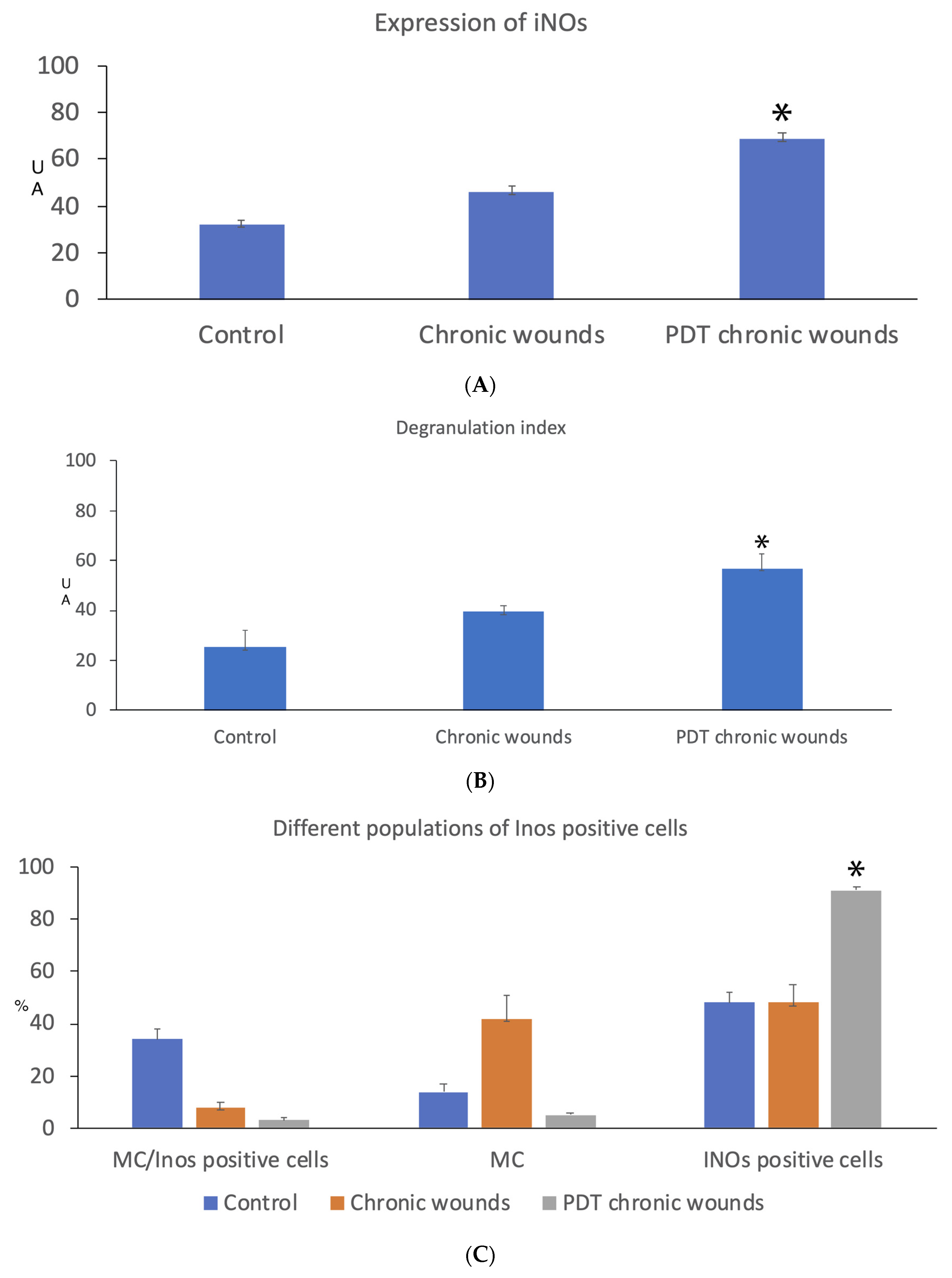
- (a)
- The morphometric evaluation of iNOs shows an increase in average surface area of about 50% in the wound and about double in the treated one, underlining the ability of PDT to influence the activity of iNOs itself (Figure 1A);
- (b)
- Avidin-positive cells, i.e., MCs, were located around vessels and nerve fibers. They increase in density as well as their degranulation index upon photodynamic therapy (Figure 1B, see page 4);
- (c)
- The percentage of MCs containing iNOs decreases significantly upon PDT (Figure 1C). Simultaneously, the % of cells containing iNOs increases significantly;
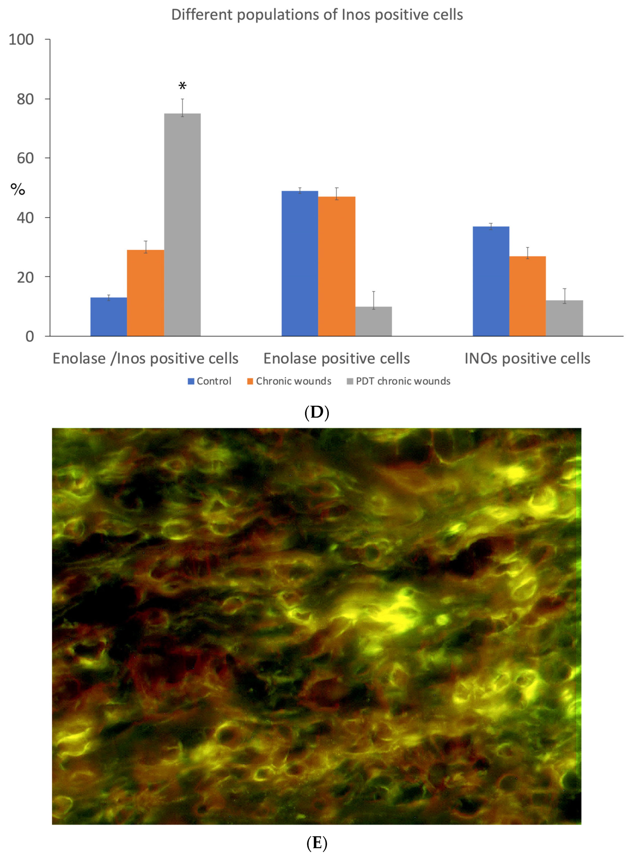
- (d)
- How the percentage of iNOs increases after treatment with PDTI is observed in the neuronal population. Surprisingly, the proportion of other cell types containing iNOS after treatment with photodynamic therapy decreases significantly compared to the control (Figure 1D, see page 5);
- (e)
- The granulocytes expressed iNOs in chronic wounds and also in those treated with PDT (Figure 1E).
4. Conclusions
Supplementary Materials
Author Contributions
Funding
Institutional Review Board Statement
Informed Consent Statement
Data Availability Statement
Conflicts of Interest
References
- Grandi, V.; Corsi, A.; Pimpinelli, N.; Bacci, S. Cellular Mechanisms in Acute and Chronic Wounds after PDT Therapy: An Update. Biomedicines 2022, 10, 1624. [Google Scholar] [CrossRef]
- Tottoli, E.M.; Dorati, R.; Genta, I.; Chiesa, E.; Pisani, S.; Conti, B. Skin Wound Healing Process and New Emerging Technologies for Skin Wound Care and Regeneration. Pharmaceutics 2020, 12, 735. [Google Scholar] [CrossRef]
- Grandi, V.; Sessa, M.; Pisano, L.; Rossi, R.; Galvan, A.; Gattai, R.; Mori, M.; Tiradritti, L.; Bacci, S.; Zuccati, G.; et al. Photodynamic therapy with topical photosensitizers in mucosal and semimucosal areas: Review from a dermatologic perspective. Photodiaggnosis Photodyn. Ther. 2018, 23, 119–131. [Google Scholar] [CrossRef]
- Steinman, L. Elaborate interactions between the immune and nervous systems. Nature Immunol. 2004, 5, 575–581. [Google Scholar] [CrossRef]
- Grandi, V.; Paroli, G.; Puliti, E.; Bacci, S.; Pimpinelli, N. Single ALA-PDT irradiation induces increase in mast cells degranulation and neuropeptide acute response in chronic venous ulcers: A pilot study. Photodiagnosis Photodyn Ther. 2021, 34, 102222. [Google Scholar] [CrossRef]
- Lee, M.; Rey, K.; Besler, K.; Wang, C.; Choy, J. Immunobiology of Nitric Oxide and Regulation of Inducible Nitric Oxide Synthase. Results Probl. Cell Diff. 2017, 62, 181–207. [Google Scholar] [CrossRef]
- Shi, H.P.; Most, D.; Efron, D.T.; Tantry, U.; Fischel, M.H.; Barbul, A. The role of iNOs in wound healing. Surgery 2001, 130, 225–229. [Google Scholar] [CrossRef]
- Bacci, S.; Pieri, L.; Buccoliero, A.M.; Bonelli, A.; Taddei, G.; Romagnoli, P. Smooth muscle cells, dendritic cells and mast cells are sources of TNFalpha and nitric oxide in human carotid artery atherosclerosis. Thrombosis Res. 2008, 122, 657–667. [Google Scholar] [CrossRef]


Disclaimer/Publisher’s Note: The statements, opinions and data contained in all publications are solely those of the individual author(s) and contributor(s) and not of MDPI and/or the editor(s). MDPI and/or the editor(s) disclaim responsibility for any injury to people or property resulting from any ideas, methods, instructions or products referred to in the content. |
© 2023 by the authors. Licensee MDPI, Basel, Switzerland. This article is an open access article distributed under the terms and conditions of the Creative Commons Attribution (CC BY) license (https://creativecommons.org/licenses/by/4.0/).
Share and Cite
Notari, L.; Nardini, P.; Grandi, V.; Corsi, A.; Pimpinelli, N.; Bacci, S. Neuroimmunomodulation in Chronic Wound Healing after Treatment with Photodynamic Therapy: The Role of iNOs. Med. Sci. Forum 2023, 21, 44. https://doi.org/10.3390/ECB2023-14135
Notari L, Nardini P, Grandi V, Corsi A, Pimpinelli N, Bacci S. Neuroimmunomodulation in Chronic Wound Healing after Treatment with Photodynamic Therapy: The Role of iNOs. Medical Sciences Forum. 2023; 21(1):44. https://doi.org/10.3390/ECB2023-14135
Chicago/Turabian StyleNotari, Lorenzo, Patrizia Nardini, Vieri Grandi, Alessandro Corsi, Nicola Pimpinelli, and Stefano Bacci. 2023. "Neuroimmunomodulation in Chronic Wound Healing after Treatment with Photodynamic Therapy: The Role of iNOs" Medical Sciences Forum 21, no. 1: 44. https://doi.org/10.3390/ECB2023-14135