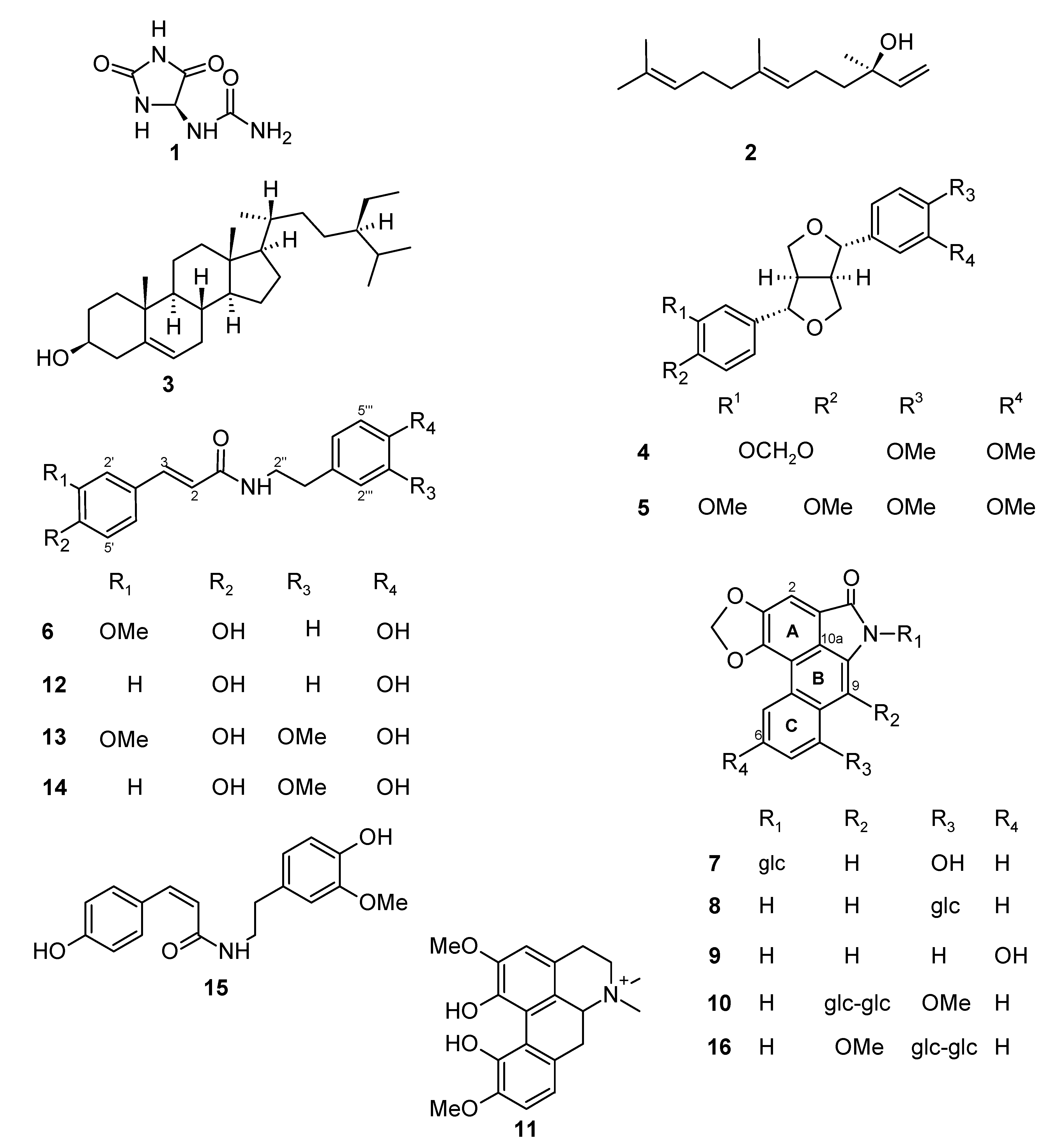
Aristolactams and Alkamides of Aristolochia gigantea

Instituto de Química, Universidade Estadual Paulista, CP 355, 14801-970 Araraquara-SP, Brazil
*
Author to whom correspondence should be addressed.
Molecules 2010, 15(12), 9462-9472; https://doi.org/10.3390/molecules15129462
Submission received: 24 November 2010
/
Revised: 16 December 2010
/
Accepted: 17 December 2010
/
Published: 21 December 2010
(This article belongs to the Section Natural Products Chemistry)
Abstract
:A new aristolactam, aristolactam 9-O-β-D-glucopyranosyl-(1→2)-β-D-glucoside, and two alkamides, N-cis- and N-trans-p-coumaroyl-3-O-methyldopamine, were isolated from stems of Aristolochia gigantea, together with the known compounds allantoin, E-nerolidol, β-sitosterol, (+)-kobusin, (+)-eudesmin, trans-N-feruloyltyramine, trans-N-coumaroyltyramine, trans-N-feruloyl-3-O-methyldopamine, aristolactam Ia-N-β-D-glucoside, aristolactam Ia 8-β-D-glucoside, aristolactam IIIa, and magnoflorine. Their structures were determined by spectroscopic analyses.
1. Introduction
The Aristolochiaceae family consists of 450 to 600 species, among which more than 200 have been at least partially studied [1]. Most of these studies have focused on a characteristic group of phenanthrenic compounds, which includes the aristolochic acids (AAs) and the aristolactams (ALs), the former of which occur mainly in species of the genus Aristolochia.
In some European countries and until recently in Brazil, Aristolochia herbs have been used in weight-loss regimens. The clinical application of aristolochic acid (AA) has been limited due to its severe nephrotoxic activity. Recent studies have revealed that AA-I can cause direct damage to renal tubular cells, and its carcinigenicity is associated with the formation of promutagenic AA-DNA adducts [2,3]. The cytotoxic potency of AL-I is higher than that of AA-I, and the cytotoxic effects of these molecules are mediated through the induction of apoptosis in a caspase 3-dependent pathway [3]. Consequently many countries have now banned the use of herbs containing AAs and ALs and the US Food and Drug Administration has banned the sale of all products that contain AAs and ALs in their formulations [4].
Recently, the aristolactams have received much attention due to an interesting array of biological properties, including anti-inflammatory, antiplatelet, antimycobacterial, and neuro-protective activities [5]. Naturally occurring aristolactams and several synthetic aristolactam derivatives have been shown to have potent antitumor activities against a broad array of human cancer cell lines. Several aristolactams which may possess postcoital antifertility activity have been isolated from Aristolochia indica. In addition, neurological disorders, especially Parkinson’s disease, have been treated by administration of the aristolactam taliscanine [6]. Brazilian Aristolochia species, including Aristolochia gigantea, have been used in traditional medicine as abortifacients and in the treatment of wounds and skin diseases [7].
Aristolochia gigantea develops a strong system of subterranean stems and roots (tuberous or rhizomatous roots). α-Phellandrene (60.9%) and linalool (16.6%) are the major constituents of the essential oil obtained from these plant parts [8], whereas germacrene D and γ-elemene are the most abundant compounds in the leaf oils. trans-Nerolidol and geraniol are the major constituents in the stem and flower oils, respectively [9]. Previous studies on the leaves of this plant have also led to the isolation of allantoin and sitosterol [7], which are also found in significant quantities in other Aristolochiaceae species. In addition, salsolinol, higenamine, and pinitol have been isolated together with several bisbenzylisoquinolinic and 8-benzylberberinic alkaloids from A. gigantea. These latter compounds have an unusual carbon skeleton [7,10,11]. As part of our continuing studies on the Aristolochiaceae family, we report here the isolation and structural elucidation of aristolactams and alkamides, among other compounds, from aerial and ground (rhizomes) stems of A. gigantea.
2. Results and Discussion
Compounds 1–15 (Figure 1) were isolated by chromatography and partition procedures from the ethanol extracts of the stems and analyzed by spectrometric methods (IR, UV, MS, 1D- and 2D-NMR).Phytochemical studies on the ethanol extract from rhizomes of A. gigantea led to the isolation of 10 known compounds: allantoin (1) [12], E-nerolidol (2) [13,14], β-sitosterol (3) [15,16], (+)-kobusin (4) [17], (+)-eudesmin (5) [17], trans-N-feruloyltyramine (6) [18], aristolactam Ia N-β-D-glucoside (7) [19], aristolactam Ia 8-β-D-glucoside (8) [20,21], aristolactam IIIa (9) [22], and magnoflorine (11) [23], together with a new aristolactam (10). In addition, four known compounds, (+)-kobusin (4), trans-N-feruloyltyramine (6), trans-N-coumaroyltyramine (12) [18], and trans-N-feruloyl-3-O-methyldopamine (13) [24], and a mixture of cis and trans new alkamides (14 + 15) were obtained from the aerial stems. The structures of the known compounds were determined by analyses of their physical and spectroscopic data and comparison of these data to those reported in the literature and to those of authentic samples available in our laboratory, which were previously isolated from Aristolochia spp.
A molecular formula of C29H31O15N was determined for compound 10 based on its HRMS spectra, which showed quasi-molecular ions at m/z 632.1614 [M – H]−. The IR spectrum of compound 10 showed characteristic absorption bands of a lactam group at 1,654 cm−1 and hydroxyl groups at 3,442 and 1,088 cm−1. The DEPT and 13C-NMR spectra of 10 (Table 1) showed signals for 14 aromatic carbons, and acyl (δC 167.4), methylenedioxy (δC 103.0), and methoxyl (δC 56.0) groups.
Figure 1.
Chemical structures of compounds 1–16.

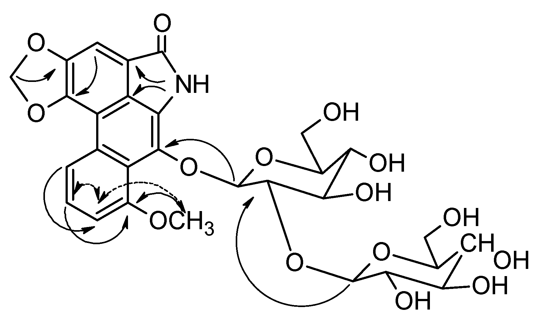
The 1H-NMR spectrum showed signals characteristic of CONH at δ 10.18 and only four aromatic hydrogens at δ 8.26 (dd, J = 8.5, 1.0), 7.56 (dd, J = 8.5, 8.0), 7.23 (dd, J = 8.0, 1.0), and 7.65 (s). In addition, signals for carbons and hydrogens for a diglycosyl were observed. These data suggested that compound 10 was an aristolactam. 1H-1H COSY and 1D-TOCSY experiments allowed us to determine that the glycosyl units were β-glucosyl-β-glucosyl (1→2). Furthermore, the negative ESI-MS/MS of the ions at m/z 632.1 gave rise to ions at m/z 308.1 and 469.9 that suggested C9-O and O-C1'' fragmentations, respectively. The substituent positions on the aristolactam structure were assigned with the help of gHMBC experiments (Figure 2). These experiments showed correlations between C-9 (δC 132.6) and H-1' (δH 5.08); C-3(δC 148.2) and CH2O2 (δH 6.46); C-8 (δC 157.2) and OCH3 (δH 3.94) and H-6 (δH 7.56), as well as between C-2′ (δC 81.0) and H-1″ (δH 4.65).
Figure 2.
Selected gHMBC (→) correlations and nOe (↔) interactions for aristolactam 10.

The 1H- and 13C-NMR, IR, and UV spectroscopic data of 10 were very similar to those reported in the literature for triangularine (16; Figure 1) [25], the main difference being due to interchange of the substituents at C-8 and C-9. The location of the methoxyl group at C-9 in 10 was corroborated by gNOESY experiments that showed an interaction between CH3O and H-7. This new aristolactam was named aristolactam 9-O-β-D-glucopyranosyl-(1→2)-β-D-glucoside.

| Position | δH | δC | Position | δH | δC |
|---|---|---|---|---|---|
| 1 | 119.1 | OCH2O | 6.46 s | 103.0 | |
| 2 | 7.65 s | 105.7 | OCH3 | 3.94 s | 56.0 |
| 3 | 148.2 | 1′ | 5.08 d (6.5) | 103.0 | |
| 4 | 146.9 | 2′ | 3.92 dd (8.5, 6.5) | 81.0 | |
| 4a | 109.0 | 3′ | 3.54 t (8.5) | 75.8 | |
| 4b | 127.9 | 4′ | 3.47 t (8.5) | 69.4 | |
| 5 | 8.26 dd (8.5, 1.0) | 118.4 | 5′ | 3.18 m | 76.9 |
| 6 | 7.56 dd (8.0, 8.5) | 126.1 | 6′α, 6′β | 3.8 − 3.6 m | 60.5 |
| 7 | 7.23 dd (8.0, 1.0) | 110.8 | 1″ | 4.65 d (7.5) | 102.4 |
| 8 | 157.2 | 2″ | 3.06 dd (7.5, 8.5) | 74.1 | |
| 8a | 120.0 | 3″ | 3.10 t (8.5) | 76.1 | |
| 9 | 132.6 | 4″ | 3.17 t (8.5) | 69.3 | |
| 10 | b | 5″ | 2.99 ddd (8.5, 4.7, 2.5) | 76.4 | |
| 10a | 124.4 | 6″α, 6″β | 3.35 m | 60.3 | |
| 3.8 − 3.6 m | |||||
| CO | 167.4 | NH | 10.18 s |
a The 1H- and 13C-NMR data were assigned with the assistance of gHMQC, gHMBC, and 1H-1H COSY experiments (11.7 T); recorded in DMSO-d6; J in Hz; b Signal not observed.
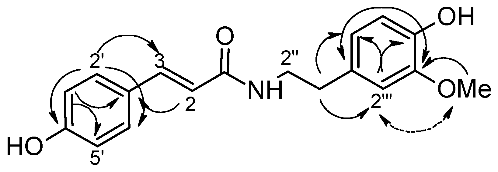
The 1H- and 13C-NMR spectra of 14 + 15 (Table 2) were very similar to those of 13, except for the absence of a methoxyl group at C-3' in 13, and suggested that it consisted of cis- and trans-alkamides with p-disubstituted and trisubstituted aromatic rings. The molecular formula (C18H19O4N) deduced from the HRMS spectra was also consistent with the lack of an OCH3 substituent. Based on the integration of the signals corresponding to the olefinic hydrogens [cis: δH-2 5.75 (d, J = 13.0) and δH-3 6.48 (d, J = 13.0); trans: δH-2 6.38 (d, J = 15.5) and δH-3 7.28 (d, J = 15.5)] it was possible to determine that the isolated mixture was in a 1:2 cis/trans proportion. Although cis and trans isomers can isomerize under UV light, both alkamide isomers may be natural compounds [26,27]. To assign with confidence all of the chemical shifts for carbons and hydrogens in the structures, this mixture was exposed to daylight for four hours. The subsequent 1H-NMR spectrum revealed that the cis/trans proportion had changed to 2:1. The linkage of the methoxyl group to C-3''' was established based on the observation of a correlation between this carbon and the methoxyl hydrogens by gHMBC experiments, as well as by the spatial interactions of methoxyl hydrogens with H-2''', as observed by 1D-NOESY experiments (Figure 3). Correlations observed by gHMBC experiments also assisted to determine the carbon skeleton. These alkamides 14 and 15 were named N-cis- and N-trans-p-coumaroyl-3-O-methyldopamine, respectively.
| Position | 14 δH | 15 δH |
|---|---|---|
| 2 | 6.38 d (15.5) | 5.75 d (13.0) |
| 3 | 7.28 d (15.5) | 6.48 d (13.0) |
| 2′, 6′ | 7.36 d (8.5) | 7.56 d (8.5) |
| 3′, 5′ | 6.76 d (8.5) | 6.68 d (8.5) |
| 2′ | 3.34 m b | 3.34 m b |
| 3′ | 2.62 t (5.5) | 2.62 t (5.5) |
| 2′′′ | 6.75 d (2.0) | 6.74 d (2.0) |
| 5′′′ | 6.66 d (8.0) | 6.66 d (8.0) |
| 6′′′ | 6.59 dd (8.0, 2.0) | 6.58 dd (8.0, 2.0) |
| OCH3 | 3.72 s | 3.71 s |
| NH | 8.00 t (5.5) | 7.98 t (5.5) |
a Recorded in DMSO-d6, 500 MHz, J in Hz; b Signals assigned with the assistance of 1H-1H COSY experiments.
Figure 3.
Select gHMBC (→) correlations and nOe (↔) interactions for alkamide 14.

Allantoin (1) is a product of purine metabolism and is widely distributed in biological systems. It has been isolated from marine sponges, animals, and numerous plants, including Aristolochia species. It is used as an anti-inflammatory, antipsoriatic (disputed), and topical vulnerary agent [28]. Allantoin is one of the best-known wound-healing agents, and exerts keratolytic and astringent effects and stimulates new tissue formation [29]. Other well-known compounds that were isolated from A. gigantea include E-nerolidol, which has been shown to possess larvicidal activity against Aedes aegypti [30] and antifungal activity against Microsporum gypseum [31], and magnoflorine, which exhibits insecticidal activity against Spodoptera frugiperda [32], among others activities [33].
3. Experimental
3.1. General
One-dimensional (1H, 13C, DEPT, and gNOESY) and two-dimensional (1H–1H gCOSY, gNOESY, gHMQC, and 1H–13C gHMBC) NMR experiments were performed on a Varian INOVA 500 spectrometer (11.7 T) at 500 MHz (1H) and 126 MHz (13C), using deuterated solvents (CDCl3, DMSO-d6) (99.98% D) as an internal standard for 13C-NMR chemical shifts, and residual solvent as an internal standard for 1H NMR. δ values are reported relative to TMS. Mass spectra (ESI-MS and ESI-MS/MS) were obtained on a Thermo LCQ, and flow injection into the electrospray source was used for ESI-MS. High-resolution mass spectra (HRMS) were obtained on a Bruker Daltonics MicroTOF Ic (ESI-TOFMS). IR spectra were obtained on a Perkin Elmer FT-IR Spectrum 2000 spectrometer using KBr discs. Optical rotations were measured on a Perkin–Elmer 341-LC polarimeter. Ultraviolet (UV) absorptions were measured on a Perkin–Elmer UV–vis Lambda 14P diode array spectrophotometer. HPLC analyses were performed using a Shimadzu liquid chromatograph (SPD-10 Avp) equipped with UV–Vis and 341-LC polarimeter detectors. RP-18 columns were used (Varian, C18, with a particle size of 5 µm, 250 by 4.6 mm for analytical analysis and 250 by 20 mm for semi-preparative analysis), and chromatograms were acquired at 336 and 254 nm. Melting points were recorded on a Microquímica MQAPF-302 melting point apparatus and are uncorrected.
3.2. Plant material
The plant material was collected in Araraquara, SP, Brazil, in February, 2004, and identified as Aristolochia gigantea Mart. (Aristolochiaceae) by Dr. Lindolpho Capellari Júnior (Escola Superior de Agricultura “Luiz de Queiroz” (ESALQ), Piracicaba, SP, Brazil). A voucher specimen (ESA 88281) was deposited at the herbarium of the ESALQ, Piracicaba, SP, Brazil. The material was separated according to the plant parts and dried (ca. 45 °C). The stems were further separated into aerial stems and rhizomes.
3.3. Extraction and isolation of the chemical constituents
The rhizomes (433.6 g) and aerial stems (379.4 g) were ground and exhaustively extracted successively at room temperature with hexane, acetone, and ethanol. The residues were extracted with ethanol in a Soxhlet apparatus and the extracts were individually concentrated. A portion of the crude ethanol extract of rhizomes (8.10 g) was washed with CH3OH. Compound 1 (43.0 mg) was isolated from the insoluble fraction. The methanol-soluble fraction was subjected to CC (6.0 by 40.0 cm, silica gel 60H, 127.3 g, hexane/CH3OH gradient, 19:1 to 100% CH3OH) to give 25 fractions (ca. 125 mL each). Fractions 7, 9, 12, 14, and 15 gave 2 (23.3 mg), 3 (25.0 mg), 4 (409.2 mg), 5 (158.3 mg), and 6 (26.2 mg), respectively. Fraction 23 after HPLC [Varian RP C18 semi-preparative column, eluted with CH3OH–H2O + 0.5% NH4OH, 3:2, flow = 8 mL min−1; λ = 254 nm] gave 1 (16.5 mg) and 11 (17.5 mg). Fraction 21 (1.10 g) was subjected to RP CC (3.0 by 45.0 cm, silica gel C18, 43.5 g, CH3OH–H2O gradient 9:1 to 100% CH3OH) to give 17 subfractions (ca. 125 mL each). Subfractions 10 and 11 (98.4 mg) after HPLC [Varian RP C18 column, eluted with CH3OH–H2O + 0.5% NH4OH 3:2, flow = 8 mL min−1, λ = 254 nm] gave 7 (4.0 mg), 8 (9.6 mg), 9 (1.8 mg), and 10 (10.1 mg). The stem crude ethanol extract (10.0 g) was fractionated over Sephadex LH-20 (120.0 g, 2.5 by 95.0 cm, MeOH) to give 17 fractions. Fraction 6 (166 mg) was subjected to two HPLC runs [Varian RP C18 semi-preparative column, eluted with CH3OH–H2O 3:2, flow = 8 mL min−1, λ = 254 nm; followed by Varian RP C18 analytical column, eluted with CH3OH–H2O 2:3, flow = 0.8 mL min−1, λ = 254 nm] to give 4 (2.0 mg), 12 (2.0 mg), 13 (1.5 mg), and 14+15 (1.0 mg)].
3.4. Spectral data
Allantoin (1). Yellow needles. mp 233–234 °C [lit. 232–235 °C] [12]. IR, 1H-NMR, and 13C-NMR data were consistent with those previously reported [12].
(−)E-Nerolidol (2). Yellow oil. ![Molecules 15 09462 i001]() −17 ° (CHCl3, c 0.2) [lit. −12.5 ° (CHCl3, c 0.02)] [13]. 1H-NMR (CDCl3) δ 5.15 (1H, dd, J 17.5, 1.5 Hz, H-1α), 5.00 (1H, dd, J 10.5, 1.5 Hz, H-1β), 5.86 (1H, dd, J 17.5, 10.5 Hz, H-2), 1.54–1.51 (2H, m, H-4), 2.00–1.92 (6H, m, H-5, H-8, H-9), 5.08–5.01 (2H, m, H-6, H-10), 1.60 (3H, s, H-12), 1.22 (3H, s, H-15), 1.54 (6H, s, H-13, H-14). 13C-NMR data were consistent with those previously reported [14].
−17 ° (CHCl3, c 0.2) [lit. −12.5 ° (CHCl3, c 0.02)] [13]. 1H-NMR (CDCl3) δ 5.15 (1H, dd, J 17.5, 1.5 Hz, H-1α), 5.00 (1H, dd, J 10.5, 1.5 Hz, H-1β), 5.86 (1H, dd, J 17.5, 10.5 Hz, H-2), 1.54–1.51 (2H, m, H-4), 2.00–1.92 (6H, m, H-5, H-8, H-9), 5.08–5.01 (2H, m, H-6, H-10), 1.60 (3H, s, H-12), 1.22 (3H, s, H-15), 1.54 (6H, s, H-13, H-14). 13C-NMR data were consistent with those previously reported [14].
 −17 ° (CHCl3, c 0.2) [lit. −12.5 ° (CHCl3, c 0.02)] [13]. 1H-NMR (CDCl3) δ 5.15 (1H, dd, J 17.5, 1.5 Hz, H-1α), 5.00 (1H, dd, J 10.5, 1.5 Hz, H-1β), 5.86 (1H, dd, J 17.5, 10.5 Hz, H-2), 1.54–1.51 (2H, m, H-4), 2.00–1.92 (6H, m, H-5, H-8, H-9), 5.08–5.01 (2H, m, H-6, H-10), 1.60 (3H, s, H-12), 1.22 (3H, s, H-15), 1.54 (6H, s, H-13, H-14). 13C-NMR data were consistent with those previously reported [14].
β-Sitosterol (3). Colorless crystals. ![Molecules 15 09462 i001]() −15.2 ° (CHCl3, c 0.2) [lit. −26.1 ° (CHCl3, c 0.1)] [15]. 13C- NMR data were consistent with those previously reported [16].
−15.2 ° (CHCl3, c 0.2) [lit. −26.1 ° (CHCl3, c 0.1)] [15]. 13C- NMR data were consistent with those previously reported [16].
−15.2 ° (CHCl3, c 0.2) [lit. −26.1 ° (CHCl3, c 0.1)] [15]. 13C- NMR data were consistent with those previously reported [16].(+)-Kobusin (4). Yellow solid. ![Molecules 15 09462 i001]() +51.4 ° (CHCl3, c 0.21) [lit. +58.0 ° (CHCl3, c 0.03)] [17]. 1H- NMR (CDCl3) δ 6.83 (1H, d, J 2.0 Hz, H-2), 6.78 (1H, d, J 2.0 Hz, H-2′), 6.71 (1H, d, J 8.0 Hz, H-5), 6.77 (1H, d, J 8.0 Hz, H-5′), 6.81 (1H, dd, J 2.0, 8.0 Hz, H-6), 6.74 (1H, dd, J 2.0, 8.0 Hz, H-6′), 4.67 (2H, d, J 5.5 Hz, H-7β, H-7′β), 3.02 (2H, m, H-8α, H-8′α), 4.18 (2H, dd, J 7.0, 14.0 Hz, H-9β, H-9′β), 3.82 (2H, m, H-9α, H-9′α), 3.83 (3H, s, OCH3), 3.80 (3H, s, OCH3), 5.85 (2H, s, OCH2O).
+51.4 ° (CHCl3, c 0.21) [lit. +58.0 ° (CHCl3, c 0.03)] [17]. 1H- NMR (CDCl3) δ 6.83 (1H, d, J 2.0 Hz, H-2), 6.78 (1H, d, J 2.0 Hz, H-2′), 6.71 (1H, d, J 8.0 Hz, H-5), 6.77 (1H, d, J 8.0 Hz, H-5′), 6.81 (1H, dd, J 2.0, 8.0 Hz, H-6), 6.74 (1H, dd, J 2.0, 8.0 Hz, H-6′), 4.67 (2H, d, J 5.5 Hz, H-7β, H-7′β), 3.02 (2H, m, H-8α, H-8′α), 4.18 (2H, dd, J 7.0, 14.0 Hz, H-9β, H-9′β), 3.82 (2H, m, H-9α, H-9′α), 3.83 (3H, s, OCH3), 3.80 (3H, s, OCH3), 5.85 (2H, s, OCH2O).
+51.4 ° (CHCl3, c 0.21) [lit. +58.0 ° (CHCl3, c 0.03)] [17]. 1H- NMR (CDCl3) δ 6.83 (1H, d, J 2.0 Hz, H-2), 6.78 (1H, d, J 2.0 Hz, H-2′), 6.71 (1H, d, J 8.0 Hz, H-5), 6.77 (1H, d, J 8.0 Hz, H-5′), 6.81 (1H, dd, J 2.0, 8.0 Hz, H-6), 6.74 (1H, dd, J 2.0, 8.0 Hz, H-6′), 4.67 (2H, d, J 5.5 Hz, H-7β, H-7′β), 3.02 (2H, m, H-8α, H-8′α), 4.18 (2H, dd, J 7.0, 14.0 Hz, H-9β, H-9′β), 3.82 (2H, m, H-9α, H-9′α), 3.83 (3H, s, OCH3), 3.80 (3H, s, OCH3), 5.85 (2H, s, OCH2O).(+)-Eudesmin (5). Yellow oil. ![Molecules 15 09462 i001]() +17.5 ° (CHCl3, c 0.12) [lit. +61 ° (CHCl3, c 0.4)] [17]. 1H-NMR and 13C-NMR data were consistent with those previously reported [17].
+17.5 ° (CHCl3, c 0.12) [lit. +61 ° (CHCl3, c 0.4)] [17]. 1H-NMR and 13C-NMR data were consistent with those previously reported [17].
+17.5 ° (CHCl3, c 0.12) [lit. +61 ° (CHCl3, c 0.4)] [17]. 1H-NMR and 13C-NMR data were consistent with those previously reported [17].trans-N-Feruloyl tyramine (6). Amorphous solid. 1H-NMR (DMSO-d6) δ 6.43 (1H, d, J 15.9, H-2), 7.31 (1H, d, J 15.9, H-3), 7.11 (1H, d, J 1.8, H-2°), 6.79 (1H, d, J 8.1, H-5′), 6.98 (1H, dd, J 8.1, 1.8, H-6′), 3.32 (2H, m, H-2″), 2.65 (2H, t, J 7.2, H-3″), 6.68 (2H, d, J 8.5, H-2′′′, H-6′′′), 7.00 (2H, d, J 8.5, H-3′′′, H-5′′′), 3.80 (3H, s, OCH3), 7.95 (1H, t, J 5.7, NH).
Aristolactam Ia N-β-D-glucoside (7). Amorphous solid. ![Molecules 15 09462 i002]() −7.9 ° (MeOH, c 0.1) [lit. −9.9 ° (MeOH, c 0.07)] [19]. 1H-NMR data were consistent with those previously reported [19].
−7.9 ° (MeOH, c 0.1) [lit. −9.9 ° (MeOH, c 0.07)] [19]. 1H-NMR data were consistent with those previously reported [19].
 −7.9 ° (MeOH, c 0.1) [lit. −9.9 ° (MeOH, c 0.07)] [19]. 1H-NMR data were consistent with those previously reported [19].
Aristolactam Ia 8-β-D-glucoside (8). Amorphous solid. ![Molecules 15 09462 i002]() −8.2 ° (MeOH, c 0.1) [lit. −10.5 ° (MeOH, c 0.2)] [20]. 1H-NMR data were consistent with those previously reported [20].
−8.2 ° (MeOH, c 0.1) [lit. −10.5 ° (MeOH, c 0.2)] [20]. 1H-NMR data were consistent with those previously reported [20].
−8.2 ° (MeOH, c 0.1) [lit. −10.5 ° (MeOH, c 0.2)] [20]. 1H-NMR data were consistent with those previously reported [20].Aristolactam IIIa (9). Amorphous solid. 1H-NMR (DMSO-d6) δ 7.62 (1H, s, H-2), 7.97 (1H, d, J 2.5, H-5), 7.10 (1H, dd, J 2.5, 8.5, H-7), 7.79 (1H, d, J 8.5, H-8), 7.05 (1H, s, H-9), 6.48 (2H, s, OCH2O), 10.65 (1H, s, NH).
Aristolactam 9-O-β-D-glucopyranosyl-(1→2)-β-D-glucoside (10). Amorphous solid. ![Molecules 15 09462 i002]() −3.9 ° (c 0.5, MeOH). 1H-NMR (CDCl3) and 13C-NMR (CDCl3) spectra see Table 1; ESI-HR-TOF-MS (probe), 4000 V, m/z (rel. int.): 632. 1614 [M − H]− (100) (calculated for C29H31O15N − H = 632.1615); ESI-MS/MS (probe) 4,500 V from ions at m/z 632.1 (100), m/z (rel. int.): 308.1 [M − glc-glc]− (92).
−3.9 ° (c 0.5, MeOH). 1H-NMR (CDCl3) and 13C-NMR (CDCl3) spectra see Table 1; ESI-HR-TOF-MS (probe), 4000 V, m/z (rel. int.): 632. 1614 [M − H]− (100) (calculated for C29H31O15N − H = 632.1615); ESI-MS/MS (probe) 4,500 V from ions at m/z 632.1 (100), m/z (rel. int.): 308.1 [M − glc-glc]− (92).
−3.9 ° (c 0.5, MeOH). 1H-NMR (CDCl3) and 13C-NMR (CDCl3) spectra see Table 1; ESI-HR-TOF-MS (probe), 4000 V, m/z (rel. int.): 632. 1614 [M − H]− (100) (calculated for C29H31O15N − H = 632.1615); ESI-MS/MS (probe) 4,500 V from ions at m/z 632.1 (100), m/z (rel. int.): 308.1 [M − glc-glc]− (92).Magnoflorine (11). Amorphous solid. ![Molecules 15 09462 i001]() +164.4 ° (MeOH, c 0.03) [lit. +150.0 ° (MeOH, c 0.1)] [23]. 1H-NMR (DMSO-d6) δ 6.49 (1H, s, H-3), 2.7–2.8 (2H, m, H-4α, H-4β), 3.7–3.6 (2H, m, H-5α, H-5β), 4.34 (1H, br d, J 13.0, H-6a), 2.59 (1H, t, J 13.0, H-7α), 3.10 (1H, br d, J 13.0, H-7β), 6.35 (1H, br d, J 8.0, H-8), 6.59 (1H, d, J 8.0, H-9), 3.65 (3H, s, OCH3-10), 3.68 (3H, s, OCH3-2), 2.88 (3H, s, N-CH3), 3.29 (3H, s, N-CH3). 13C-NMR (DMSO-d6) δ 152.1 (C-1), 151.2 (C-2), 108.8 (C-3), 111.6 (C-3a), 23.2 (C-4), 60.5 (C-5), 69.2 (C-6a), 30.4 (C-7), 125.1 (C-7a), 112.4 (C-8), 109.9 (C-9), 150.3 (C-10), 152.5 (C-11), 122.6 (C-11a), 123.1 (C-1a), 120.0 (C-1b), 55.2, 55.7 (OCH3), 42.5, 52.6 (N-CH3).
+164.4 ° (MeOH, c 0.03) [lit. +150.0 ° (MeOH, c 0.1)] [23]. 1H-NMR (DMSO-d6) δ 6.49 (1H, s, H-3), 2.7–2.8 (2H, m, H-4α, H-4β), 3.7–3.6 (2H, m, H-5α, H-5β), 4.34 (1H, br d, J 13.0, H-6a), 2.59 (1H, t, J 13.0, H-7α), 3.10 (1H, br d, J 13.0, H-7β), 6.35 (1H, br d, J 8.0, H-8), 6.59 (1H, d, J 8.0, H-9), 3.65 (3H, s, OCH3-10), 3.68 (3H, s, OCH3-2), 2.88 (3H, s, N-CH3), 3.29 (3H, s, N-CH3). 13C-NMR (DMSO-d6) δ 152.1 (C-1), 151.2 (C-2), 108.8 (C-3), 111.6 (C-3a), 23.2 (C-4), 60.5 (C-5), 69.2 (C-6a), 30.4 (C-7), 125.1 (C-7a), 112.4 (C-8), 109.9 (C-9), 150.3 (C-10), 152.5 (C-11), 122.6 (C-11a), 123.1 (C-1a), 120.0 (C-1b), 55.2, 55.7 (OCH3), 42.5, 52.6 (N-CH3).
+164.4 ° (MeOH, c 0.03) [lit. +150.0 ° (MeOH, c 0.1)] [23]. 1H-NMR (DMSO-d6) δ 6.49 (1H, s, H-3), 2.7–2.8 (2H, m, H-4α, H-4β), 3.7–3.6 (2H, m, H-5α, H-5β), 4.34 (1H, br d, J 13.0, H-6a), 2.59 (1H, t, J 13.0, H-7α), 3.10 (1H, br d, J 13.0, H-7β), 6.35 (1H, br d, J 8.0, H-8), 6.59 (1H, d, J 8.0, H-9), 3.65 (3H, s, OCH3-10), 3.68 (3H, s, OCH3-2), 2.88 (3H, s, N-CH3), 3.29 (3H, s, N-CH3). 13C-NMR (DMSO-d6) δ 152.1 (C-1), 151.2 (C-2), 108.8 (C-3), 111.6 (C-3a), 23.2 (C-4), 60.5 (C-5), 69.2 (C-6a), 30.4 (C-7), 125.1 (C-7a), 112.4 (C-8), 109.9 (C-9), 150.3 (C-10), 152.5 (C-11), 122.6 (C-11a), 123.1 (C-1a), 120.0 (C-1b), 55.2, 55.7 (OCH3), 42.5, 52.6 (N-CH3).trans-N-Coumaroyltyramine (12). 1H-NMR (DMSO-d6) δ 6.38 (1H, d, J 15.9, H-2), 7.30 (1H, d, J 15.9, H-3), 7.37 (2H, d, J 8.7, H-2′, H-6′), 6.78 (2H, d, J 8.7, H-3′, H-5′), 3.31 (2H, m, H-2″), 2.64 (2H, t, J 7.2, H-3″), 7.00 (2H, d, J 8.4, H-2′′′, H-6′′′), 6.67 (2H, d, J 8.4, H-3′′′, H-5′′′), 7.94 (1H, m, NH).
trans-N-Feruloyl-3-O-methyldopamine (13). 1H-NMR (DMSO-d6) δ 6.43 (1H, d, J 15.3, H-2), 7.30 (1H, d, J 15.3, H-3), 7.10 (1H, d, J 2.1, H-2′), 6.78 (1H, d, J 7.8, H-5′), 6.97 (1H, dd, J 7.8, 2.1, H-6°), 3.32 (2H, m, H-2″), 2.65 (2H, t, J 6.9, H-3″), 6.77 (1H, d, J 2.1, H-2′′′), 6.68 (1H, d, J 7.8, H-5′′′), 6.60 (1H, dd, J 7.8, 2.1, H-6′′′), 3.79 (3H, s, OCH3), 3.74 (3H, s, OCH3), 7.94 (1H, m, NH).
N-cis- and N-trans-p-Coumaroyl-3-O-methyldopamine (14 + 15). Colorless oil. 1H-NMR (CDCl3) spectra see Table 1. 13C-NMR (DMSO-d6) δ 137.8 (C-3), 127.0 (C-2′, 6′), 116.0 (C-3′, 5′), 113.2 (C-2′′′), 120.9 (C-6′′′), 147.4 (C-3′′′), 145.1 (C-4′′′), 130.6 (C-1′′′), 126.3 (C-1′), 159.0 (C-4′), 56.0 (OCH3). ESI-HR-TOF-MS (probe) 4,000V, m/z (rel. int.): 312.1233 [M − H]− (100) (calculated for C18H19O4N − H = 312.1236).
4. Conclusions
Extracts from different parts of A. gigantea showed a diverse chemical composition. As previously observed, the characteristic chemical constituents of the leaves of this species arebisbenzylisoquinolinic and 8-benzylberberinic alkaloids, whereas stems contain lignans at high concentrations, alkamides, and aristolactams. Among the compounds that were isolated from stems, two alkamides and an aristolactam are described here for the first time.
Acknowledgements
The authors thank Condorcet Aranha and Lindolpho Capellari Jr. for plant identification. Thank the Fundação de Amparo à Pesquisa do Estado de São Paulo (FAPESP) and Conselho Nacional de Desenvolvimento Científico e Tecnológico (CNPq/MCT/MS/PRONEX, Brazil) for financial support, and the Coordenação de Aperfeiçoamento de Nível Superior (CAPES) for providing a fellowship to J.C.H.
- Sample Availability: Samples of the compounds are available from the authors.
References and Notes
- Lopes, L.M.X.; Nascimento, I.R.; Silva, T. Phytochemistry of the Aristolochiaceae Family. In Research Advances in Phytochemistry; Mohan, R.M.M., Ed.; Global Research Network: Kerala, India, 2001; Volume 2, pp. 19–108. [Google Scholar]
- Li, Y.; Liu, Z.; Guo, X.; Shu, J.; Chen, Z.; Li, L. Aristolochic Acid I-induced DNA Damage and Cell Cycle Arrest in Renal Tubular Epithelial Cells in Vitro. Arch. Toxicol. 2006, 80, 524–532. [Google Scholar] [CrossRef]
- Li, J.; Zhang, L.; Jiang, Z.; Shu, B.; Li, F.; Bao, Q.; Zhang, L. Toxicities of Aristolochic Acid I and Aristololactam I in Cultured Renal Epithelial Cells. Toxicol. In Vitro 2010, 24, 1092–1097. [Google Scholar] [CrossRef]
- Zhang, J.; Xiao, Y.; Feng, J.; Wu, S.L.; Xue, X.; Zhang, X.; Liang, X. Selectively Preparative Purification of Aristolochic Acids and Aristololactams from Aristolochia Plants. J. Pharm. Biomed. Anal. 2010, 52, 446–451. [Google Scholar] [CrossRef]
- Choi, Y.L.; Kim, J. K.; Choi, S.-U.; Min, Y.-K.; Bae, M.-A.; Kim, B.T.; Heo, J.-N. Synthesis of Aristolactam Analogues and Evaluation of Their Antitumor Activity. Bioorg. Med. Chem. Lett. 2009, 19, 3036–3040. [Google Scholar]
- Kumar, V.; Prasad, A.K.; Parmar, V.S. Naturally Occurring Aristolactams, Aristolochic Acids and Dioxoaporphines and Their Biological Activities. Nat. Prod. Rep. 2003, 20, 565–583. [Google Scholar] [CrossRef]
- Lopes, L.M.X.; Humpfer, E. 8-Benzylberbine and N-Oxide Alkaloids from Aristolochia gigantea. Phytochemistry 1997, 45, 431–435. [Google Scholar]
- Francisco, C.S.; Messiano, G.B.; Lopes, L.M.X.; Tininis, A.G.; de Oliveira, J.E.; Capellari, L., Jr. Classification of Aristolochia Species Based on GC-MS and Chemometric Analyses of Essential Oils. Phytochemistry 2008, 69, 168–175. [Google Scholar]
- Leitão, G.G.; Lopes, D.; Menezes, F.D.S.; Kaplan, M.A.C.; Craveiro, A.A.; Alencar, J.W. Essential Oils from Brazilian Aristolochia. J. Essent. Oil Res. 1991, 3, 403–408. [Google Scholar] [CrossRef]
- Cortes, D.; Dadoun, H.; Paiva, R.L.R.; de Oliveira, A.B. Nouveaux Alcaloïdes Bis-benzylisoquinoleiques Isoles des Feuilles de Aristolochia gigantea. J. Nat. Prod. 1987, 50, 910–914. [Google Scholar] [CrossRef]
- Lopes, L.M.X. 8-Benzylberbine Alkaloids from Aristolochia gigantea. Phytochemistry 1992, 31, 4005–4009. [Google Scholar] [CrossRef]
- Sang, S.; Lao, A.; Wang, H.; Chen, Z.; Uzawa, J.; Fujimoto, Y. A Phenylpropanoid Glycoside from Vaccaria segetalis. Phytochemistry 1998, 48, 569–571. [Google Scholar]
- Cane, D.E.; Ha, H.-J.; McIlwaine, D.B.; Pascoe, K.O. The Synthesis of (3R)-Nerolidol. Tetrahedron Lett. 1990, 31, 7553–7554. [Google Scholar] [CrossRef]
- Suarez, L.E.C.; Menichini, F.; Monache, F.D. Tetranortriterpenoids and Dihydrocinnamic Acid Derivatives from Hortia colombiana. J. Braz. Chem. Soc. 2002, 13, 339–344. [Google Scholar] [CrossRef]
- Nakabayashi, R.; Kusano, M.; Kobayashi, M.; Tohge, T.; Yonekura-Sakakibara, K.; Kogure, N.; Yamazaki, M.; Kitajima, M.; Saito, K.; Takayama, H. Metabolomics-oriented Isolation and Structure Elucidation of 37 Compounds Including Two Anthocyanins from Arabidopsis thaliana. Phytochemistry 2009, 70, 1017–1029. [Google Scholar]
- Nes, W.D.; Norton, R.A.; Benson, M. Carbon-13 NMR Studies on Sitosterol Biosynthesized from [13C] Mevalonates. Phytochemistry 1992, 31, 805–811. [Google Scholar]
- Latip, J.; Hartley, T.G.; Waterman, P.G. Lignans and Coumarins Metabolites from Melico pehayesii. Phytochemistry 1999, 51, 107–110. [Google Scholar]
- Chang, Y.-C.; Chen, C.-Y.; Chang, F.-R.; Wub, Y.-C. Alkaloids from Lindera glauca. J. Chin. Chem. Soc. 2001, 48, 811–815. [Google Scholar]
- Leu, Y.-L.; Chan, Y.-Y.; Hsu, M.-Y.; Chen, I.-S.; Wu, T.-S. The Constituents of the Stem and Roots of Aristolochia foveolata. J. Chin. Chem. Soc. 1998, 45, 539–541. [Google Scholar]
- Zhang, Y.T.; Jiang, J.Q. Alkaloids from Aristolochia manshuriensis (Aristolochiaceae). Helv. Chim. Acta 2006, 89, 2665–2670. [Google Scholar] [CrossRef]
- Hegde, V.R.; Borges, S.; Patel, M.; Das, P.R.; Wu, B.; Gullo, V.P.; Chan, T.-M. New Potential Antitumor Compounds from the Plant Aristolochia manshuriensis as Inhibitors of the CDK2 Enzyme. Bioorg. Med. Chem. Lett. 2010, 20, 1344–1346. [Google Scholar] [CrossRef]
- Priestap, H.A. Seven Aristolactams from Aristolochia argentina. Phytochemistry 1985, 24, 849–852. [Google Scholar]
- Chen, J.-H.; Du, Z.-Z.; Shen, Y.-M.; Yang, Y.-P. Aporphine Alkaloids from Clematis parviloba and their Antifungal Activity. Arch. Pharm. Res. 2009, 32, 3–5. [Google Scholar] [CrossRef]
- Cutillo, F.; D’Abrosca, B.; DellaGreca, M.; Di Marino, C.; Golino, A.; Previtera, L.; Zarrelli, A. Cinnamic Acid Amides from Chenopodium album: Effects on Seeds Germination and Plant Growth. Phytochemistry 2003, 64, 1381–1387. [Google Scholar]
- Lin, W.-H.; Fu, H.-Z.; Hano, Y.; Nomura, T. Alkaloids from the Roots of Aristolochia Triangularis (I). J. Chin. Pharm. Sci. 1997, 6, 8–13. [Google Scholar]
- Pedersen, H.A.; Steffensen, S.K.; Christophersen, C. Cinnamoylphenethylamine 1H-NMR Chemical Shifts: A Concise Reference for Ubiquitous Compounds. Nat. Prod. Commun. 2010, 5, 1259–1262. [Google Scholar]
- Navickiene, H.M.D.; Lopes, L.M.X. Alkamides and Phenethyl Derivatives from Aristolochia gehrtii. J. Braz. Chem. Soc. 2001, 12, 467–472. [Google Scholar]
- Ferreira, D.T.; Alvares, P.S.M.; Houghton, P.J.; Braz-Filho, R. Constituintes Químicos das Raízes de Pyrostegia Venusta e Considerações Sobre a sua Importância Medicinal. Quim. Nova 2000, 23, 42–46. [Google Scholar] [CrossRef]
- Lee, M.-Y.; Lee, N.-H.; Jung, D.; Lee, J.-A.; Seo, C.-S.; Lee, H.; Kim, J.-H.; Shin, H.-K. Protective Effects of Allantoin Against Ovalbumin (OVA)-Induced Lung Inflammation in a Murine Model of Asthma. Int. Immunopharmacol. 2010, 10, 474–480. [Google Scholar] [CrossRef]
- Simas, N.K.; Lima, E.C.; Conceição, S.R.; Kuster, R.M.; Oliveira, A.M. Produtos Naturais para o Controle da Transmissão da Dengue: Atividade Larvicida de Myroxylon balsamum (óleo vermelho) e de Terpenóides e Fenilpropanóides. Quim. Nova 2004, 27, 46–49. [Google Scholar]
- Shen, Y.; Li, C.G.; Zhou, S.F.; Pang, E.C.K.; Story, D.F.; Xue, C.C.L. Chemistry and Bioactivity of Flos Magnoliae, a Chinese Herb for Rhinitis and Sinusitis. Curr. Med. Chem. 2008, 15, 1616–1627. [Google Scholar] [CrossRef]
- Tringali, C.; Spatafora, C.; Calì, V.; Simmonds, M.S.J. Antifeedant Constituents from Fagara macrophylla. Fitoterapia 2001, 72, 538–543. [Google Scholar] [CrossRef]
- Yokozawa, T.; Satoh, A.; Cho, E.J.; Kashiwada, Y.; Ikeshiro, Y. Protective Role of Coptidis Rhizoma Alkaloids Against Peroxynitrite-induced Damage to Renal Tubular Epithelial Cells. J. Pharm.Pharmacol. 2005, 57, 367–374. [Google Scholar]
© 2010 by the authors; licensee MDPI, Basel, Switzerland. This article is an open access article distributed under the terms and conditions of the Creative Commons Attribution license (http://creativecommons.org/licenses/by/3.0/).
Share and Cite
MDPI and ACS Style
Holzbach, J.C.; Lopes, L.M.X. Aristolactams and Alkamides of Aristolochia gigantea. Molecules 2010, 15, 9462-9472. https://doi.org/10.3390/molecules15129462
AMA Style
Holzbach JC, Lopes LMX. Aristolactams and Alkamides of Aristolochia gigantea. Molecules. 2010; 15(12):9462-9472. https://doi.org/10.3390/molecules15129462
Chicago/Turabian StyleHolzbach, Juliana C., and Lucia M. X. Lopes. 2010. "Aristolactams and Alkamides of Aristolochia gigantea" Molecules 15, no. 12: 9462-9472. https://doi.org/10.3390/molecules15129462