
Effects of Apatite Cement Containing Atelocollagen on Attachment to and Proliferation and Differentiation of MC3T3-E1 Osteoblastic Cells

Abstract
:1. Introduction
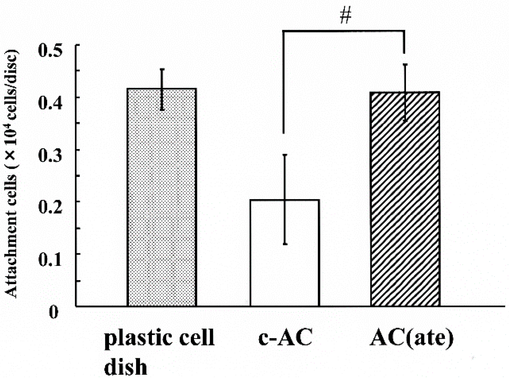
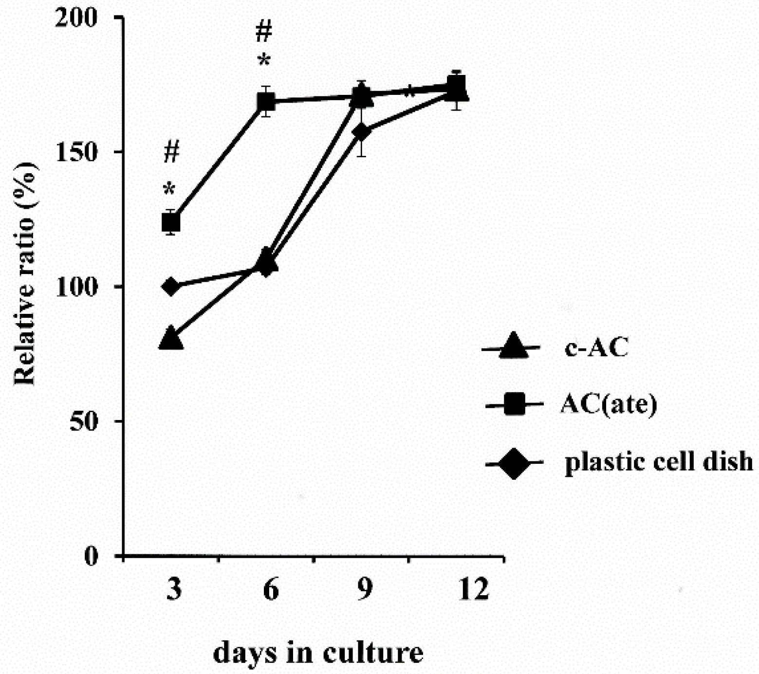
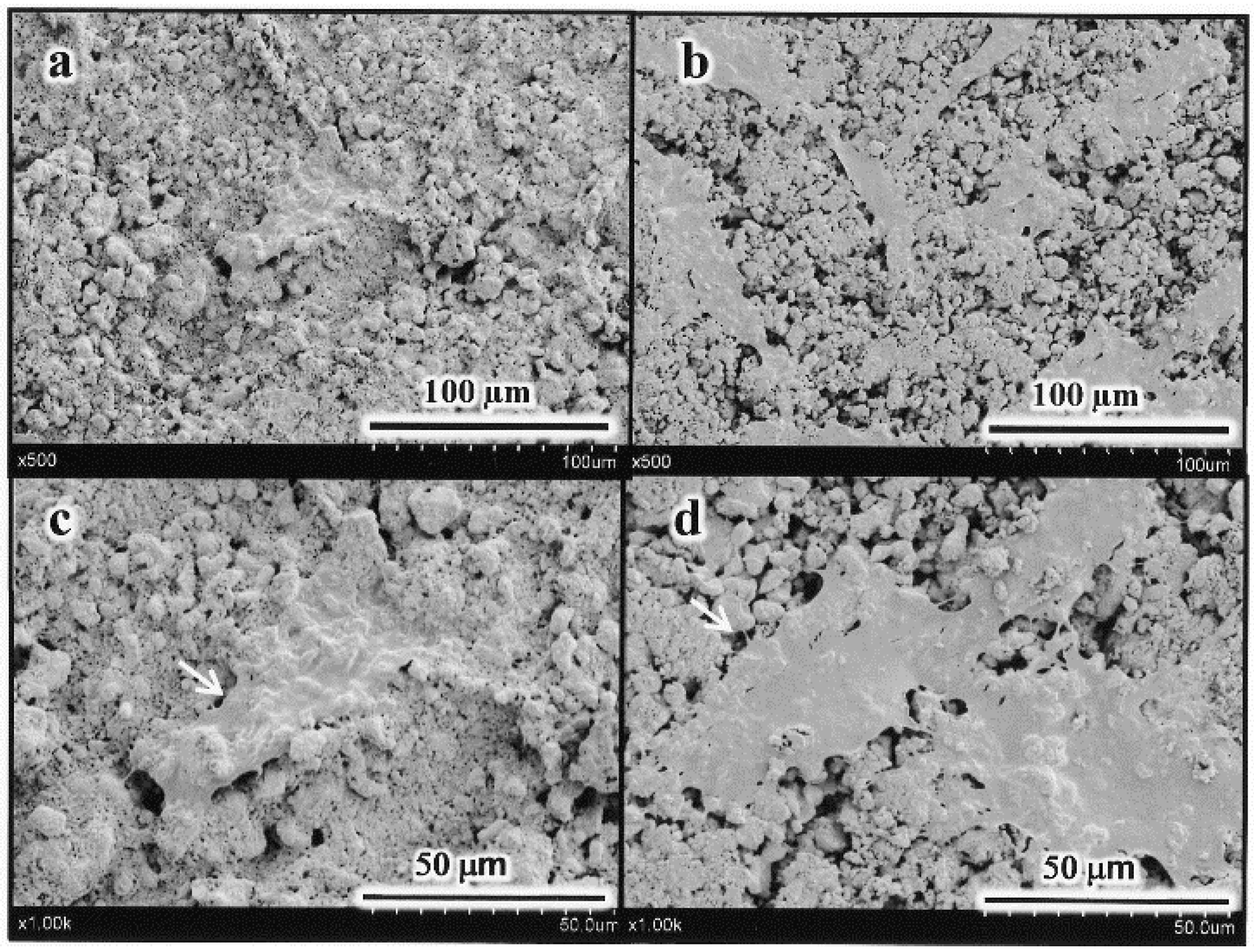
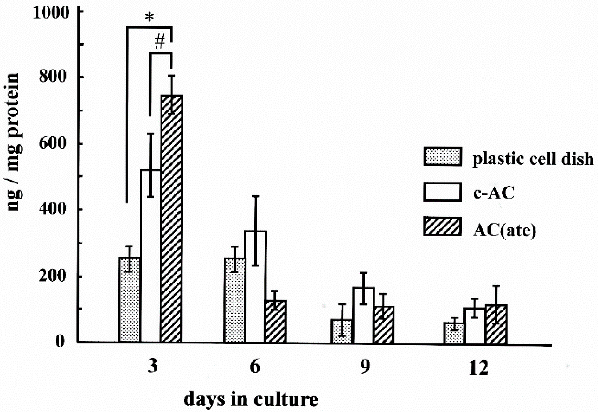
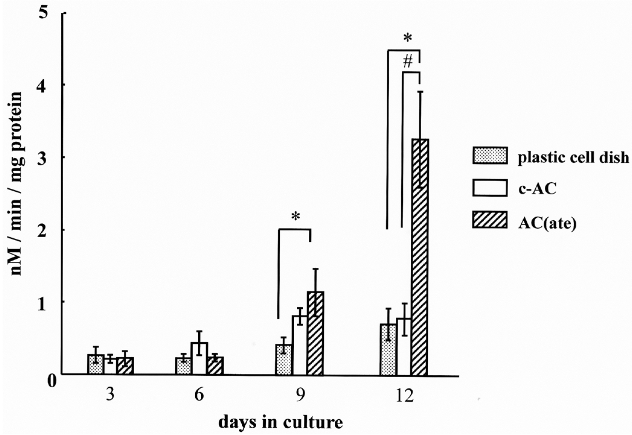
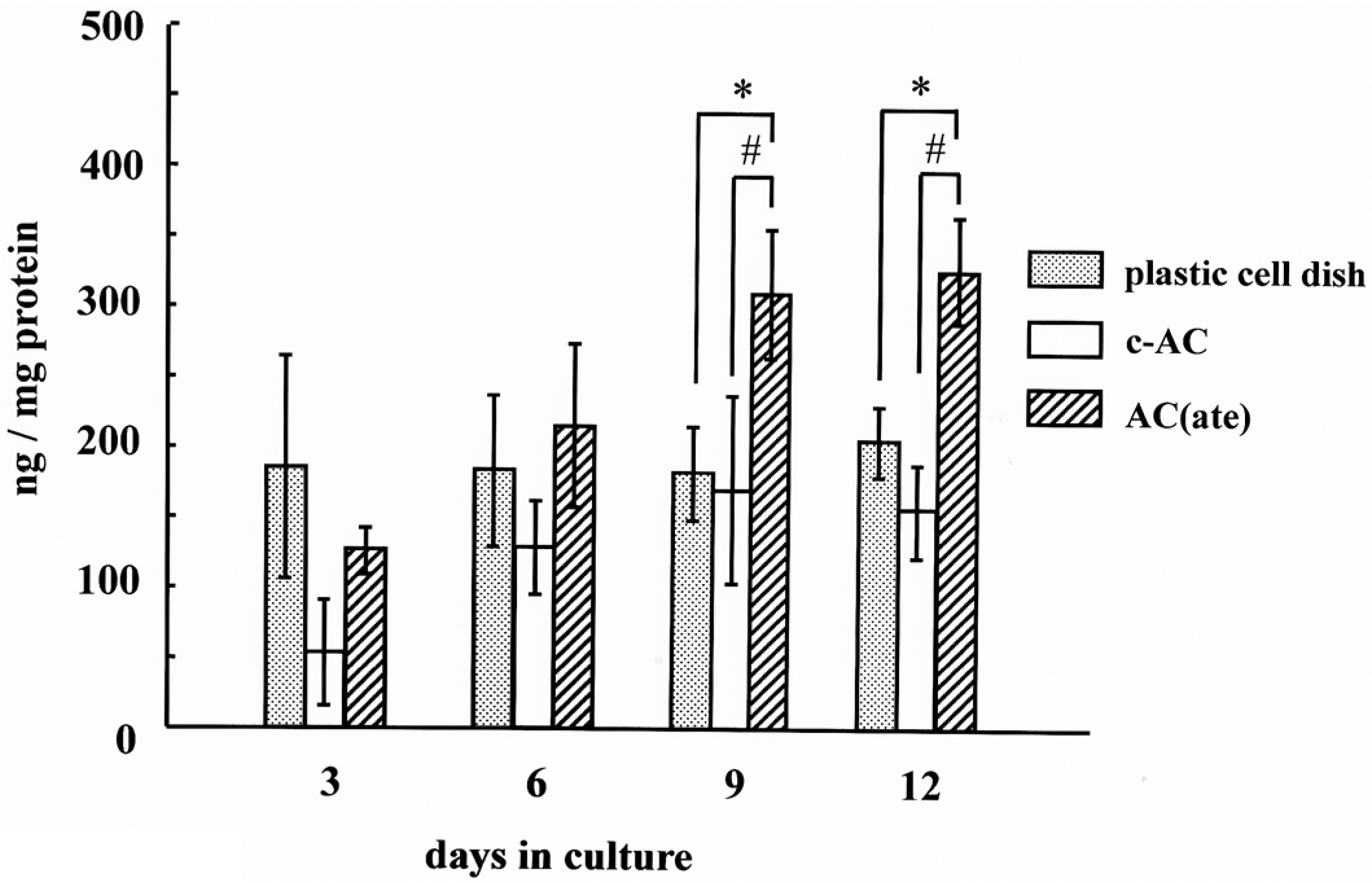
2. Results
3. Discussion
4. Materials and Methods
4.1. Preparation of AC Containing Atelocollagen
4.2. Cells and Incubation Condition
4.3. Initial Cell Attachment and Proliferation of MC3T3-E1 Cells with c-AC and AC(ate)
4.4. Alkaline Phosphatase Activity in MC3T3-E1 Cells with c-AC and AC(ate)
4.5. Production of Procollagen Type I Carboxy-Terminal Peptide and Osteocalcin in MC3T3-E1 Cells with c-AC and AC(ate)
4.6. Statistical Analysis
5. Conclusions
Acknowledgments
Author Contributions
Conflicts of Interest
References
- Kent, J.N.; Quinn, J.H.; Zide, M.F.; Guerra, L.R.; Boyne, P.J. Alveolar ridge augmentation using nonresorbable hydroxylapatite with or without autogenous cancellous bone. J. Oral Maxillofac. Surg. 1983, 41, 629–642. [Google Scholar] [CrossRef]
- Brook, I.M.; Lamb, D.J. The use of particulate and block forms of hydroxylapatite for alveolar augmentation. Int. J. Oral Maxillofac. Implant. 1987, 2, 85–89. [Google Scholar]
- Yuasa, T.; Miyamoto, Y.; Ishikawa, K.; Takechi, M.; Momota, Y.; Tatehara, S.; Nagayama, M. Effect of apatite cements on proliferation and differentiation of human osteoblasts in vitro. Biomaterials 2004, 25, 1159–1166. [Google Scholar] [CrossRef] [PubMed]
- Brown, W.E.; Chow, L.C. A new calcium phosphate, water-setting cement. In Cements Research Progress; Brown, P.W., Ed.; American Ceramic Society: Westerville, OH, USA, 1986; pp. 351–379. [Google Scholar]
- Brown, W.E.; Chow, L.C. Combinations of Sparingly Soluble Calcium Phosphates in Slurries and Paste as Mineralizers and Cements. U.S. Patent US RE33161 E, 6 February 1990. [Google Scholar]
- Chow, L.C. Development of self-setting calcium phosphate cements. J. Ceram. Soc. Jpn. 1991, 99, 954–964. [Google Scholar] [CrossRef]
- Miyamoto, Y.; Ishikawa, K.; Takechi, M.; Toh, T.; Yoshida, Y.; Nagayama, M.; Kon, M.; Asaoka, K. Tissue responses to fast setting calcium phosphate cement in bone. J. Biomed. Mater. Res. 1997, 37, 457–464. [Google Scholar] [CrossRef]
- Miyamoto, Y.; Ishikawa, K.; Fukao, H.; Sawada, M.; Nagayama, M.; Kon, M.; Asaoka, K. In vivo setting behaviour of fast-setting calcium phosphate cement. Biomaterials 1995, 16, 855–860. [Google Scholar] [CrossRef]
- Miyamoto, Y.; Ishikawa, K.; Takechi, M.; Yuasa, M.; Kon, M.; Nagayama, M.; Asaoka, K. Non-decay type fast-setting calcium phosphate cement: Setting behaviour in calf serum and its tissue response. Biomaterials 1996, 17, 1429–1435. [Google Scholar] [CrossRef]
- Takechi, M.; Miyamoto, Y.; Ishikawa, K.; Yuasa, M.; Nagayama, M.; Kon, M.; Asaoka, K. Non-decay type fast-setting calcium phosphate cement using chitosan. J. Mater. Sci. Mater. Med. 1996, 7, 317–322. [Google Scholar] [CrossRef]
- Miyata, T.; Taira, T.; Noishiki, Y. Collagen engineering for biomaterial use. Clin. Mater. 1992, 9, 139–148. [Google Scholar] [CrossRef]
- Lynch, M.P.; Stein, J.L.; Stein, G.S.; Lian, J.B. The influence of type collagen on the development and maintenance of the osteoblast phenotype in primary and passaged rat calvarial osteoblasts: Modiþcation on expression of genes supporting cell growth, adhesion, and extracellular matrix mineralization. Exp. Cell Res. 1995, 216, 35–45. [Google Scholar] [CrossRef] [PubMed]
- Hanai, K.; Kojima, T.; Ota, M.; Onodera, J.; Sawada, N. Effects of atelocollagen formulation containing oligonucleotide on endothelial permeability. Drug Deliv. 2012, 2012. [Google Scholar] [CrossRef] [PubMed]
- Takechi, M.; Miyamoto, Y.; Ishikawa, K.; Momota, Y.; Yuasa, T.; Tatehara, S.; Takano, H.; Minamiguchi, S.; Nagayama, M. Histological evaluation of apatite cement containing atelocollagen. Dent. Mater. J. 2007, 26, 194–200. [Google Scholar] [CrossRef] [PubMed]
- Sugaya, A.; Minabe, M.; Tamura, T.; Hori, T.; Tatsumi, J.; Watanabe, Y.; Ikeda, K.; Numabe, Y.; Hayashi, H.; Kamoi, K. Effects on wound healing of tricalcium phosphate-collagen complex implants in periodontal osseous defects in the dog. J. Periodontal Res. 1990, 25, 60–63. [Google Scholar] [CrossRef] [PubMed]
- Miyamoto, Y.; Ishikawa, K.; Takechi, M.; Toh, T.; Yuasa, T.; Nagayama, M.; Suzuki, K. Basic properties of calcium phosphate cement containing atelocollagen in its liquid or powder phases. Biomaterials 1998, 19, 707–715. [Google Scholar] [CrossRef]
- Takeuchi, Y.; Nakayama, K.; Matsumoto, T. Differentiation and cell surface expression of transforming growth factor-b receptors are regulated by interactions with matrix collagen in murine osteoblastic cells. J. Biol. Chem. 1996, 271, 3938–3944. [Google Scholar] [PubMed]
- Stein, G.S.; Lian, J.B.; Owen, T.A. Relationship of cell growth to the regulation of tissue-specific gene expression during osteoblast differentiation. FASEB J. 1990, 4, 111–123. [Google Scholar]
- Shu, R.; McMullen, R.; Baumann, M.J.; McCabe, L.R. Hydroxyapatite accelerates differentiation and suppresses growth of MC3T3-E1 osteoblasts. J. Biomed. Mater. Res. 2003, 67, 1196–1204. [Google Scholar] [CrossRef] [PubMed]
- Gilles, J.A.; Carnes, D.L.; Windeler, A.S. Development of an in vitro culture system for the study of osteoclast activity and function. J. Endod. 1994, 20, 327–331. [Google Scholar] [CrossRef]
- De Bruijn, J.D.; Bovell, Y.P.; Davies, J.E.; van Blitterswijk, C.A. Osteoclastic resorption of calcium phosphates is potentiated in postosteogenic culture conditions. J. Biomed. Mater. Res. 1994, 28, 105–112. [Google Scholar] [CrossRef] [PubMed]
- Bradford, M.M. A rapid and sensitive method for the quantitation of microgram quantities of protein utilizing the principle of protein-dye binding. Anal. Biochem. 1976, 72, 248–254. [Google Scholar] [CrossRef]


© 2016 by the authors; licensee MDPI, Basel, Switzerland. This article is an open access article distributed under the terms and conditions of the Creative Commons by Attribution (CC-BY) license (http://creativecommons.org/licenses/by/4.0/).
Share and Cite
Takechi, M.; Ninomiya, Y.; Ohta, K.; Tada, M.; Sasaki, K.; Rahman, M.Z.; Ohta, A.; Tsuru, K.; Ishikawa, K. Effects of Apatite Cement Containing Atelocollagen on Attachment to and Proliferation and Differentiation of MC3T3-E1 Osteoblastic Cells. Materials 2016, 9, 283. https://doi.org/10.3390/ma9040283
Takechi M, Ninomiya Y, Ohta K, Tada M, Sasaki K, Rahman MZ, Ohta A, Tsuru K, Ishikawa K. Effects of Apatite Cement Containing Atelocollagen on Attachment to and Proliferation and Differentiation of MC3T3-E1 Osteoblastic Cells. Materials. 2016; 9(4):283. https://doi.org/10.3390/ma9040283
Chicago/Turabian StyleTakechi, Masaaki, Yoshiaki Ninomiya, Kouji Ohta, Misato Tada, Kazuki Sasaki, Mohammad Zeshaan Rahman, Akira Ohta, Kanji Tsuru, and Kunio Ishikawa. 2016. "Effects of Apatite Cement Containing Atelocollagen on Attachment to and Proliferation and Differentiation of MC3T3-E1 Osteoblastic Cells" Materials 9, no. 4: 283. https://doi.org/10.3390/ma9040283