
Extracellular Vesicles as a Novel Liquid Biopsy-Based Diagnosis for the Central Nervous System, Head and Neck, Lung, and Gastrointestinal Cancers: Current and Future Perspectives


Abstract
:Simple Summary
Abstract
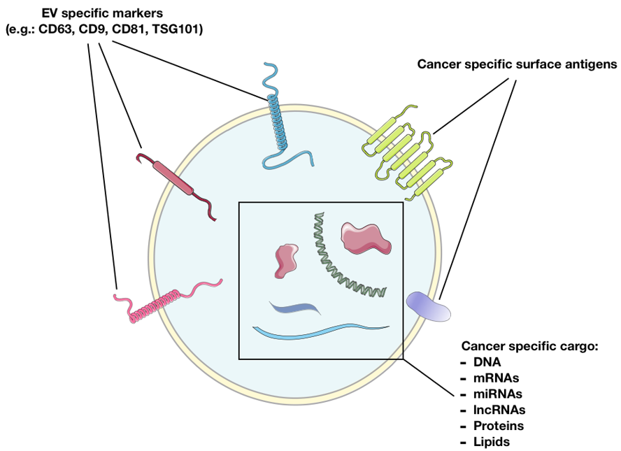
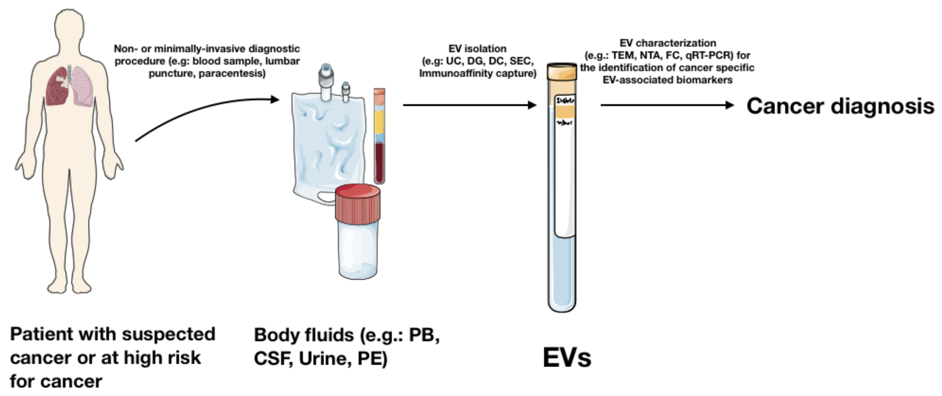
1. Introduction
2. Isolation and Purification Methods for EV Clinical Application
3. Tumours of the Central Nervous System (CNS)
3.1. Gliomas
3.2. Brain Metastases, Nonfunctional Pituitary Adenomas, Pediatric Brain Tumours
4. Head and Neck Cancers
4.1. Oral Cancer
4.2. Nasopharyngeal and Oropharyngeal Squamous Cell Carcinoma
4.3. Laryngeal Squamous Cell Carcinoma
5. Lung Cancers
6. Cancers of the Gastrointestinal Tract
6.1. Oesophageal Cancer
6.2. Gastric Cancer (GC)
6.3. Colorectal Cancer
6.4. Pancreatic Cancer
6.5. Hepatocellular Carcinoma
7. Conclusions
Author Contributions
Funding
Institutional Review Board Statement
Informed Consent Statement
Data Availability Statement
Conflicts of Interest
References
- Mattiuzzi, C.; Lippi, G. Current Cancer Epidemiology. JEGH 2019, 9, 217. [Google Scholar] [CrossRef] [PubMed] [Green Version]
- Alexander, B.M.; Cloughesy, T.F. Adult Glioblastoma. J. Clin. Oncol. 2017, 35, 2402–2409. [Google Scholar] [CrossRef]
- Lemjabbar-Alaoui, H.; Hassan, O.U.; Yang, Y.-W.; Buchanan, P. Lung Cancer: Biology and Treatment Options. Biochim. Biophys. Acta BBA Rev. Cancer 2015, 1856, 189–210. [Google Scholar] [CrossRef] [PubMed] [Green Version]
- Al-Mahmood, S.; Sapiezynski, J.; Garbuzenko, O.B.; Minko, T. Metastatic and Triple-Negative Breast Cancer: Challenges and Treatment Options. Drug Deliv. Transl. Res. 2018, 8, 1483–1507. [Google Scholar] [CrossRef] [PubMed] [Green Version]
- Poonyam, P.; Aumpan, N.; Vilaichone, R. Prognostic Factors for Survival in Patients with Gastric Adenocarcinoma. Cancer Rep. 2021, 4, e1305. [Google Scholar] [CrossRef]
- McGuigan, A.; Kelly, P.; Turkington, R.C.; Jones, C.; Coleman, H.G.; McCain, R.S. Pancreatic Cancer: A Review of Clinical Diagnosis, Epidemiology, Treatment and Outcomes. WJG 2018, 24, 4846–4861. [Google Scholar] [CrossRef] [PubMed]
- Neal, R.D.; Tharmanathan, P.; France, B.; Din, N.U.; Cotton, S.; Fallon-Ferguson, J.; Hamilton, W.; Hendry, A.; Hendry, M.; Lewis, R.; et al. Is Increased Time to Diagnosis and Treatment in Symptomatic Cancer Associated with Poorer Outcomes? Systematic Review. Br. J. Cancer 2015, 112, S92–S107. [Google Scholar] [CrossRef] [Green Version]
- Cree, I.A. Liquid Biopsy for Cancer Patients: Principles and Practice. Pathogenesis 2015, 2, 1–4. [Google Scholar] [CrossRef] [Green Version]
- Xu, R.; Rai, A.; Chen, M.; Suwakulsiri, W.; Greening, D.W.; Simpson, R.J. Extracellular Vesicles in Cancer—Implications for Future Improvements in Cancer Care. Nat. Rev. Clin. Oncol. 2018, 15, 617–638. [Google Scholar] [CrossRef]
- Théry, C.; Witwer, K.W.; Aikawa, E.; Alcaraz, M.J.; Anderson, J.D.; Andriantsitohaina, R.; Antoniou, A.; Arab, T.; Archer, F.; Atkin-Smith, G.K.; et al. Minimal Information for Studies of Extracellular Vesicles 2018 (MISEV2018): A Position Statement of the International Society for Extracellular Vesicles and Update of the MISEV2014 Guidelines. J. Extracell. Vesicles 2018, 7, 1535750. [Google Scholar] [CrossRef] [Green Version]
- Emilio Venturelli Using Servier Medical Art Templates, Which Are Licensed under a Creative Commons Attribution 3.0 Unported License. Available online: https://smart.servier.com (accessed on 14 April 2021).
- Cavallari, C.; Camussi, G.; Brizzi, M.F. Extracellular Vesicles in the Tumour Microenvironment: Eclectic Supervisors. Int. J. Mol. Sci. 2020, 21, 6768. [Google Scholar] [CrossRef]
- Becker, A.; Thakur, B.K.; Weiss, J.M.; Kim, H.S.; Peinado, H.; Lyden, D. Extracellular Vesicles in Cancer: Cell-to-Cell Mediators of Metastasis. Cancer Cell 2016, 30, 836–848. [Google Scholar] [CrossRef] [Green Version]
- Deregibus, M.C.; Figliolini, F.; D’Antico, S.; Manzini, P.M.; Pasquino, C.; De Lena, M.; Tetta, C.; Brizzi, M.F.; Camussi, G. Charge-based precipitation of extracellular vesicles. Int. J. Mol. Med. 2016, 38, 1359. [Google Scholar] [CrossRef] [Green Version]
- Konoshenko, M.Y.; Lekchnov, E.A.; Vlassov, A.V.; Laktionov, P.P. Isolation of Extracellular Vesicles: General Methodologies and Latest Trends. BioMed Res. Int. 2018, 2018, 1–27. [Google Scholar] [CrossRef]
- Zhang, Z.; Wang, C.; Li, T.; Liu, Z.; Li, L. Comparison of Ultracentrifugation and Density Gradient Separation Methods for Isolating Tca8113 Human Tongue Cancer Cell Line-Derived Exosomes. Oncol. Lett. 2014, 8, 1701–1706. [Google Scholar] [CrossRef] [Green Version]
- Tauro, B.J.; Greening, D.W.; Mathias, R.A.; Ji, H.; Mathivanan, S.; Scott, A.M.; Simpson, R.J. Comparison of Ultracentrifugation, Density Gradient Separation, and Immunoaffinity Capture Methods for Isolating Human Colon Cancer Cell Line LIM1863-Derived Exosomes. Methods 2012, 56, 293–304. [Google Scholar] [CrossRef]
- Sidhom, K.; Obi, P.O.; Saleem, A. A Review of Exosomal Isolation Methods: Is Size Exclusion Chromatography the Best Option? IJMS 2020, 21, 6466. [Google Scholar] [CrossRef]
- Sódar, B.W.; Kittel, Á.; Pálóczi, K.; Vukman, K.V.; Osteikoetxea, X.; Szabó-Taylor, K.; Németh, A.; Sperlágh, B.; Baranyai, T.; Giricz, Z.; et al. Low-Density Lipoprotein Mimics Blood Plasma-Derived Exosomes and Microvesicles during Isolation and Detection. Sci. Rep. 2016, 6, 24316. [Google Scholar] [CrossRef] [Green Version]
- Böing, A.N.; van der Pol, E.; Grootemaat, A.E.; Coumans, F.A.W.; Sturk, A.; Nieuwland, R. Single-Step Isolation of Extracellular Vesicles by Size-Exclusion Chromatography. J. Extracell. Vesicles 2014, 3, 23430. [Google Scholar] [CrossRef]
- Sáenz-Cuesta, M.; Arbelaiz, A.; Oregi, A.; Irizar, H.; Osorio-Querejeta, I.; Muñoz-Culla, M.; Banales, J.M.; Falcón-Pérez, J.M.; Olascoaga, J.; Otaegui, D. Methods for Extracellular Vesicles Isolation in a Hospital Setting. Front. Immunol 2015, 6, 50. [Google Scholar] [CrossRef]
- Cizmar, P.; Yuana, Y. Detection and Characterization of Extracellular Vesicles by Transmission and Cryo-Transmission Electron Microscopy. Methods Mol. Biol. 2017, 1660, 221–232. [Google Scholar] [CrossRef] [PubMed]
- Sharma, S.; Rasool, H.I.; Palanisamy, V.; Mathisen, C.; Schmidt, M.; Wong, D.T.; Gimzewski, J.K. Structural-Mechanical Characterization of Nanoparticle Exosomes in Human Saliva, Using Correlative AFM, FESEM, and Force Spectroscopy. ACS Nano 2010, 4, 1921–1926. [Google Scholar] [CrossRef] [PubMed] [Green Version]
- Wu, Y.; Deng, W.; Klinke, D.J. Exosomes: Improved Methods to Characterize Their Morphology, RNA Content, and Surface Protein Biomarkers. Analyst 2015, 140, 6631–6642. [Google Scholar] [CrossRef] [PubMed] [Green Version]
- Hartjes, T.A.; Mytnyk, S.; Jenster, G.W.; van Steijn, V.; van Royen, M.E. Extracellular Vesicle Quantification and Characterization: Common Methods and Emerging Approaches. Bioengineering 2019, 6, 7. [Google Scholar] [CrossRef] [Green Version]
- Lucchetti, D.; Fattorossi, A.; Sgambato, A. Extracellular Vesicles in Oncology: Progress and Pitfalls in the Methods of Isolation and Analysis. Biotechnol. J. 2019, 14, 1700716. [Google Scholar] [CrossRef] [Green Version]
- Nolan, J.P.; Duggan, E. Analysis of Individual Extracellular Vesicles by Flow Cytometry. Methods Mol. Biol. 2018, 1678, 79–92. [Google Scholar] [CrossRef]
- Kowal, E.J.K.; Ter-Ovanesyan, D.; Regev, A.; Church, G.M. Extracellular Vesicle Isolation and Analysis by Western Blotting. Methods Mol. Biol. 2017, 1660, 143–152. [Google Scholar] [CrossRef]
- Serrano-Pertierra, E.; Oliveira-Rodríguez, M.; Matos, M.; Gutiérrez, G.; Moyano, A.; Salvador, M.; Rivas, M.; Blanco-López, M.C. Extracellular Vesicles: Current Analytical Techniques for Detection and Quantification. Biomolecules 2020, 10, 824. [Google Scholar] [CrossRef]
- Takahashi, K.; Yan, I.K.; Kim, C.; Kim, J.; Patel, T. Analysis of Extracellular RNA by Digital PCR. Front. Oncol. 2014, 4, 129. [Google Scholar] [CrossRef] [Green Version]
- Navarro, E.; Serrano-Heras, G.; Castaño, M.J.; Solera, J. Real-Time PCR Detection Chemistry. Clin. Chim. Acta 2015, 439, 231–250. [Google Scholar] [CrossRef]
- Ji, H.; Greening, D.W.; Barnes, T.W.; Lim, J.W.; Tauro, B.J.; Rai, A.; Xu, R.; Adda, C.; Mathivanan, S.; Zhao, W.; et al. Proteome Profiling of Exosomes Derived from Human Primary and Metastatic Colorectal Cancer Cells Reveal Differential Expression of Key Metastatic Factors and Signal Transduction Components. Proteomics 2013, 13, 1672–1686. [Google Scholar] [CrossRef]
- Chitoiu, L.; Dobranici, A.; Gherghiceanu, M.; Dinescu, S.; Costache, M. Multi-Omics Data Integration in Extracellular Vesicle Biology-Utopia or Future Reality? Int. J. Mol. Sci. 2020, 21, 8550. [Google Scholar] [CrossRef]
- Jakobsen, K.R.; Paulsen, B.S.; Bæk, R.; Varming, K.; Sorensen, B.S.; Jørgensen, M.M. Exosomal Proteins as Potential Diagnostic Markers in Advanced Non-Small Cell Lung Carcinoma. J. Extracell. Vesicles 2015, 4, 26659. [Google Scholar] [CrossRef] [PubMed]
- Sun, B.; Li, Y.; Zhou, Y.; Ng, T.K.; Zhao, C.; Gan, Q.; Gu, X.; Xiang, J. Circulating Exosomal CPNE3 as a Diagnostic and Prognostic Biomarker for Colorectal Cancer. J. Cell Physiol. 2019, 234, 1416–1425. [Google Scholar] [CrossRef]
- Julich-Haertel, H.; Urban, S.K.; Krawczyk, M.; Willms, A.; Jankowski, K.; Patkowski, W.; Kruk, B.; Krasnodębski, M.; Ligocka, J.; Schwab, R.; et al. Cancer-Associated Circulating Large Extracellular Vesicles in Cholangiocarcinoma and Hepatocellular Carcinoma. J. Hepatol. 2017, 67, 282–292. [Google Scholar] [CrossRef]
- Louis, D.N.; Ohgaki, H.; Wiestler, O.D.; Cavenee, W.K.; Burger, P.C.; Jouvet, A.; Scheithauer, B.W.; Kleihues, P. The 2007 WHO Classification of Tumours of the Central Nervous System. Acta Neuropathol. 2007, 114, 97–109. [Google Scholar] [CrossRef] [Green Version]
- Shankar, G.M.; Balaj, L.; Stott, S.L.; Nahed, B.; Carter, B.S. Liquid Biopsy for Brain Tumors. Expert Rev. Mol. Diagn. 2017, 17, 943–947. [Google Scholar] [CrossRef]
- Srivastava, A.; Filant, J.; Moxley, K.M.; Sood, A.; McMeekin, S.; Ramesh, R. Exosomes: A Role for Naturally Occurring Nanovesicles in Cancer Growth, Diagnosis and Treatment. Curr. Gene Ther. 2015, 15, 182–192. [Google Scholar] [CrossRef]
- Westphal, M.; Lamszus, K. Circulating Biomarkers for Gliomas. Nat. Rev. Neurol. 2015, 11, 556–566. [Google Scholar] [CrossRef]
- Gold, B.; Cankovic, M.; Furtado, L.V.; Meier, F.; Gocke, C.D. Do Circulating Tumor Cells, Exosomes, and Circulating Tumor Nucleic Acids Have Clinical Utility? A Report of the Association for Molecular Pathology. J. Mol. Diagn. 2015, 17, 209–224. [Google Scholar] [CrossRef] [Green Version]
- Wesseling, P.; Kros, J.M.; Jeuken, J.W.M. The Pathological Diagnosis of Diffuse Gliomas: Towards a Smart Synthesis of Microscopic and Molecular Information in a Multidisciplinary Context. Diagn. Histopathol. 2011, 17, 486–494. [Google Scholar] [CrossRef] [Green Version]
- García-Romero, N.; Carrión-Navarro, J.; Esteban-Rubio, S.; Lázaro-Ibáñez, E.; Peris-Celda, M.; Alonso, M.M.; Guzmán-De-Villoria, J.; Fernández-Carballal, C.; de Mendivil, A.O.; García-Duque, S.; et al. DNA Sequences within Glioma-Derived Extracellular Vesicles Can Cross the Intact Blood-Brain Barrier and Be Detected in Peripheral Blood of Patients. Oncotarget 2017, 8, 1416–1428. [Google Scholar] [CrossRef] [PubMed] [Green Version]
- Akers, J.C.; Ramakrishnan, V.; Kim, R.; Phillips, S.; Kaimal, V.; Mao, Y.; Hua, W.; Yang, I.; Fu, C.-C.; Nolan, J.; et al. MiRNA Contents of Cerebrospinal Fluid Extracellular Vesicles in Glioblastoma Patients. J. Neuro-Oncol. 2015, 123, 205–216. [Google Scholar] [CrossRef] [PubMed] [Green Version]
- Best, M.G.; Sol, N.; Zijl, S.; Reijneveld, J.C.; Wesseling, P.; Wurdinger, T. Liquid Biopsies in Patients with Diffuse Glioma. Acta Neuropathol. 2015, 129, 849–865. [Google Scholar] [CrossRef] [PubMed] [Green Version]
- Chen, C.; Skog, J.; Hsu, C.-H.; Lessard, R.T.; Balaj, L.; Wurdinger, T.; Carter, B.S.; Breakefield, X.O.; Toner, M.; Irimia, D. Microfluidic Isolation and Transcriptome Analysis of Serum Microvesicles. Lab Chip 2010, 10, 505–511. [Google Scholar] [CrossRef] [PubMed] [Green Version]
- Lan, F.; Qing, Q.; Pan, Q.; Hu, M.; Yu, H.; Yue, X. Serum Exosomal MiR-301a as a Potential Diagnostic and Prognostic Biomarker for Human Glioma. Cell Oncol. 2018, 41, 25–33. [Google Scholar] [CrossRef] [PubMed]
- Ebrahimkhani, S.; Vafaee, F.; Hallal, S.; Wei, H.; Lee, M.Y.T.; Young, P.E.; Satgunaseelan, L.; Beadnall, H.; Barnett, M.H.; Shivalingam, B.; et al. Deep Sequencing of Circulating Exosomal MicroRNA Allows Non-Invasive Glioblastoma Diagnosis. NPJ Precis. Oncol. 2018, 2, 28. [Google Scholar] [CrossRef] [PubMed] [Green Version]
- Manterola, L.; Guruceaga, E.; Gállego Pérez-Larraya, J.; González-Huarriz, M.; Jauregui, P.; Tejada, S.; Diez-Valle, R.; Segura, V.; Samprón, N.; Barrena, C.; et al. A Small Noncoding RNA Signature Found in Exosomes of GBM Patient Serum as a Diagnostic Tool. Neuro-Oncology 2014, 16, 520–527. [Google Scholar] [CrossRef]
- Akers, J.C.; Hua, W.; Li, H.; Ramakrishnan, V.; Yang, Z.; Quan, K.; Zhu, W.; Li, J.; Figueroa, J.; Hirshman, B.R.; et al. A Cerebrospinal Fluid MicroRNA Signature as Biomarker for Glioblastoma. Oncotarget 2017, 8, 68769–68779. [Google Scholar] [CrossRef]
- Santangelo, A.; Imbrucè, P.; Gardenghi, B.; Belli, L.; Agushi, R.; Tamanini, A.; Munari, S.; Bossi, A.M.; Scambi, I.; Benati, D.; et al. A MicroRNA Signature from Serum Exosomes of Patients with Glioma as Complementary Diagnostic Biomarker. J. Neuro-Oncol. 2018, 136, 51–62. [Google Scholar] [CrossRef]
- Skog, J.; Würdinger, T.; van Rijn, S.; Meijer, D.H.; Gainche, L.; Sena-Esteves, M.; Curry, W.T.; Carter, B.S.; Krichevsky, A.M.; Breakefield, X.O. Glioblastoma Microvesicles Transport RNA and Proteins That Promote Tumour Growth and Provide Diagnostic Biomarkers. Nat. Cell Biol. 2008, 10, 1470–1476. [Google Scholar] [CrossRef]
- Manda, S.V.; Kataria, Y.; Tatireddy, B.R.; Ramakrishnan, B.; Ratnam, B.G.; Lath, R.; Ranjan, A.; Ray, A. Exosomes as a Biomarker Platform for Detecting Epidermal Growth Factor Receptor-Positive High-Grade Gliomas. J. Neurosurg. 2018, 128, 1091–1101. [Google Scholar] [CrossRef] [Green Version]
- Indira Chandran, V.; Welinder, C.; Månsson, A.-S.; Offer, S.; Freyhult, E.; Pernemalm, M.; Lund, S.M.; Pedersen, S.; Lehtiö, J.; Marko-Varga, G.; et al. Ultrasensitive Immunoprofiling of Plasma Extracellular Vesicles Identifies Syndecan-1 as a Potential Tool for Minimally Invasive Diagnosis of Glioma. Clin. Cancer Res. 2019, 25, 3115–3127. [Google Scholar] [CrossRef] [Green Version]
- Yang, J.-K.; Song, J.; Huo, H.-R.; Zhao, Y.-L.; Zhang, G.-Y.; Zhao, Z.-M.; Sun, G.-Z.; Jiao, B.-H. DNM3, P65 and P53 from Exosomes Represent Potential Clinical Diagnosis Markers for Glioblastoma Multiforme. Ther. Adv. Med. Oncol. 2017, 9, 741–754. [Google Scholar] [CrossRef] [Green Version]
- Chen, W.W.; Balaj, L.; Liau, L.M.; Samuels, M.L.; Kotsopoulos, S.K.; Maguire, C.A.; Loguidice, L.; Soto, H.; Garrett, M.; Zhu, L.D.; et al. BEAMing and Droplet Digital PCR Analysis of Mutant IDH1 MRNA in Glioma Patient Serum and Cerebrospinal Fluid Extracellular Vesicles. Mol. Ther. Nucleic Acids 2013, 2, e109. [Google Scholar] [CrossRef]
- D’Asti, E.; Chennakrishnaiah, S.; Lee, T.H.; Rak, J. Extracellular Vesicles in Brain Tumor Progression. Cell Mol. Neurobiol. 2016, 36, 383–407. [Google Scholar] [CrossRef]
- Jackson, H.K.; Linke, F.; Kerr, I.D.; Coyle, B. MBRS-21. Extracellular Vesicles from Metastatic Medulloblastoma Cell Lines Carry mRNAs Known to Correlate with Metastatic Disease. Neuro-Oncology 2018, 20, i132. [Google Scholar] [CrossRef]
- Shao, H.; Chung, J.; Balaj, L.; Charest, A.; Bigner, D.D.; Carter, B.S.; Hochberg, F.H.; Breakefield, X.O.; Weissleder, R.; Lee, H. Protein Typing of Circulating Microvesicles Allows Real-Time Monitoring of Glioblastoma Therapy. Nat. Med. 2012, 18, 1835–1840. [Google Scholar] [CrossRef]
- Huang, K.; Yang, C.; Wang, Q.-X.; Li, Y.-S.; Fang, C.; Tan, Y.-L.; Wei, J.-W.; Wang, Y.-F.; Li, X.; Zhou, J.-H.; et al. The CRISPR/Cas9 System Targeting EGFR Exon 17 Abrogates NF-ΚB Activation via Epigenetic Modulation of UBXN1 in EGFRwt/VIII Glioma Cells. Cancer Lett. 2017, 388, 269–280. [Google Scholar] [CrossRef]
- Osti, D.; Del Bene, M.; Rappa, G.; Santos, M.; Matafora, V.; Richichi, C.; Faletti, S.; Beznoussenko, G.V.; Mironov, A.; Bachi, A.; et al. Clinical Significance of Extracellular Vesicles in Plasma from Glioblastoma Patients. Clin. Cancer Res. 2019, 25, 266–276. [Google Scholar] [CrossRef] [Green Version]
- Huang, K.; Fang, C.; Yi, K.; Liu, X.; Qi, H.; Tan, Y.; Zhou, J.; Li, Y.; Liu, M.; Zhang, Y.; et al. The Role of PTRF/Cavin1 as a Biomarker in Both Glioma and Serum Exosomes. Theranostics 2018, 8, 1540–1557. [Google Scholar] [CrossRef] [PubMed]
- Wang, H.; Chen, K.; Yang, Z.; Li, W.; Wang, C.; Zhang, G.; Zhu, L.; Liu, P.; Yang, Y. Diagnosis of Invasive Nonfunctional Pituitary Adenomas by Serum Extracellular Vesicles. Anal. Chem. 2019, 91, 9580–9589. [Google Scholar] [CrossRef] [PubMed]
- Al-Nedawi, K.; Meehan, B.; Micallef, J.; Lhotak, V.; May, L.; Guha, A.; Rak, J. Intercellular Transfer of the Oncogenic Receptor EGFRvIII by Microvesicles Derived from Tumour Cells. Nat. Cell Biol. 2008, 10, 619–624. [Google Scholar] [CrossRef] [PubMed]
- Brennan, C.W.; Verhaak, R.G.W.; McKenna, A.; Campos, B.; Noushmehr, H.; Salama, S.R.; Zheng, S.; Chakravarty, D.; Sanborn, J.Z.; Berman, S.H.; et al. The Somatic Genomic Landscape of Glioblastoma. Cell 2013, 155, 462–477. [Google Scholar] [CrossRef]
- Graner, M.W.; Alzate, O.; Dechkovskaia, A.M.; Keene, J.D.; Sampson, J.H.; Mitchell, D.A.; Bigner, D.D. Proteomic and Immunologic Analyses of Brain Tumor Exosomes. FASEB J. 2009, 23, 1541–1557. [Google Scholar] [CrossRef] [Green Version]
- Bouwens, T.a.M.; Trouw, L.A.; Veerhuis, R.; Dirven, C.M.F.; Lamfers, M.L.M.; Al-Khawaja, H. Complement Activation in Glioblastoma Multiforme Pathophysiology: Evidence from Serum Levels and Presence of Complement Activation Products in Tumor Tissue. J. Neuroimmunol. 2015, 278, 271–276. [Google Scholar] [CrossRef] [Green Version]
- Gollapalli, K.; Ray, S.; Srivastava, R.; Renu, D.; Singh, P.; Dhali, S.; Bajpai Dikshit, J.; Srikanth, R.; Moiyadi, A.; Srivastava, S. Investigation of Serum Proteome Alterations in Human Glioblastoma Multiforme. Proteomics 2012, 12, 2378–2390. [Google Scholar] [CrossRef]
- Hunter, S.B.; Varma, V.; Shehata, B.; Nolen, J.D.L.; Cohen, C.; Olson, J.J.; Ou, C.-Y. Apolipoprotein D Expression in Primary Brain Tumors: Analysis by Quantitative RT-PCR in Formalin-Fixed, Paraffin-Embedded Tissue. J. Histochem. Cytochem. 2005, 53, 963–969. [Google Scholar] [CrossRef] [Green Version]
- Luo, D.; Chen, W.; Tian, Y.; Li, J.; Xu, X.; Chen, C.; Li, F. Serpin Peptidase Inhibitor, Clade A Member 3 (SERPINA3), Is Overexpressed in Glioma and Associated with Poor Prognosis in Glioma Patients. Onco. Targets Ther. 2017, 10, 2173–2181. [Google Scholar] [CrossRef] [Green Version]
- Nicoll, J.A.R.; Zunarelli, E.; Rampling, R.; Murray, L.S.; Papanastassiou, V.; Stewart, J. Involvement of Apolipoprotein E in Glioblastoma: Immunohistochemistry and Clinical Outcome. Neuroreport 2003, 14, 1923–1926. [Google Scholar] [CrossRef]
- Nowacki, P.; Tabaka, J. Human von Willebrand Factor (Factor VIII-Related Antigen) in Glial Neoplastic Cells of Brain Gliomas. Folia Neuropathol. 2003, 41, 23–27. [Google Scholar]
- Park, Y.E.; Yeom, J.; Kim, Y.; Lee, H.J.; Han, K.-C.; Lee, S.-T.; Lee, C.; Lee, J.E. Identification of Plasma Membrane Glycoproteins Specific to Human Glioblastoma Multiforme Cells Using Lectin Arrays and LC-MS/MS. Proteomics 2018, 18, 1700302. [Google Scholar] [CrossRef]
- Wu, T.; Li, Y.; Liu, B.; Zhang, S.; Wu, L.; Zhu, X.; Chen, Q. Expression of Ferritin Light Chain (FTL) Is Elevated in Glioblastoma, and FTL Silencing Inhibits Glioblastoma Cell Proliferation via the GADD45/JNK Pathway. PLoS ONE 2016, 11, e0149361. [Google Scholar] [CrossRef] [Green Version]
- André-Grégoire, G.; Bidère, N.; Gavard, J. Temozolomide Affects Extracellular Vesicles Released by Glioblastoma Cells. Biochimie 2018, 155, 11–15. [Google Scholar] [CrossRef]
- Müller Bark, J.; Kulasinghe, A.; Chua, B.; Day, B.W.; Punyadeera, C. Circulating Biomarkers in Patients with Glioblastoma. Br. J. Cancer 2020, 122, 295–305. [Google Scholar] [CrossRef] [Green Version]
- Haraszti, R.A.; Didiot, M.-C.; Sapp, E.; Leszyk, J.; Shaffer, S.A.; Rockwell, H.E.; Gao, F.; Narain, N.R.; DiFiglia, M.; Kiebish, M.A.; et al. High-Resolution Proteomic and Lipidomic Analysis of Exosomes and Microvesicles from Different Cell Sources. J. Extracell. Vesicles 2016, 5, 32570. [Google Scholar] [CrossRef]
- Figueroa, J.M.; Skog, J.; Akers, J.; Li, H.; Komotar, R.; Jensen, R.; Ringel, F.; Yang, I.; Kalkanis, S.; Thompson, R.; et al. Detection of Wild-Type EGFR Amplification and EGFRvIII Mutation in CSF-Derived Extracellular Vesicles of Glioblastoma Patients. Neuro-Oncology 2017, 19, 1494–1502. [Google Scholar] [CrossRef]
- Akers, J.C.; Ramakrishnan, V.; Kim, R.; Skog, J.; Nakano, I.; Pingle, S.; Kalinina, J.; Hua, W.; Kesari, S.; Mao, Y.; et al. MiR-21 in the Extracellular Vesicles (EVs) of Cerebrospinal Fluid (CSF): A Platform for Glioblastoma Biomarker Development. PLoS ONE 2013, 8, e78115. [Google Scholar] [CrossRef]
- Ozdemir-Kaynak, E.; Qutub, A.A.; Yesil-Celiktas, O. Advances in Glioblastoma Multiforme Treatment: New Models for Nanoparticle Therapy. Front. Physiol. 2018, 9, 170. [Google Scholar] [CrossRef] [Green Version]
- Curtaz, C.J.; Schmitt, C.; Blecharz-Lang, K.G.; Roewer, N.; Wöckel, A.; Burek, M. Circulating MicroRNAs and Blood-Brain-Barrier Function in Breast Cancer Metastasis. Curr. Pharm. Des. 2020, 26, 1417–1427. [Google Scholar] [CrossRef]
- Masoudi, M.S.; Mehrabian, E.; Mirzaei, H. MiR-21: A Key Player in Glioblastoma Pathogenesis. J. Cell Biochem. 2018, 119, 1285–1290. [Google Scholar] [CrossRef]
- Ramachandran, A.; Yan, H.; Bentink, S.; Noerholm, M.; Berking, C.; Flaherty, K.; Hochberg, F.; Skog, J. Abstract C139: Detection of BRAF Mutations in Serum/Plasma Microvesicles (Exosomes) of Malignant Melanoma Patients. In Proceedings of the AACR-NCI-EORTC International Conference: Molecular Targets and Cancer Therapeutics, San Francisco, CA, USA, 12 November 2011; American Association for Cancer Research: Philadelphia, PA, USA, 2011; p. C139. [Google Scholar]
- Zlotogorski-Hurvitz, A.; Dayan, D.; Chaushu, G.; Salo, T.; Vered, M. Morphological and Molecular Features of Oral Fluid-Derived Exosomes: Oral Cancer Patients versus Healthy Individuals. J. Cancer Res. Clin. Oncol. 2016, 142, 101–110. [Google Scholar] [CrossRef]
- Gallo, A.; Alevizos, I. Isolation of Circulating MicroRNA in Saliva. Methods Mol. Biol. 2013, 1024, 183–190. [Google Scholar] [CrossRef]
- Gai, C.; Camussi, F.; Broccoletti, R.; Gambino, A.; Cabras, M.; Molinaro, L.; Carossa, S.; Camussi, G.; Arduino, P.G. Salivary Extracellular Vesicle-Associated MiRNAs as Potential Biomarkers in Oral Squamous Cell Carcinoma. BMC Cancer 2018, 18, 439. [Google Scholar] [CrossRef] [Green Version]
- Ries, J.; Vairaktaris, E.; Agaimy, A.; Kintopp, R.; Baran, C.; Neukam, F.W.; Nkenke, E. MiR-186, MiR-3651 and MiR-494: Potential Biomarkers for Oral Squamous Cell Carcinoma Extracted from Whole Blood. Oncol. Rep. 2014, 31, 1429–1436. [Google Scholar] [CrossRef] [Green Version]
- He, L.; Ping, F.; Fan, Z.; Zhang, C.; Deng, M.; Cheng, B.; Xia, J. Salivary Exosomal MiR-24-3p Serves as a Potential Detective Biomarker for Oral Squamous Cell Carcinoma Screening. Biomed. Pharmacother. 2020, 121, 109553. [Google Scholar] [CrossRef]
- Sun, X.; Xiao, D.; Xu, T.; Yuan, Y. MiRNA-24-3p Promotes Cell Proliferation and Regulates Chemosensitivity in Head and Neck Squamous Cell Carcinoma by Targeting CHD5. Future Oncol. 2016, 12, 2701–2712. [Google Scholar] [CrossRef]
- Zheng, X.; Li, J.; Peng, C.; Zhao, J.; Chi, J.; Meng, X.; Yun, X.; Li, D.; Yu, Y.; Gao, M.; et al. MicroRNA-24 Induces Cisplatin Resistance by Targeting PTEN in Human Tongue Squamous Cell Carcinoma. Oral Oncol. 2015, 51, 998–1003. [Google Scholar] [CrossRef]
- Zhao, J.; Hu, C.; Chi, J.; Li, J.; Peng, C.; Yun, X.; Li, D.; Yu, Y.; Li, Y.; Gao, M.; et al. MiR-24 Promotes the Proliferation, Migration and Invasion in Human Tongue Squamous Cell Carcinoma by Targeting FBXW7. Oncol. Rep. 2016, 36, 1143–1149. [Google Scholar] [CrossRef] [Green Version]
- Momen-Heravi, F.; Trachtenberg, A.J.; Kuo, W.P.; Cheng, Y.S. Genomewide Study of Salivary MicroRNAs for Detection of Oral Cancer. J. Dent. Res. 2014, 93, 86S–93S. [Google Scholar] [CrossRef]
- Liu, C.-J.; Lin, S.-C.; Yang, C.-C.; Cheng, H.-W.; Chang, K.-W. Exploiting Salivary MiR-31 as a Clinical Biomarker of Oral Squamous Cell Carcinoma. Head Neck 2012, 34, 219–224. [Google Scholar] [CrossRef] [PubMed]
- Zahran, F.; Ghalwash, D.; Shaker, O.; Al-Johani, K.; Scully, C. Salivary MicroRNAs in Oral Cancer. Oral Dis. 2015, 21, 739–747. [Google Scholar] [CrossRef] [PubMed]
- Wang, J.; Zhou, Y.; Lu, J.; Sun, Y.; Xiao, H.; Liu, M.; Tian, L. Combined Detection of Serum Exosomal MiR-21 and HOTAIR as Diagnostic and Prognostic Biomarkers for Laryngeal Squamous Cell Carcinoma. Med. Oncol. 2014, 31, 148. [Google Scholar] [CrossRef] [PubMed]
- Zou, X.; Zhu, D.; Zhang, H.; Zhang, S.; Zhou, X.; He, X.; Zhu, J.; Zhu, W. MicroRNA Expression Profiling Analysis in Serum for Nasopharyngeal Carcinoma Diagnosis. Gene 2020, 727, 144243. [Google Scholar] [CrossRef]
- Liu, L.; Zuo, L.; Yang, J.; Xin, S.; Zhang, J.; Zhou, J.; Li, G.; Tang, J.; Lu, J. Exosomal Cyclophilin A as a Novel Noninvasive Biomarker for Epstein-Barr Virus Associated Nasopharyngeal Carcinoma. Cancer Med. 2019, 8, 3142–3151. [Google Scholar] [CrossRef] [Green Version]
- Nguyen, B.; Meehan, K.; Pereira, M.R.; Mirzai, B.; Lim, S.H.; Leslie, C.; Clark, M.; Sader, C.; Friedland, P.; Lindsay, A.; et al. A Comparative Study of Extracellular Vesicle-Associated and Cell-Free DNA and RNA for HPV Detection in Oropharyngeal Squamous Cell Carcinoma. Sci. Rep. 2020, 10, 6083. [Google Scholar] [CrossRef] [Green Version]
- Shimada, Y.; Matsubayashi, J.; Saito, A.; Ohira, T.; Kuroda, M.; Ikeda, N. Small RNA Sequencing to Differentiate Lung Squamous Cell Carcinomas from Metastatic Lung Tumors from Head and Neck Cancers. PLoS ONE 2021, 16, e0248206. [Google Scholar] [CrossRef]
- Siegel, R.L.; Miller, K.D.; Fuchs, H.E.; Jemal, A. Cancer Statistics, 2021. CA Cancer J. Clin. 2021, 71, 7–33. [Google Scholar] [CrossRef]
- Travis, W.D.; Brambilla, E.; Nicholson, A.G.; Yatabe, Y.; Austin, J.H.M.; Beasley, M.B.; Chirieac, L.R.; Dacic, S.; Duhig, E.; Flieder, D.B.; et al. The 2015 World Health Organization Classification of Lung Tumors: Impact of Genetic, Clinical and Radiologic Advances Since the 2004 Classification. J. Thorac. Oncol. 2015, 10, 1243–1260. [Google Scholar] [CrossRef] [Green Version]
- Taverna, S.; Giallombardo, M.; Gil-Bazo, I.; Carreca, A.P.; Castiglia, M.; Chacártegui, J.; Araujo, A.; Alessandro, R.; Pauwels, P.; Peeters, M.; et al. Exosomes Isolation and Characterization in Serum Is Feasible in Non-Small Cell Lung Cancer Patients: Critical Analysis of Evidence and Potential Role in Clinical Practice. Oncotarget 2016, 7, 28748–28760. [Google Scholar] [CrossRef]
- Aushev, V.N.; Zborovskaya, I.B.; Laktionov, K.K.; Girard, N.; Cros, M.-P.; Herceg, Z.; Krutovskikh, V. Comparisons of MicroRNA Patterns in Plasma before and after Tumor Removal Reveal New Biomarkers of Lung Squamous Cell Carcinoma. PLoS ONE 2013, 8, e78649. [Google Scholar] [CrossRef]
- Zhou, X.; Wen, W.; Shan, X.; Zhu, W.; Xu, J.; Guo, R.; Cheng, W.; Wang, F.; Qi, L.-W.; Chen, Y.; et al. A Six-MicroRNA Panel in Plasma Was Identified as a Potential Biomarker for Lung Adenocarcinoma Diagnosis. Oncotarget 2017, 8, 6513–6525. [Google Scholar] [CrossRef]
- Cazzoli, R.; Buttitta, F.; Di Nicola, M.; Malatesta, S.; Marchetti, A.; Rom, W.N.; Pass, H.I. MicroRNAs Derived from Circulating Exosomes as Noninvasive Biomarkers for Screening and Diagnosing Lung Cancer. J. Thorac. Oncol. 2013, 8, 1156–1162. [Google Scholar] [CrossRef] [Green Version]
- Fang, H.; Liu, Y.; He, Y.; Jiang, Y.; Wei, Y.; Liu, H.; Gong, Y.; An, G. Extracellular Vesicle-delivered MiR-505-5p, as a Diagnostic Biomarker of Early Lung Adenocarcinoma, Inhibits Cell Apoptosis by Targeting TP53AIP1. Int. J. Oncol. 2019, 54, 1821–1832. [Google Scholar] [CrossRef]
- Zhong, Y.; Ding, X.; Bian, Y.; Wang, J.; Zhou, W.; Wang, X.; Li, P.; Shen, Y.; Wang, J.-J.; Li, J.; et al. Discovery and Validation of Extracellular Vesicle-Associated MiRNAs as Noninvasive Detection Biomarkers for Early-Stage Non-Small-Cell Lung Cancer. Mol. Oncol. 2020. [Google Scholar] [CrossRef]
- Fan, T.W.M.; Zhang, X.; Wang, C.; Yang, Y.; Kang, W.-Y.; Arnold, S.; Higashi, R.M.; Liu, J.; Lane, A.N. Exosomal Lipids for Classifying Early and Late Stage Non-Small Cell Lung Cancer. Anal. Chim. Acta 2018, 1037, 256–264. [Google Scholar] [CrossRef]
- Porcel, J.M. Malignant Pleural Effusions Because of Lung Cancer. Curr. Opin. Pulm. Med. 2016, 22, 356–361. [Google Scholar] [CrossRef]
- Lin, J.; Wang, Y.; Zou, Y.-Q.; Chen, X.; Huang, B.; Liu, J.; Xu, Y.-M.; Li, J.; Zhang, J.; Yang, W.-M.; et al. Differential MiRNA Expression in Pleural Effusions Derived from Extracellular Vesicles of Patients with Lung Cancer, Pulmonary Tuberculosis, or Pneumonia. Tumor Biol. 2016, 37, 15835–15845. [Google Scholar] [CrossRef]
- Roman-Canal, B.; Moiola, C.P.; Gatius, S.; Bonnin, S.; Ruiz-Miró, M.; González, E.; Ojanguren, A.; Recuero, J.L.; Gil-Moreno, A.; Falcón-Pérez, J.M.; et al. EV-Associated MiRNAs from Pleural Lavage as Potential Diagnostic Biomarkers in Lung Cancer. Sci. Rep. 2019, 9, 15057. [Google Scholar] [CrossRef] [Green Version]
- Wang, Y.; Xu, Y.-M.; Zou, Y.-Q.; Lin, J.; Huang, B.; Liu, J.; Li, J.; Zhang, J.; Yang, W.-M.; Min, Q.-H.; et al. Identification of Differential Expressed PE Exosomal MiRNA in Lung Adenocarcinoma, Tuberculosis, and Other Benign Lesions. Medicine 2017, 96, e8361. [Google Scholar] [CrossRef]
- Tamiya, H.; Mitani, A.; Saito, A.; Ishimori, T.; Saito, M.; Isago, H.; Jo, T.; Yamauchi, Y.; Tanaka, G.; Nagase, T. Exosomal MicroRNA Expression Profiling in Patients with Lung Adenocarcinoma-Associated Malignant Pleural Effusion. Anticancer Res. 2018, 38, 6707–6714. [Google Scholar] [CrossRef]
- The National Lung Screening Trial Research Team. Reduced Lung-Cancer Mortality with Low-Dose Computed Tomographic Screening. N. Engl. J. Med. 2011, 365, 395–409. [Google Scholar] [CrossRef] [Green Version]
- Lam, A.K.-Y. Updates on World Health Organization Classification and Staging of Esophageal Tumors: Implications for Future Clinical Practice. Hum. Pathol. 2021, 108, 100–112. [Google Scholar] [CrossRef]
- Zhang, Y. Epidemiology of Esophageal Cancer. World J. Gastroenterol. 2013, 19, 5598–5606. [Google Scholar] [CrossRef]
- Nagai, K.; Ishihara, R.; Ishiguro, S.; Ohta, T.; Kanzaki, H.; Yamashina, T.; Aoi, K.; Matsuura, N.; Ito, T.; Fujii, M.; et al. Endoscopic Optical Diagnosis Provides High Diagnostic Accuracy of Esophageal Squamous Cell Carcinoma. BMC Gastroenterol. 2014, 14, 141. [Google Scholar] [CrossRef] [Green Version]
- Domper Arnal, M.J.; Ferrández Arenas, Á.; Lanas Arbeloa, Á. Esophageal Cancer: Risk Factors, Screening and Endoscopic Treatment in Western and Eastern Countries. World J. Gastroenterol. 2015, 21, 7933–7943. [Google Scholar] [CrossRef]
- Zhao, A.; Guo, L.; Xu, J.; Zheng, L.; Guo, Z.; Ling, Z.; Wang, L.; Mao, W. Identification and Validation of Circulating Exosomes-Based Liquid Biopsy for Esophageal Cancer. Cancer Med. 2019, 8, 3566–3574. [Google Scholar] [CrossRef]
- Takeshita, N.; Hoshino, I.; Mori, M.; Akutsu, Y.; Hanari, N.; Yoneyama, Y.; Ikeda, N.; Isozaki, Y.; Maruyama, T.; Akanuma, N.; et al. Serum MicroRNA Expression Profile: MiR-1246 as a Novel Diagnostic and Prognostic Biomarker for Oesophageal Squamous Cell Carcinoma. Br. J. Cancer 2013, 108, 644–652. [Google Scholar] [CrossRef]
- Zhou, X.; Wen, W.; Zhu, J.; Huang, Z.; Zhang, L.; Zhang, H.; Qi, L.-W.; Shan, X.; Wang, T.; Cheng, W.; et al. A Six-MicroRNA Signature in Plasma Was Identified as a Potential Biomarker in Diagnosis of Esophageal Squamous Cell Carcinoma. Oncotarget 2017, 8, 34468–34480. [Google Scholar] [CrossRef] [PubMed] [Green Version]
- Warnecke-Eberz, U.; Chon, S.-H.; Hölscher, A.H.; Drebber, U.; Bollschweiler, E. Exosomal Onco-MiRs from Serum of Patients with Adenocarcinoma of the Esophagus: Comparison of MiRNA Profiles of Exosomes and Matching Tumor. Tumour Biol. 2015, 36, 4643–4653. [Google Scholar] [CrossRef]
- Rana, S.; Maples, P.B.; Senzer, N.; Nemunaitis, J. Stathmin 1: A Novel Therapeutic Target for Anticancer Activity. Expert Rev. Anticancer Ther. 2008, 8, 1461–1470. [Google Scholar] [CrossRef] [PubMed]
- Biaoxue, R.; Xiguang, C.; Hua, L.; Shuanying, Y. Stathmin-Dependent Molecular Targeting Therapy for Malignant Tumor: The Latest 5 Years’ Discoveries and Developments. J. Transl. Med. 2016, 14, 279. [Google Scholar] [CrossRef] [PubMed] [Green Version]
- Mao, Q.; Chen, Z.; Wang, K.; Xu, R.; Lu, H.; He, X. Prognostic Role of High Stathmin 1 Expression in Patients with Solid Tumors: Evidence from a Meta-Analysis. Cell Physiol. Biochem. 2018, 50, 66–78. [Google Scholar] [CrossRef] [PubMed]
- Askeland, C.; Wik, E.; Finne, K.; Birkeland, E.; Arnes, J.B.; Collett, K.; Knutsvik, G.; Krüger, K.; Davidsen, B.; Aas, T.; et al. Stathmin Expression Associates with Vascular and Immune Responses in Aggressive Breast Cancer Subgroups. Sci. Rep. 2020, 10, 2914. [Google Scholar] [CrossRef] [Green Version]
- Yan, L.; Dong, X.; Gao, J.; Liu, F.; Zhou, L.; Sun, Y.; Zhao, X. A Novel Rapid Quantitative Method Reveals Stathmin-1 as a Promising Marker for Esophageal Squamous Cell Carcinoma. Cancer Med. 2018, 7, 1802–1813. [Google Scholar] [CrossRef]
- Lin, Y.; Dong, H.; Deng, W.; Lin, W.; Li, K.; Xiong, X.; Guo, Y.; Zhou, F.; Ma, C.; Chen, Y.; et al. Evaluation of Salivary Exosomal Chimeric GOLM1-NAA35 RNA as a Potential Biomarker in Esophageal Carcinoma. Clin. Cancer Res. 2019, 25, 3035–3045. [Google Scholar] [CrossRef]
- Liu, X.; Chen, X.; Zeng, K.; Xu, M.; He, B.; Pan, Y.; Sun, H.; Pan, B.; Xu, X.; Xu, T.; et al. DNA-Methylation-Mediated Silencing of MiR-486-5p Promotes Colorectal Cancer Proliferation and Migration through Activation of PLAGL2/IGF2/β-Catenin Signal Pathways. Cell Death Dis. 2018, 9, 1037. [Google Scholar] [CrossRef] [Green Version]
- Ogata-Kawata, H.; Izumiya, M.; Kurioka, D.; Honma, Y.; Yamada, Y.; Furuta, K.; Gunji, T.; Ohta, H.; Okamoto, H.; Sonoda, H.; et al. Circulating Exosomal MicroRNAs as Biomarkers of Colon Cancer. PLoS ONE 2014, 9, e92921. [Google Scholar] [CrossRef]
- Min, L.; Zhu, S.; Chen, L.; Liu, X.; Wei, R.; Zhao, L.; Yang, Y.; Zhang, Z.; Kong, G.; Li, P.; et al. Evaluation of Circulating Small Extracellular Vesicles Derived MiRNAs as Biomarkers of Early Colon Cancer: A Comparison with Plasma Total MiRNAs. J. Extracell. Vesicles 2019, 8, 1643670. [Google Scholar] [CrossRef] [Green Version]
- Que, R.; Ding, G.; Chen, J.; Cao, L. Analysis of Serum Exosomal MicroRNAs and Clinicopathologic Features of Patients with Pancreatic Adenocarcinoma. World J. Surg. Oncol. 2013, 11, 219. [Google Scholar] [CrossRef] [Green Version]
- Lai, X.; Wang, M.; McElyea, S.D.; Sherman, S.; House, M.; Korc, M. A MicroRNA Signature in Circulating Exosomes Is Superior to Exosomal Glypican-1 Levels for Diagnosing Pancreatic Cancer. Cancer Lett. 2017, 393, 86–93. [Google Scholar] [CrossRef] [Green Version]
- Melo, S.A.; Luecke, L.B.; Kahlert, C.; Fernandez, A.F.; Gammon, S.T.; Kaye, J.; LeBleu, V.S.; Mittendorf, E.A.; Weitz, J.; Rahbari, N.; et al. Glypican-1 Identifies Cancer Exosomes and Detects Early Pancreatic Cancer. Nature 2015, 523, 177–182. [Google Scholar] [CrossRef] [Green Version]
- Ostenfeld, M.S.; Jensen, S.G.; Jeppesen, D.K.; Christensen, L.-L.; Thorsen, S.B.; Stenvang, J.; Hvam, M.L.; Thomsen, A.; Mouritzen, P.; Rasmussen, M.H.; et al. MiRNA Profiling of Circulating EpCAM+ Extracellular Vesicles: Promising Biomarkers of Colorectal Cancer. J. Extracell. Vesicles 2016, 5, 31488. [Google Scholar] [CrossRef] [Green Version]
- Karimi, N.; Ali Hosseinpour Feizi, M.; Safaralizadeh, R.; Hashemzadeh, S.; Baradaran, B.; Shokouhi, B.; Teimourian, S. Serum Overexpression of MiR-301a and MiR-23a in Patients with Colorectal Cancer. J. Chin. Med. Assoc. 2019, 82, 215–220. [Google Scholar] [CrossRef]
- Machida, T.; Tomofuji, T.; Maruyama, T.; Yoneda, T.; Ekuni, D.; Azuma, T.; Miyai, H.; Mizuno, H.; Kato, H.; Tsutsumi, K.; et al. MiR-1246 and MiR-4644 in Salivary Exosome as Potential Biomarkers for Pancreatobiliary Tract Cancer. Oncol. Rep. 2016, 36, 2375–2381. [Google Scholar] [CrossRef] [Green Version]
- Xu, Y.-F.; Hannafon, B.N.; Zhao, Y.D.; Postier, R.G.; Ding, W.-Q. Plasma Exosome MiR-196a and MiR-1246 Are Potential Indicators of Localized Pancreatic Cancer. Oncotarget 2017, 8, 77028–77040. [Google Scholar] [CrossRef]
- Kumata, Y.; Iinuma, H.; Suzuki, Y.; Tsukahara, D.; Midorikawa, H.; Igarashi, Y.; Soeda, N.; Kiyokawa, T.; Horikawa, M.; Fukushima, R. Exosome-encapsulated MicroRNA-23b as a Minimally Invasive Liquid Biomarker for the Prediction of Recurrence and Prognosis of Gastric Cancer Patients in Each Tumor Stage. Oncol. Rep. 2018, 40, 319–330. [Google Scholar] [CrossRef]
- Li, J.; Chen, Y.; Guo, X.; Zhou, L.; Jia, Z.; Peng, Z.; Tang, Y.; Liu, W.; Zhu, B.; Wang, L.; et al. GPC1 Exosome and Its Regulatory MiRNAs Are Specific Markers for the Detection and Target Therapy of Colorectal Cancer. J. Cell Mol. Med. 2017, 21, 838–847. [Google Scholar] [CrossRef]
- Zou, S.-L.; Chen, Y.-L.; Ge, Z.-Z.; Qu, Y.-Y.; Cao, Y.; Kang, Z.-X. Downregulation of Serum Exosomal MiR-150-5p Is Associated with Poor Prognosis in Patients with Colorectal Cancer. Cancer Biomark. 2019, 26, 69–77. [Google Scholar] [CrossRef]
- Zhang, Y.; Han, T.; Feng, D.; Li, J.; Wu, M.; Peng, X.; Wang, B.; Zhan, X.; Fu, P. Screening of Non-Invasive MiRNA Biomarker Candidates for Metastasis of Gastric Cancer by Small RNA Sequencing of Plasma Exosomes. Carcinogenesis 2020, 41, 582–590. [Google Scholar] [CrossRef]
- Ohzawa, H.; Saito, A.; Kumagai, Y.; Kimura, Y.; Yamaguchi, H.; Hosoya, Y.; Lefor, A.K.; Sata, N.; Kitayama, J. Reduced Expression of Exosomal MiR-29s in Peritoneal Fluid Is a Useful Predictor of Peritoneal Recurrence after Curative Resection of Gastric Cancer with Serosal Involvement. Oncol. Rep. 2020, 43, 1081–1088. [Google Scholar] [CrossRef]
- Yan, S.; Jiang, Y.; Liang, C.; Cheng, M.; Jin, C.; Duan, Q.; Xu, D.; Yang, L.; Zhang, X.; Ren, B.; et al. Exosomal MiR-6803-5p as Potential Diagnostic and Prognostic Marker in Colorectal Cancer. J. Cell Biochem. 2018, 119, 4113–4119. [Google Scholar] [CrossRef]
- Wang, J.; Yan, F.; Zhao, Q.; Zhan, F.; Wang, R.; Wang, L.; Zhang, Y.; Huang, X. Circulating Exosomal MiR-125a-3p as a Novel Biomarker for Early-Stage Colon Cancer. Sci. Rep. 2017, 7, 4150. [Google Scholar] [CrossRef] [Green Version]
- Goto, T.; Fujiya, M.; Konishi, H.; Sasajima, J.; Fujibayashi, S.; Hayashi, A.; Utsumi, T.; Sato, H.; Iwama, T.; Ijiri, M.; et al. An Elevated Expression of Serum Exosomal MicroRNA-191, -21, -451a of Pancreatic Neoplasm Is Considered to Be Efficient Diagnostic Marker. BMC Cancer 2018, 18, 116. [Google Scholar] [CrossRef] [Green Version]
- Zhang, Y.; Xi, H.; Nie, X.; Zhang, P.; Lan, N.; Lu, Y.; Liu, J.; Yuan, W. Assessment of MiR-212 and Other Biomarkers in the Diagnosis and Treatment of HBV-Infection-Related Liver Diseases. CDM 2019, 20, 785–798. [Google Scholar] [CrossRef]
- Wang, H.; Hou, L.; Li, A.; Duan, Y.; Gao, H.; Song, X. Expression of Serum Exosomal MicroRNA-21 in Human Hepatocellular Carcinoma. BioMed Res. Int. 2014, 2014, 1–5. [Google Scholar] [CrossRef]
- Kitagawa, T.; Taniuchi, K.; Tsuboi, M.; Sakaguchi, M.; Kohsaki, T.; Okabayashi, T.; Saibara, T. Circulating Pancreatic Cancer Exosomal RNAs for Detection of Pancreatic Cancer. Mol. Oncol. 2019, 13, 212–227. [Google Scholar] [CrossRef] [PubMed] [Green Version]
- Tian, Y.; Ma, L.; Gong, M.; Su, G.; Zhu, S.; Zhang, W.; Wang, S.; Li, Z.; Chen, C.; Li, L.; et al. Protein Profiling and Sizing of Extracellular Vesicles from Colorectal Cancer Patients via Flow Cytometry. ACS Nano 2018, 12, 671–680. [Google Scholar] [CrossRef] [PubMed]
- Caruso Bavisotto, C.; Cappello, F.; Macario, A.J.L.; Conway de Macario, E.; Logozzi, M.; Fais, S.; Campanella, C. Exosomal HSP60: A Potentially Useful Biomarker for Diagnosis, Assessing Prognosis, and Monitoring Response to Treatment. Expert Rev. Mol. Diagn. 2017, 17, 815–822. [Google Scholar] [CrossRef] [PubMed]
- Zheng, J.; Hernandez, J.M.; Doussot, A.; Bojmar, L.; Zambirinis, C.P.; Costa-Silva, B.; van Beek, E.J.A.H.; Mark, M.T.; Molina, H.; Askan, G.; et al. Extracellular Matrix Proteins and Carcinoembryonic Antigen-Related Cell Adhesion Molecules Characterize Pancreatic Duct Fluid Exosomes in Patients with Pancreatic Cancer. HPB 2018, 20, 597–604. [Google Scholar] [CrossRef] [Green Version]
- Jin, H.; Liu, P.; Wu, Y.; Meng, X.; Wu, M.; Han, J.; Tan, X. Exosomal Zinc Transporter ZIP4 Promotes Cancer Growth and Is a Novel Diagnostic Biomarker for Pancreatic Cancer. Cancer Sci. 2018, 109, 2946–2956. [Google Scholar] [CrossRef] [Green Version]
- Lin, L.-Y.; Yang, L.; Zeng, Q.; Wang, L.; Chen, M.-L.; Zhao, Z.-H.; Ye, G.-D.; Luo, Q.-C.; Lv, P.-Y.; Guo, Q.-W.; et al. Tumor-Originated Exosomal LncUEGC1 as a Circulating Biomarker for Early-Stage Gastric Cancer. Mol. Cancer 2018, 17, 84. [Google Scholar] [CrossRef]
- Zhao, R.; Zhang, Y.; Zhang, X.; Yang, Y.; Zheng, X.; Li, X.; Liu, Y.; Zhang, Y. Exosomal Long Noncoding RNA HOTTIP as Potential Novel Diagnostic and Prognostic Biomarker Test for Gastric Cancer. Mol. Cancer 2018, 17, 68. [Google Scholar] [CrossRef]
- Oehme, F.; Krahl, S.; Gyorffy, B.; Muessle, B.; Rao, V.; Greif, H.; Ziegler, N.; Lin, K.; Thepkaysone, M.-L.; Polster, H.; et al. Low Level of Exosomal Long Non-Coding RNA HOTTIP Is a Prognostic Biomarker in Colorectal Cancer. RNA Biol. 2019, 16, 1339–1345. [Google Scholar] [CrossRef]
- Guo, X.; Lv, X.; Ru, Y.; Zhou, F.; Wang, N.; Xi, H.; Zhang, K.; Li, J.; Chang, R.; Xie, T.; et al. Circulating Exosomal Gastric Cancer-Associated Long Noncoding RNA1 as a Biomarker for Early Detection and Monitoring Progression of Gastric Cancer: A Multiphase Study. JAMA Surg. 2020, 155, 572–579. [Google Scholar] [CrossRef]
- Barbagallo, C.; Brex, D.; Caponnetto, A.; Cirnigliaro, M.; Scalia, M.; Magnano, A.; Caltabiano, R.; Barbagallo, D.; Biondi, A.; Cappellani, A.; et al. LncRNA UCA1, Upregulated in CRC Biopsies and Downregulated in Serum Exosomes, Controls MRNA Expression by RNA-RNA Interactions. Mol. Ther. Nucleic Acids 2018, 12, 229–241. [Google Scholar] [CrossRef] [Green Version]
- Liu, L.; Meng, T.; Yang, X.-H.; Sayim, P.; Lei, C.; Jin, B.; Ge, L.; Wang, H.-J. Prognostic and Predictive Value of Long Non-Coding RNA GAS5 and MircoRNA-221 in Colorectal Cancer and Their Effects on Colorectal Cancer Cell Proliferation, Migration and Invasion. Cancer Biomark. 2018, 22, 283–299. [Google Scholar] [CrossRef]
- Wang, L.; Duan, W.; Yan, S.; Xie, Y.; Wang, C. Circulating Long Non-Coding RNA Colon Cancer-Associated Transcript 2 Protected by Exosome as a Potential Biomarker for Colorectal Cancer. Biomed. Pharmacother. 2019, 113, 108758. [Google Scholar] [CrossRef]
- Liang, Z.-X.; Liu, H.-S.; Wang, F.-W.; Xiong, L.; Zhou, C.; Hu, T.; He, X.-W.; Wu, X.-J.; Xie, D.; Wu, X.-R.; et al. LncRNA RPPH1 Promotes Colorectal Cancer Metastasis by Interacting with TUBB3 and by Promoting Exosomes-Mediated Macrophage M2 Polarization. Cell Death Dis. 2019, 10, 829. [Google Scholar] [CrossRef] [Green Version]
- Hu, D.; Zhan, Y.; Zhu, K.; Bai, M.; Han, J.; Si, Y.; Zhang, H.; Kong, D. Plasma Exosomal Long Non-Coding RNAs Serve as Biomarkers for Early Detection of Colorectal Cancer. Cell Physiol. Biochem. 2018, 51, 2704–2715. [Google Scholar] [CrossRef]
- Chen, Y.; Chen, J.; Liu, Y.; Li, S.; Huang, P. Plasma MiR-15b-5p, MiR-338-5p, and MiR-764 as Biomarkers for Hepatocellular Carcinoma. Med. Sci. Monit. 2015, 21, 1864–1871. [Google Scholar] [CrossRef] [PubMed] [Green Version]
- Sexton, R.E.; Al Hallak, M.N.; Diab, M.; Azmi, A.S. Gastric Cancer: A Comprehensive Review of Current and Future Treatment Strategies. Cancer Metastasis Rev. 2020, 39, 1179–1203. [Google Scholar] [CrossRef] [PubMed]
- Alfaro, E.E.; Lauwers, G.Y. Early Gastric Neoplasia: Diagnosis and Implications. Adv. Anat. Pathol. 2011, 18, 268–280. [Google Scholar] [CrossRef] [PubMed]
- Lin, J.-P.; Lin, J.-X.; Ma, Y.-B.; Xie, J.-W.; Yan, S.; Wang, J.-B.; Lu, J.; Chen, Q.-Y.; Ma, X.-F.; Cao, L.-L.; et al. Prognostic Significance of Pre- and Post-Operative Tumour Markers for Patients with Gastric Cancer. Br. J. Cancer 2020, 123, 418–425. [Google Scholar] [CrossRef]
- Yang, H.; Fu, H.; Wang, B.; Zhang, X.; Mao, J.; Li, X.; Wang, M.; Sun, Z.; Qian, H.; Xu, W. Exosomal MiR-423-5p Targets SUFU to Promote Cancer Growth and Metastasis and Serves as a Novel Marker for Gastric Cancer. Mol. Carcinog. 2018, 57, 1223–1236. [Google Scholar] [CrossRef]
- Arnold, M.; Sierra, M.S.; Laversanne, M.; Soerjomataram, I.; Jemal, A.; Bray, F. Global Patterns and Trends in Colorectal Cancer Incidence and Mortality. Gut 2017, 66, 683–691. [Google Scholar] [CrossRef] [Green Version]
- Elsafi, S.H.; Alqahtani, N.I.; Zakary, N.Y.; Al Zahrani, E.M. The Sensitivity, Specificity, Predictive Values, and Likelihood Ratios of Fecal Occult Blood Test for the Detection of Colorectal Cancer in Hospital Settings. Clin. Exp. Gastroenterol. 2015, 8, 279–284. [Google Scholar] [CrossRef] [Green Version]
- Liu, T.; Zhang, X.; Gao, S.; Jing, F.; Yang, Y.; Du, L.; Zheng, G.; Li, P.; Li, C.; Wang, C. Exosomal Long Noncoding RNA CRNDE-h as a Novel Serum-Based Biomarker for Diagnosis and Prognosis of Colorectal Cancer. Oncotarget 2016, 7, 85551–85563. [Google Scholar] [CrossRef]
- Chen, Y.; Xie, Y.; Xu, L.; Zhan, S.; Xiao, Y.; Gao, Y.; Wu, B.; Ge, W. Protein Content and Functional Characteristics of Serum-Purified Exosomes from Patients with Colorectal Cancer Revealed by Quantitative Proteomics. Int. J. Cancer 2017, 140, 900–913. [Google Scholar] [CrossRef]
- Lydic, T.A.; Townsend, S.; Adda, C.G.; Collins, C.; Mathivanan, S.; Reid, G.E. Rapid and Comprehensive “shotgun” Lipidome Profiling of Colorectal Cancer Cell Derived Exosomes. Methods 2015, 87, 83–95. [Google Scholar] [CrossRef] [Green Version]
- Ryan, D.P.; Hong, T.S.; Bardeesy, N. Pancreatic Adenocarcinoma. N. Engl. J. Med. 2014, 371, 1039–1049. [Google Scholar] [CrossRef]
- Tao, L.; Zhou, J.; Yuan, C.; Zhang, L.; Li, D.; Si, D.; Xiu, D.; Zhong, L. Metabolomics Identifies Serum and Exosomes Metabolite Markers of Pancreatic Cancer. Metabolomics 2019, 15, 86. [Google Scholar] [CrossRef]
- Villanueva, A. Hepatocellular Carcinoma. N. Engl. J. Med. 2019, 380, 1450–1462. [Google Scholar] [CrossRef] [Green Version]
- Bialecki, E.S.; Di Bisceglie, A.M. Diagnosis of Hepatocellular Carcinoma. HPB 2005, 7, 26–34. [Google Scholar] [CrossRef] [Green Version]
- Wang, W.; Li, H.; Zhou, Y.; Jie, S. Peripheral Blood Microvesicles Are Potential Biomarkers for Hepatocellular Carcinoma. Cancer Biomark. 2013, 13, 351–357. [Google Scholar] [CrossRef]
- Wang, X.; Kwak, K.J.; Yang, Z.; Zhang, A.; Zhang, X.; Sullivan, R.; Lin, D.; Lee, R.L.; Castro, C.; Ghoshal, K.; et al. Extracellular MRNA Detected by Molecular Beacons in Tethered Lipoplex Nanoparticles for Diagnosis of Human Hepatocellular Carcinoma. PLoS ONE 2018, 13, e0198552. [Google Scholar] [CrossRef]
- Yao, Z.; Jia, C.; Tai, Y.; Liang, H.; Zhong, Z.; Xiong, Z.; Deng, M.; Zhang, Q. Serum Exosomal Long Noncoding RNAs Lnc-FAM72D-3 and Lnc-EPC1-4 as Diagnostic Biomarkers for Hepatocellular Carcinoma. Aging 2020, 12, 11843–11863. [Google Scholar] [CrossRef]
- Chapuy-Regaud, S.; Dubois, M.; Plisson-Chastang, C.; Bonnefois, T.; Lhomme, S.; Bertrand-Michel, J.; You, B.; Simoneau, S.; Gleizes, P.-E.; Flan, B.; et al. Characterization of the Lipid Envelope of Exosome Encapsulated HEV Particles Protected from the Immune Response. Biochimie 2017, 141, 70–79. [Google Scholar] [CrossRef]


| Method | Brief Technique Description | Advantages | Disadvantages | Ref. |
|---|---|---|---|---|
| UC | The sample is centrifuged (300× g for 10 min; 2000× g for 20 min to eliminate dead cells; and 10,000× g for 30 min to remove debris). EVs are isolated by UC at 100,000× g for 60 min at 4 °C. The pellet is resuspended in PBS and re-centrifuged at 100,000× g for 60 min | It represents the most widely used technique for EV isolation in basic and translational research. For these reasons it is highly standardized and reproducible | Generation of EV aggregates, contamination of the EV sample with smaller size particles. The whole isolation process is time consuming | [16] [17] [18] [19] |
| DG | A density gradient is generated by adding decreasing concentration of iodixanol or sucrose in a tube. The sample is then added on the top of the gradient and is centrifuged at 100,000× g for 18 h at 4 °C. After centrifugation, the recovered gradient fractions are diluted in PBS. An additional 3 h centrifugation is performed at 100,000× g | It represents a highly efficient method allowing to isolate purified EVs | It is more time consuming and more labour- intensive than standard UC. This consideration can make its introduction in the clinical practice difficult | [16] [17] |
| SEC | SEC separates molecules based on their size by filtration through a resin-packed column. After sample centrifugation (1100× g for 10 min) and filtration, samples are loaded in size exclusion chromatography columns | SEC isolates high purity, functionally and structurally preserved EVs. It has been defined as one of the best methods for EV isolation | Compared to other methods, the total EV yield is lower and in the clinical setting, higher volumes of biological fluid may be needed to overcome this issue | [18,20] |
| Polymer-based precipitation (e.g.,: Exoquick) | The method is based on the use of PEG to capture EVs. After PEG preparation, the sample undergoes isolation using 10% PEG solution at 4 °C for 2 h. After incubation, samples are centrifuged at low-speed × 10 min. To ensure major purity a double-step approach can be followed | It represents a fast, easy, and widespread method for EV isolation. It also easily allows a simultaneous isolation from multiple samples | EV obtained with this method have higher chances of being contaminated and therefore less suitable for omics-based analysis | [14,20,21] |
| Immunoaffinity capture | The immunoaffinity capture method relies on the isolation of EVs based on the expression of surface markers. It commonly uses antibodies against specific EV surface proteins, e.g., tetraspanin: CD9, CD63, and CD81 | The immunoaffinity capture isolation allows EV-specific isolation based on their surface markers. It can also be used as an additional step after UC to enhance EV purity | The removal of antibodies from EV surface could damage EVs. The selection of specific EVs could eventually not reflect the characteristics of all separated EVs. It is more expensive compared to other methods. | [17,20] |
| Microfluidic isolation | It consists in a high-throughput method using microfluidic devices to isolate EVs based on several principles: immunoaffinity, size, and density. The most used is the immuno-microfluidic technique, which is similar to the immune-affinity-capture isolation method. Antibodies immobilized on microfluidic devices serve to EV isolation | Relatively fast and high purity EV isolation can be obtained with this technique. It is a new and promising method for EV isolation | Shares with immunoaffinity the same disadvantages. It is expensive and standardization is still lacking | [18] |
| Method | EV Information | Disadvantages | Ref. |
|---|---|---|---|
| TEM |
|
| [21,22,23] |
| AFM |
|
| [22,23] |
| DLS |
|
| [24] |
| NTA |
|
| [24] |
| WB |
|
| [27] |
| PCR |
|
| [25,28,29] |
| FC |
|
| [25,26] |
| OMICS based techniques |
|
| [25,30,31] |
| Biomarker Types | Specific Markers | Tumour | Expression | Ref. |
|---|---|---|---|---|
| miRNAs | miR-301a | GBM | Upregulated | [47] |
| miR-182-5p | GBM | Upregulated | [48] | |
| miR-328-3p | GBM | Downregulated | [48] | |
| miR-339-5p | GBM | Downregulated | [49] | |
| miR-340-5p | GBM | Downregulated | [48] | |
| miR-485-3p | GBM | Downregulated | [48] | |
| miR-486-5p | GBM | Upregulated | [48] | |
| miR-543 | GBM | Downregulated | [48] | |
| miR-320 | GBM | Upregulated | [49] | |
| miR-301a | GBM | Upregulated | [47] | |
| miR-182-5p | GBM | Upregulated | [48] | |
| miR-328-3p | GBM | Downregulated | [48] | |
| miR-339-5p | GBM | Downregulated | [48] | |
| miR-340-5p | GBM | Downregulated | [48] | |
| miR-485-3p | GBM | Downregulated | [48] | |
| miR-486-5p | GBM | Upregulated | [48] | |
| miR-543 | GBM | Downregulated | [48] | |
| miR-320 | GBM | Upregulated | [49] | |
| miR-574-3p | GBM | Upregulated | [49] | |
| miR-21 miR-21 | GBM GBM/Brain metastases | Upregulated Upregulated | [50] [51] | |
| miR-218 | GBM | Upregulated | [50] | |
| miR-193b | GBM | Upregulated | [50] | |
| miR-331 | GBM | Upregulated | [50] | |
| miR-374a | GBM | Upregulated | [50] | |
| miR-548c | GBM | Downregulated | [50] | |
| miR-520f | GBM | Downregulated | [50] | |
| miR-27 | GBM | Downregulate | [50] | |
| miR-130b | GBM | Downregulated | [50] | |
| miR-222 | GBM/Brain metastases | Upregulated | [51] | |
| miR-124-3p | GBM/Brain metastases | Upregulated | [51] | |
| mRNAs | EGFRvIII | GBM | Upregulated | [52,53] |
| Syndecan-1 (SDC1) | GBM | Upregulated | [54] | |
| p65 | GBM | Upregulated | [55] | |
| DNM3 | GBM | Upregulated | [55] | |
| CD117 | GBM | Upregulated | [55] | |
| PTEN | GBM | Downregulated | [55] | |
| p53 | GBM | Downregulated | [55] | |
| APC | GBM | Downregulated | [55] | |
| RNU6-1 | GBM | Upregulated | [49] | |
| IDH1 | GBM | Upregulated | [56] | |
| KIAA1549/BRAF | Pilocytic astrocytoma | Upregulated | [57] | |
| c-MET | Medulloblastoma | Upregulated | [58] | |
| ABCB1 | Medulloblastoma | Upregulated | [58] | |
| MMP2 | Medulloblastoma | Upregulated | [58] | |
| BSG | Medulloblastoma | Upregulated | [58] | |
| ITG-A9 | Medulloblastoma | Upregulated | [58] | |
| Proteins | EGFR EGFRvIII | GBM GBM | Overexpressed Overexpressed | [59] [59] |
| PDPN | GBM | Overexpressed | [59] | |
| IDH1 R132H | GBM | Overexpressed | [59] | |
| PTRF | GBM | Ratio | [60] | |
| vWF | GBM | Overexpressed | [61] | |
| AZGP1 | GBM | Overexpressed | [61] | |
| Serpin3 | GBM | Overexpressed | [61] | |
| FTL | GBM | Overexpressed | [61] | |
| C3 | GBM | Overexpressed | [61] | |
| APOE | GBM | Overexpressed | [61] | |
| CD63 | GBM | Ratio | [62] | |
| FOLR1 | NFPA | Downregulated | [63] | |
| EpCAM | NFPA | Downregulated | [63] |
| Biomarker Types | Specific Markers | Tumour | Expression | Ref. |
|---|---|---|---|---|
| miRNAs | miR-21 miR-21 | LSCC OSCC | Upregulated Upregulated | [95] [94] |
| miR-27b | OSCC | Upregulated | [92] | |
| miR-27a-3p | OSCC | Upregulated | [86] | |
| miR-412-3p | OSCC | Upregulated | [86] | |
| miR-512-3p | OSCC | Upregulated | [86] | |
| miR-302b-3p | OSCC | Upregulated | [86] | |
| miR-517b-3p | OSCC | Upregulated | [86] | |
| miR-494-3p | OSCC | Upregulated | [86] | |
| miR-24-3p miR-24-3p | OSCC NPC | Upregulated Upregulated | [88] [96] | |
| miR-31 | OSCC | Upregulated | [93] | |
| miR-184 | OSCC | Upregulated | [94] | |
| miR-145 | OSCC | Downregulated | [94] | |
| Let-7b-5p | NPC | Upregulated | [96] | |
| miR-140-3p | NPC | Upregulated | [96] | |
| miR-192-5p | NPC | Upregulated | [96] | |
| miR-223-3p | NPC | Upregulated | [96] | |
| LncRNA | HOTAIR | LSCC | Upregulated | [95] |
| Proteins | CD9 | OC | Downregulated | [84] |
| CD81 | OC | Downregulated | [84] | |
| CYPA | EBV-associated NPC | Overexpressed | [97] |
| Biomarker Types | Specific Markers | Tumour | Expression | Ref. |
|---|---|---|---|---|
| miRNAs | miR-182 | LC | Upregulated | [113] |
| miR-21-5p | LAC | Upregulated | [104] | |
| miR-520c-3p | LC | Upregulated | [107] | |
| miR-19b-3p | LAC | Upregulated | [104] | |
| miR-221-3p | LAC | Upregulated | [104] | |
| miR-502-5p | LAC | Upregulated | [105] | |
| miR-376a-5p | LAC | Upregulated | [105] | |
| miR-1974 | LAC | Upregulated | [105] | |
| miR-378a | LAC | Upregulated | [105] | |
| miR-379 | LAC | Upregulated | [105] | |
| miR-151a-5p | LAC | Upregulated | [105] | |
| miR-139-5p | LAC | Upregulated | [105] | |
| miR-200b-5p | LAC | Upregulated | [105] | |
| miR-190b | LAC | Upregulated | [105] | |
| miR-30a-3p | LAC | Upregulated | [105] | |
| miR-629 | LAC | Upregulated | [105] | |
| miR-17 | LAC | Upregulated | [105] | |
| miR-100 | LAC | Upregulated | [105] | |
| miR-154-3p | LAC | Upregulated | [105] | |
| miR-505-5p | LAC | Upregualated | [106] | |
| miR-382-3p | LAC | Downregulated | [106] | |
| miR-1274b | LC | Upregulated | [107] | |
| miR-1-3p | LC | Downregulated | [111] | |
| miR-144-5p | LC | Downregulated | [111] | |
| miR-150-5p | LC | Upregulated | [112] | |
| miR-210 | LC | Upregulated | [113] | |
| Proteins | EGFRvIII | LC | Downregulated | [36] |
| CD9 | LC | Overexpressed | [36] | |
| TAG72 | LC | Overexpressed | [36] | |
| MUC | LC | Overexpressed | [36] | |
| CD142 | LC | Overexpressed | [36] | |
| N-cadherin | LC | Overexpressed | [36] | |
| CD163 | LC | Overexpressed | [36] | |
| CD63 | LC | Overexpressed | [36] | |
| CD81 | LC | Downregulated | [36] | |
| TSG101 | LC | Downregulated | [36] | |
| Hsp90 | LC | Downregulated | [36] | |
| EpCam | LC | Downregulated | [36] |
| Biomarker Types | Specific Markers | Tumour | Expression | Ref. |
|---|---|---|---|---|
| miRNAs | miR-486-5p | CRC | Upregulated | [129] |
| miR-21 | CRC PC HCC | Upregulated Upregulated Upregulated | [130] [131,132,133] [134] | |
| miR-27a-3p | CRC | Upregulated | [135] | |
| miR-27b-3p | CRC EAC | Upregulated Downregulated | [135] [122] | |
| miR-222-3p | CRC | Upregulated | [135] | |
| miR-31 | CRC | Upregulated | [136] | |
| miR-145-3p | CRC | Upregulated | [131] | |
| Let-7a | PC CRC | Downregulated Upregulated | [133] [130] | |
| miR-192-5p | EAC | Upregulated | [122] | |
| miR-223 | CRC EAC | Upregulated Upregulated | [130] [122] | |
| miR-223-3p | ESCC EAC | Downregulated Upregulated | [121] [122] | |
| miR-223-5p | EAC | Upregulated | [122] | |
| miR-1246 | ESCC CRC PC | Upregulated Upregulated Upregulated | [120] [130] [137,138] | |
| miR-106a | ESCC | Upregulated | [121] | |
| miR-18 | PC | Upregulated | [133] | |
| miR-18a | ESCC | Upregulated | [121] | |
| miR-20b | ESCC | Downregulated | [121] | |
| miR-584 | ESCC | Upregulated | [121] | |
| miR-126-5p | EAC | Upregulated | [122] | |
| miR-146a-5p | EAC | Upregulated | [122] | |
| miR-196 | PC | Upregulated | [138] | |
| miR-196b-5p | EAC | Upregulated | [122] | |
| miR-409-3p | EAC | Upregulated | [122] | |
| miR-483-5p | EAC | Upregulated | [122] | |
| miR-22-3p | EAC | Downregulated | [122] | |
| miR-23a | CRC | Upregulated | [130] | |
| miR-23a-3p | CRC | Upregulated | [135] | |
| miR-23b | GC | Downregulated | [139] | |
| miR-23b-3p | CRC | Upregulated | [135] | |
| miR-23b-5p | EAC | Downregulated | [122] | |
| miR-149-5p | EAC | Downregulated | [122] | |
| miR-203-5p | EAC | Downregulated | [122] | |
| miR-224-5p | EAC | Downregulated | [122] | |
| miR-452-5p | EAC | Downregulated | [122] | |
| miR-671-3p | EAC | Downregulated | [122] | |
| miR-944-5p | EAC | Downregulated | [122] | |
| miR-1201-5p | EAC | Downregulated | [122] | |
| miR-423-5p | GC | Upregulated | [122] | |
| miR-139-3p | CRC | Upregulated | [140] | |
| miR-30b-5p | CRC | Upregulated | [135] | |
| miR-30c | PC | Upregulated | [133] | |
| miR-30c-5p | CRC | Upregulated | [135] | |
| miR-17-5p | PC | Upregulated | [132] | |
| miR-150 | CRC | Upregulated | [130] | |
| miR-150-5p | CRC | Downregulated | [141] | |
| miR-10b | PC | Upregulated | [133] | |
| miR-10b-5p | GC | Upregulated | [142] | |
| miR-101-3p | GC | Upregulated | [142] | |
| miR-143-5p | GC | Upregulated | [142] | |
| miR-29 | GC | Downregulated | [143] | |
| miR-29a-3p | GC | Downregulated | [143] | |
| miR-29b-3p | GC | Downregulated | [143] | |
| miR-29c-3p | GC | Downregulated | [143] | |
| miR-16-5p | CRC | Upregulated | [135] | |
| miR-1229 | CRC | Upregulated | [130] | |
| miR-6803-5p | CRC | Upregulated | [144] | |
| miR-125a-3p | CRC | Upregulated | [145] | |
| miR-155 | PC | Downregulated | [132] | |
| miR-4644 | PC | Upregulated | [137] | |
| miR-122 | PC | Downregulated | [133] | |
| miR-191 | PC/IPMN | Upregulated | [146] | |
| miR-451a | PC/IPMN | Upregulated | [146] | |
| miR-212 | HBV-related HCC | Upregulated | [147] | |
| miR-15b-5p | HCC | Upregulated | [148] | |
| miR-338-5p | HCC | Upregulated | [148] | |
| miR-764 | HCC | Upregulated | [148] | |
| mRNAs | ARF6 | PC | Upregulated | [149] |
| Vav3 | PC | Upregulated | [149] | |
| WASF2 | PC | Upregulated | [149] | |
| Proteins | EpCAM | HCC/Colangiocarcinoma | Overexpressed | [36] |
| Stathmin 1 | ESCC | Overexpressed | [127] | |
| CD147 | CRC HCC/Colangiocarcinoma | Overexpressed Overexpressed | [150] [36] | |
| Hsp60 | CRC | Overexpressed | [151] | |
| Glypican-1 (GPC1) | CRC | Overexpressed | [140] | |
| CopineIII (CPNE3) | CRC | Overexpressed | [35] | |
| CEACAMs | PC | Overexpressed | [152] | |
| Tenascin C | PC | Overexpressed | [152] | |
| Glypcan-1 (GCP-1) | PC | Overexpressed | [134] | |
| ZIP-4 | PC | Overexpressed | [153] | |
| AnnexinV | HCC/Colangiocarcinoma | Overexpressed | [36] | |
| lncRNAs | lncUEGC1 | GC | Upregulated | [154] |
| HOTTIP | GC CRC | Upregulated Downregulated | [155] [156] | |
| lncRNAGC1 | GC | Upregulated | [157] | |
| UCA1 | CRC | Downregulated | [158] | |
| GAS5 | CRC | Downregulated | [159] | |
| CCAT2 | CRC | Upregulated | [160] | |
| RPPH1 | CRC | Upregulated | [161] | |
| CRNDEh | CRC | Upregulated | [132] | |
| LNCV6_116109 | CRC | Upregulated | [162] | |
| LNCV6_98390 | CRC | Upregulated | [162] | |
| LNCV6_38772 | CRC | Upregulated | [162] | |
| LNCV_108266 | CRC | Upregulated | [162] | |
| LNCV6_84003 | CRC | Upregulated | [162] | |
| LNCV6_98602 | CRC | Upregulated | [162] | |
| GPR89B | HCC | Upregulated | [163] | |
| FAM72D-3 | HCC | Upregulated | [163] | |
| EPC1-4 | HCC | Downregulated | [163] | |
| ZEB2-19 | HCC | Downregulated | [163] |
Publisher’s Note: MDPI stays neutral with regard to jurisdictional claims in published maps and institutional affiliations. |
© 2021 by the authors. Licensee MDPI, Basel, Switzerland. This article is an open access article distributed under the terms and conditions of the Creative Commons Attribution (CC BY) license (https://creativecommons.org/licenses/by/4.0/).
Share and Cite
Testa, A.; Venturelli, E.; Brizzi, M.F. Extracellular Vesicles as a Novel Liquid Biopsy-Based Diagnosis for the Central Nervous System, Head and Neck, Lung, and Gastrointestinal Cancers: Current and Future Perspectives. Cancers 2021, 13, 2792. https://doi.org/10.3390/cancers13112792
Testa A, Venturelli E, Brizzi MF. Extracellular Vesicles as a Novel Liquid Biopsy-Based Diagnosis for the Central Nervous System, Head and Neck, Lung, and Gastrointestinal Cancers: Current and Future Perspectives. Cancers. 2021; 13(11):2792. https://doi.org/10.3390/cancers13112792
Chicago/Turabian StyleTesta, Anna, Emilio Venturelli, and Maria Felice Brizzi. 2021. "Extracellular Vesicles as a Novel Liquid Biopsy-Based Diagnosis for the Central Nervous System, Head and Neck, Lung, and Gastrointestinal Cancers: Current and Future Perspectives" Cancers 13, no. 11: 2792. https://doi.org/10.3390/cancers13112792