Human umbilical cord mesenchymal stem cell-derived exosomes promote murine skin wound healing by neutrophil and macrophage modulations revealed by single-cell RNA sequencing
 Yuanyuan Liu1,2†
 Mingwang Zhang3†
 Yong Liao2Hongbo Chen4Dandan Su4Yuandong Tao5
 Jiangbo Li6Kai Luo7Lihua Wu7Xingyue Zhang2‡
 Rongya Yang2*‡
- 1Medical School of Chinese People’s Liberation Army, Beijing, China
- 2Department of Dermatology, the Seventh Medical Center of Chinese People’s Liberation Army (PLA) General Hospital, Beijing, China
- 3Department of Dermatology, Southwest Hospital, Army Medical University, Chongqing, China
- 4School of Pharmaceutical Sciences, Sun Yat-sen University, Shenzhen, China
- 5Department of Pediatric Urology, the Seventh Medical Center of Chinese People’s Liberation Army (PLA) General Hospital, Beijing, China
- 6Bioinformatics Center of Academy of Military Medical Sciences, Beijing, China
- 7Biomedical Treatment Center, the Seventh Medical Center of Chinese People’s Liberation Army (PLA) General Hospital, Beijing, China
by Liu Y, Zhang M, Liao Y, Chen H, Su D, Tao Y, Li J, Luo K, Wu L, Zhang X and Yang R (2023) Front. Immunol. 14:1142088. doi: 10.3389/fimmu.2023.1142088
In the published article, there was an error in the legend for Figure 1 as published. The legend for Figure 1A was missing and the legend for Figures 1A–F was misaligned. The corrected legend appears below.
FIGURE 1
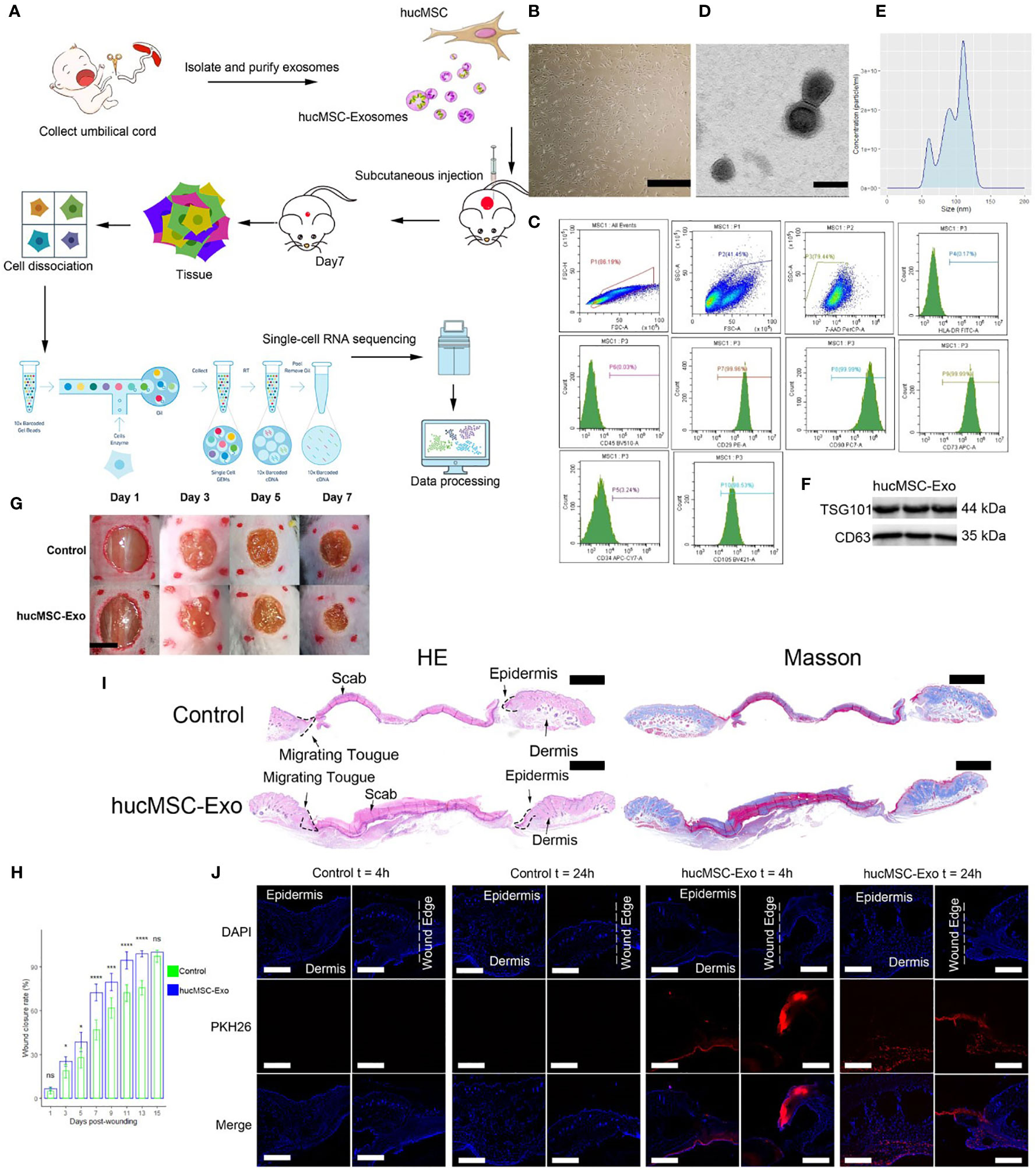
Figure 1 HucMSC-Exosomes accelerate wound healing. (A) Schematic overview of the study design. (B) Microscope image of huc-MSCs. The scale bar is 500 µm. (C) Flow cytometry analysis of HLA, CD29, CD34, CD45, CD73, CD90 and CD105 expression on the huc-MSC surface. (D) Transmission electron microscopy image of hucMSC-Exosomes. Scale bar, 100 nm. (E) Density plot of hucMSC-Exosomes. (F) Western blot of hucMSC-Exosome markers (G) Representative images of wound closure process of hucMSC-Exosomes treatment and PBS control. The scale bar is 4 mm. (H) Wound closure rate of hucMSC-Exosomes treatment and PBS control group. Two-tailed unpaired t-test (n=6). Error bar: mean standard deviation. Ns, not significant, *p < 0.05, ***p < 0.001, ****p < 0.0001. (I) H & E and Masson staining of wounds seven days after injury. Scale bar = 1mm. (J) Uptake of hucMSC-Exosome by skin wound in vivo. Scale bar, 500μm.
“HucMSC-Exosomes accelerate wound healing. (A) Schematic overview of the study design. (B) Microscope image of huc-MSCs. The scale bar is 500 µm. (C) Flow cytometry analysis of HLA, CD29, CD34, CD45, CD73, CD90 and CD105 expression on the huc-MSC surface. (D) Transmission electron microscopy image of hucMSC-Exosomes. Scale bar, 100 nm. (E) Density plot of hucMSC-Exosomes. (F) Western blot of hucMSC-Exosome markers (G) Representative images of wound closure process of hucMSC-Exosomes treatment and PBS control. The scale bar is 4 mm. (H) Wound closure rate of hucMSC-Exosomes treatment and PBS control group. Two-tailed unpaired t-test (n=6). Error bar: mean standard deviation. ns, not significant, *p < 0.05, ***p < 0.001, ****p < 0.0001. (I) H & E and Masson staining of wounds seven days after injury. Scale bar = 1mm. (J) Uptake of hucMSC-Exosome by skin wound in vivo. Scale bar, 500μm.”
The authors apologize for this error and state that this does not change the scientific conclusions of the article in any way. The original article has been updated.
Publisher’s note
All claims expressed in this article are solely those of the authors and do not necessarily represent those of their affiliated organizations, or those of the publisher, the editors and the reviewers. Any product that may be evaluated in this article, or claim that may be made by its manufacturer, is not guaranteed or endorsed by the publisher.
Keywords: exosomes, wound healing, single-cell RNA sequencing, cellular heterogeneity, neutrophils, macrophages
Citation: Liu Y, Zhang M, Liao Y, Chen H, Su D, Tao Y, Li J, Luo K, Wu L, Zhang X and Yang R (2023) Corrigendum: Human umbilical cord mesenchymal stem cell-derived exosomes promote murine skin wound healing by neutrophil and macrophage modulations revealed by single-cell RNA sequencing. Front. Immunol. 14:1181215. doi: 10.3389/fimmu.2023.1181215
Received: 07 March 2023; Accepted: 08 March 2023;
Published: 14 March 2023.
Approved by:
Frontiers Editorial Office, Frontiers Media SA, SwitzerlandCopyright © 2023 Liu, Zhang, Liao, Chen, Su, Tao, Li, Luo, Wu, Zhang and Yang. This is an open-access article distributed under the terms of the Creative Commons Attribution License (CC BY). The use, distribution or reproduction in other forums is permitted, provided the original author(s) and the copyright owner(s) are credited and that the original publication in this journal is cited, in accordance with accepted academic practice. No use, distribution or reproduction is permitted which does not comply with these terms.
*Correspondence: Rongya Yang, rongyayang960@outlook.com
†These authors have contributed equally to this work and share first authorship
‡These authors have contributed equally to this work and share last authorship