Assessing the Potential Association Between Microbes and Corrosion of Intra-Oral Metallic Alloy-Based Dental Appliances Through a Systematic Review of the Literature
 Umarevathi Gopalakrishnan1
  A. Sumathi Felicita2 Lodd Mahendra1 Masroor Ahmed Kanji3
  Saranya Varadarajan4
  A. Thirumal Raj4 Shaikh Mohammed Abdul Feroz5
  Deepak Mehta6 Hosam Ali Baeshen7
  Shankargouda Patil8*
- 1Department of Orthodontics, Sri Venkateswara Dental College and Hospital, Chennai, India
- 2Department of Orthodontics, Saveetha Dental College, Saveetha Institute of Medical and Technical Sciences, Saveetha University, Chennai, India
- 3Department of Prosthodontics, College of Applied Sciences, King Khalid University, Abha, Saudi Arabia
- 4Department of Oral Pathology and Microbiology, Sri Venkateswara Dental College and Hospital, Chennai, India
- 5Department of Prosthetic Dental Sciences, College of Dentistry, Jazan University, Jazan, Saudi Arabia
- 6Department of Preventive and Restorative Dentistry, College of Dental Medicine, University of Sharjah, Sharjah, United Arab Emirates
- 7Department of Orthodontics, College of Dentistry, King Abdulaziz University, Jeddah, Saudi Arabia
- 8Division of Oral Pathology, Department of Maxillofacial Surgery and Diagnostic Sciences, College of Dentistry, Jazan University, Jazan, Saudi Arabia
Objective: Systematic review assessing the association between oral microorganisms and corrosion of intra-oral metallic alloy-based dental appliances.
Design: PubMed, Scopus, and Web of Science were searched using keyword combinations such as microbes and oral and corrosion; microbes and dental and corrosion; microorganisms and oral and corrosion; microorganisms and dental and corrosion.
Results: Out of 141 articles, only 25 satisfied the selection criteria. Lactobacillus reuteri, Streptococcus mutans, Streptococcus sanguis, Streptococcus mitis, Streptococcus sobrinus, Streptococcus salivarius, sulfate-reducing bacteria, sulfate oxidizing bacteria, Veilonella, Actinomyces, Candida albicans were found to have a potential association with corrosion of intraoral metallic alloys such as stainless steel, titanium, nickel, cobalt-chromium, neodymium-iron-boron magnets, zirconia, amalgam, copper aluminum, and precious metal alloys.
Conclusion: The included studies inferred an association between oral microorganisms and intra-oral metallic alloys-based dental appliances, although, it is vital to acknowledge that most studies in the review employed an in-vitro simulation of the intra-oral condition.
Introduction
Metals in their pure or alloy forms are commonly used in dentistry despite the introduction of advanced materials like resins and ceramics, which can be largely attributed to the mechanical properties of metallic alloys (Upadhyay et al., 2006). The intra-oral environment has several factors that could predispose such metal alloy-based dental appliances to corrosion. These factors include varying temperature, oxygenation, mechanical forces, acidity, and alkalinity of external agents (foods, drugs), microorganisms, local anaerobic environments (e.g., subgingival). Some of the metals used in dentistry are amalgams of silver-tin, copper, noble metal alloys of gold and silver palladium, base metal alloys of nickel, cobalt, iron, and titanium alloys. Though most of the alloys are passivized and resistant to corrosion, the susceptibility still exists because of the predisposing factors in the oral environment (Bayramoglu et al., 2000; Karov and Hinberg, 2001). The clinical relevance of corrosion of dental appliances in the oral environment is due to some major clinical implications. The first is the potential toxic risk posed by the corrosion by-products. The second is that the corroded dental appliance could lose its functional integrity. The risk of allergy to the unbounded metal elements when released by corrosion should also be considered. A study by zora et al. suggested that corrosion products may pose a risk in immunologically susceptible patients (Venclíková et al., 2007).
The role of microorganisms in corrosion is extensively discussed in the sewage and pipeline industry, although the literature is relatively scanty when it comes to the biological environment, including the oral cavity (Mystkowska et al., 2018). Thus, the present systematic review was formulated to assess the association between microorganisms and corrosion of intra-oral dental appliances.
Materials and Methods
The present systematic review adhered strictly to the Preferred Reporting Items for Systematic Reviews and Meta-Analyses (PRISMA) statement (Moher et al., 2009; Hutton et al., 2015) (Figure 1).
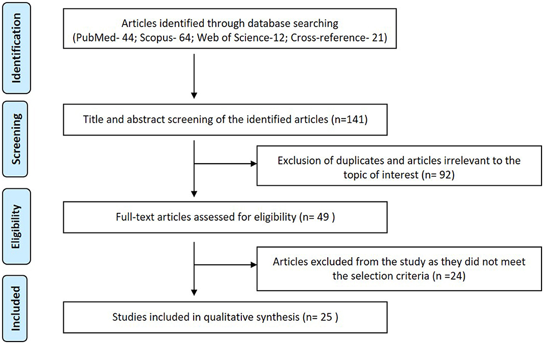
Figure 1. PRISMA flow diagram summarizing the study selection.
Inclusion Criteria
In-vitro studies in the English language assessing the potential effect of intra oral micro organisms on corrosion of metallic alloy-based dental appliances.
Exclusion Criteria
In-vivo studies, reviews, letters, case reports/series, editorials. Articles not in the English language. Articles without sufficient details on either the microbe or the dental appliance, for invivo studies, due consideration to antibiotic use during sampling was checked. In-vivo studies were excluded as the research design could potentially play a major role in determining the final outcome. In addition, a prilimanary literature search revealed that at present, there are were no in-vivo studies which have assessed the effect of intra oral micro organisms on corrosion of metallic alloy-based dental appliances.
Focus Question
What is the effect of an intra-oral microorganism on the corrosion of intraoral metallic alloy-based dental appliances? (population –metallic alloy-based dental appliances, intervention–intra oral microorganisms, comparator- metallic alloy-based dental appliances without oral microorganisms, outcome–corrosion).
Search Strategy
PubMed, Scopus, and Web of Science were searched using various combinations of the following keywords: microbes and oral and corrosion; microbes and dental and corrosion; microorganisms and oral and corrosion; microorganisms and dental and corrosion. The identified articles were manually cross-referenced to identify further potential articles.
Study Selection and Data Extraction
• Identified articles were screened for relevance to the topic and potential duplicates using their titles and abstracts.
• The full text of the screened articles was assessed using the selection criteria
Two reviewers (UG and SV) independently performed steps 1 and 2. Kappa coefficient (κ) was calculated to assess inter-observer reliability. Only studies satisfying the selection criteria were included in the qualitative analysis. Data including the study characteristics, design, assessment tools, the microbe, the metallic alloy assessed, results, and inference were extracted from these included articles. Due to the lack of a standard risk of bias tool for in-vitro studies, a customized risk of bias tool was formulated. The categorization in to high, medium, and low risk was based on Joanna Brigg's critical appraisal tool (The Joanna Briggs Institute, 2014; Normando et al., 2017).
Results
Study Selection
Hundred and forty-one articles (PubMed- 44; Scopus- 64; Web of Science-12; Cross-reference- 21) were identified in the search. Title and abstract screening led to the exclusion of 92 articles as they were either duplicate or lacked relevance to the topic of interest. Of the 49 articles subjected to full-text review, 24 articles were excluded as they did not fulfill the inclusion criteria (Supplementary Table 1). Only 25 articles met the eligibility criteria and were included in this review. Figure 1 summarizes the selection strategy employed in the qualitative analysis. Table 1 summarizes the data extracted from the studies included in the systematic review. Kappa coefficient (κ) for 1st and 2nd step of the review was 0.97 and 0.94, respectively indicating a good interreviewer reliability.
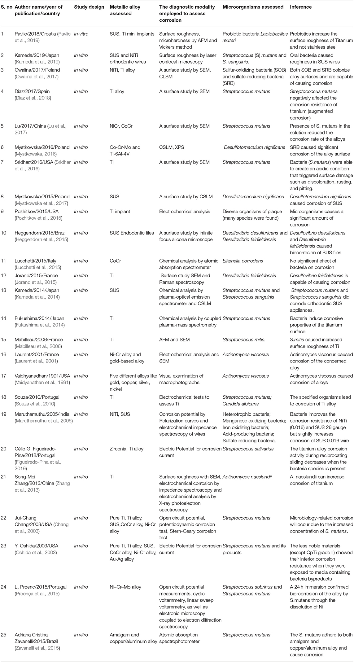
Table 1. Data extracted from the studies included in the systematic review.
Risk of Bias
Of the 25 studies included, 4 studies had a low risk of bias and 4 studies had a high risk of bias. The rest of the studies (n = 16) had moderate risk of bias. The summary of the risk of bias assessment is presented in Table 2.
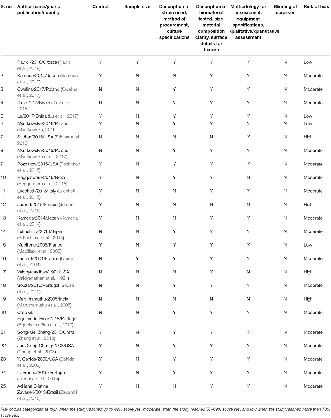
Table 2. Summary of the risk of bias assessment of the studies included in the systematic review.
Study Characteristics
Of the 25 articles selected 5 were from the USA (Vaidyanathan et al., 1991; Chang et al., 2003; Oshida et al., 2003; Pozhitkov et al., 2015; Sridhar et al., 2016), 3 each were from Japan (Fukushima et al., 2014; Kameda et al., 2014, 2019), Portugal (Souza et al., 2010; Proença et al., 2015; Figueiredo-Pina et al., 2019), France (Laurent et al., 2001; Mabilleau et al., 2006; Jorand et al., 2015), Poland (Mystkowska, 2016; Cwalina et al., 2017; Mystkowska et al., 2017), 2 each from Brazil (Heggendorn et al., 2015; Zavanelli et al., 2015) and China (Zhang et al., 2013; Lu et al., 2017), 1 each from Croatia (Pavlic et al., 2019), Spain (Díaz et al., 2018), Italy (Lucchetti et al., 2015), and India (Maruthamuthu et al., 2005).
Main Findings
Vital data including the assessed metallic alloy, the microorganisms, the methodology employed for detecting the microbe, and for assessing the corrosion, the statistical data, and the inferences drawn were extracted from all the included studies (Table 1). Titanium (Ti) was assessed in 16 studies (Oshida et al., 2003; Maruthamuthu et al., 2005; Mabilleau et al., 2006; Souza et al., 2010; Fukushima et al., 2014; Kameda et al., 2014; Jorand et al., 2015; Pozhitkov et al., 2015; Mystkowska, 2016; Sridhar et al., 2016; Díaz et al., 2018; Figueiredo-Pina et al., 2019), stainless steel (SUS) in five studies (Chang et al., 2003; Oshida et al., 2003; Maruthamuthu et al., 2005; Kameda et al., 2014; Heggendorn et al., 2015; Mystkowska et al., 2017), nickel (NiCr) and cobalt-chromium (CoCr) alloys in seven studies (Laurent et al., 2001; Chang et al., 2003; Oshida et al., 2003; Lucchetti et al., 2015; Proença et al., 2015; Mystkowska, 2016; Lu et al., 2017), neodymium-iron, zirconia (Figueiredo-Pina et al., 2019), amalgam and copper aluminum alloy (Zavanelli et al., 2015) and precious metal alloys (Vaidyanathan et al., 1991) were each assessed on one study. The bacteria that were studied include probiotic bacteria Lactobacillus reuteri (Pavlic et al., 2019), Streptococcus (S.) mutans (Chang et al., 2003; Oshida et al., 2003; Souza et al., 2010; Fukushima et al., 2014; Kameda et al., 2014, 2019; Zavanelli et al., 2015; Sridhar et al., 2016; Lu et al., 2017), S.sanguis (Kameda et al., 2014, 2019), S.mitis (Mabilleau et al., 2006), S.obrinus (Proença et al., 2015), S.salivarius (Figueiredo-Pina et al., 2019) sulfate-reducing bacteria (SRB) (Maruthamuthu et al., 2005; Heggendorn et al., 2015; Jorand et al., 2015; Mystkowska, 2016; Cwalina et al., 2017; Mystkowska et al., 2017), sulfate oxidizing bacteria (SOB) (Cwalina et al., 2017), Actinomyces (Vaidyanathan et al., 1991; Laurent et al., 2001; Zhang et al., 2013), Eikenella (Lucchetti et al., 2015), Candida albicans (Souza et al., 2010) and non-specific oral bacteria (Maruthamuthu et al., 2005; Pozhitkov et al., 2015). The corrosion property was studied with scanning electron microscope (SEM) (Laurent et al., 2001; Mabilleau et al., 2006; Zhang et al., 2013; Jorand et al., 2015; Proença et al., 2015; Sridhar et al., 2016; Cwalina et al., 2017; Lu et al., 2017), confocal laser scanning microscopy (CSLM) (Kameda et al., 2014, 2019; Mystkowska, 2016; Cwalina et al., 2017; Mystkowska et al., 2017), microhardness with atomic force microscopy (AFM) (Mabilleau et al., 2006; Pavlic et al., 2019), atomic absorption spectrophotometry (Zavanelli et al., 2015), mass spectroscopy (Lucchetti et al., 2015; Pozhitkov et al., 2015), corrosion potential measurement (Chang et al., 2003; Oshida et al., 2003; Maruthamuthu et al., 2005; Proença et al., 2015; Figueiredo-Pina et al., 2019), impedance spectroscopy (Maruthamuthu et al., 2005; Zhang et al., 2013), and electrochemical analysis (Souza et al., 2010). Only one study by Vaidhyanadhan et al. (26) used visual examination by macro photography to assess the corrosion. Probiotics-like Lactobacillus Reuteri (Pavlic et al., 2019) increases the surface roughness of Ti mini-implants while less effect is seen on SUS. In a study by Kameda et al. (2019), oral bacteria like S.mutans and S.sanguinis were shown to corrode stainless steel orthodontic wires. S.mutans has been shown to increase the corrosion of both SUS and Ti alloys (Chang et al., 2003; Oshida et al., 2003; Souza et al., 2010; Fukushima et al., 2014; Kameda et al., 2014, 2019; Proença et al., 2015; Zavanelli et al., 2015; Sridhar et al., 2016; Díaz et al., 2018). S.mitis has also been shown to be corrosive toward Ti alloy (Mabilleau et al., 2006). Sulfate-reducing bacteria (Heggendorn et al., 2015; Jorand et al., 2015; Mystkowska, 2016; Cwalina et al., 2017; Mystkowska et al., 2017) have been shown to corrode SUS and Ti alloys. Actinomyces (Vaidyanathan et al., 1991; Laurent et al., 2001; Zhang et al., 2013) caused corrosion of NiCr and other precious metal alloys. Eikenella (Lucchetti et al., 2015) did not show any association with the corrosion of metals. Candida albicans has been shown to have a corrosive influence on Ti (Souza et al., 2010). Maruthamuthu et al. (2005) reported that bacteria improve the corrosion resistance of NiTi (0.016) and SUS 26 gauge but slightly increases corrosion of SUS 0.016 wire. Figueiredo-Pina et al. (2019) reported that the presence of S.salivarious in the lubricant reduces the corrosion wear of Ti. Table 3 summarizes the effect of oral-microorganisms on the corrosion of metal alloys.
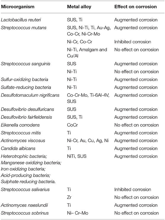
Table 3. Effect of oral microorganism on the corrosion of metal alloy in the included studies.
Discussion
Microbial Corrosion of metal is induced by activities of microorganisms like bacteria, fungi, and algae (Wilson et al., 1997; Daubert et al., 2018). The bacteria more commonly attributed to corrosion are SRB, SOB, iron-oxidizing/ reducing bacteria, manganese-oxidizing bacteria, Pseudomonas, bacteria secreting organic acids, and slime. Among the fungi, Cladosporium, Aspergillus, Penicillium, and Paecilomyces (Iverson, 1987), and Candida albicans (Souza et al., 2010) are associated with metallic alloy corrosion. Bluegreen algae and a species of red algae (Graciollasia sp.) are the algae associated with corrosion (Iverson, 1987). In the present article, the published literature was reviewed to assess the association between oral microbes and corrosion in intra-oral dental materials.
It was observed that various species like Streptococcus, Actinomyces, Veilonella, SRB, SOB, were reported to cause corrosion intraorally. There can be two categories of microbial corrosion based on the involvement of oxygen, anaerobic, and aerobic corrosion. SRB is a classic example of anaerobic corrosion while SOB is a prime example of aerobic corrosion. The basic process of corrosion involves a flow of electricity between certain areas of a metal surface through a solution that can conduct an electric current. Organisms like SOB secrete organic acids as part of their fermentation process which in turn stimulates anodic reactions. Sulphuric acid produced by SOB reduces the pH which in turn favors the growth of iron and manganese-oxidizing bacteria. These microbes oxidize manganese and iron metal alloys and cause their corrosion (Maruthamuthu et al., 2005). Pavlic et al. (2019), Sridhar et al. (2016), Pozhitkov et al. (2015), and Vaidyanathan et al. (1991) reported that a difference in pH could have contributed to the microbial corrosion. Literature suggests that the lowering pH, although may not corrode as the pH does not reach the depassivation point, it is plausible that it may favor the process (Nash and Kelly, 1993; Schiff et al., 2002). Mabilleau et al. (2006) suggested that S.mitis releases lactic acid in the microenvironment and it is likely that this compound is the main candidate to explain Ti corrosion. Some organisms stimulate cathodic reactions by consuming hydrogen. Sulfate-reducing bacteria (SRB) consume hydrogen through hydrogenase enzymes thereby depolarizing the cathode enhancing the process of corrosion (Mystkowska, 2016). SRB also utilize lactate produced by other bacteria in the biofilm as a carbon source and reduce sulfate to sulfide. Sulfide combines with iron in SUS alloys to form ferrous sulfide as the corrosion product. Cwalina et al. (2017) found that both groups of bacteria of sulfur cycle, SRB, and SOB colonize NiTi and Ti alloys, with a lower pH favoring the growth of SRB and causing further corrosion. Both SRB and SOB are capable of corroding NiTi and Ti alloys even though Kameda et al. (2014) found a higher degree of corrosion in SUS and none in Ti. This in turn could be because Ti is more resistant to corrosion by electric current.
Corrosion cells also occur when two areas are in contact with different concentrations of the same solution, like a difference in concentration of oxygen. The less-aerated zone acts as an anode, which undergoes corrosion. One of the factors causing such oxygenation difference is the heterogeneous layer of a biofilm with bacteria like Streptococcus mutans which use oxygen and create a difference in degrees of oxygen concentration based on their presence or absence in the biofilm (Alasvand Zarasvand and Rai, 2014). This is the main mechanism behind the corrosion of S.mutans. Fukushima et al. (2014) also suggested a similar mechanism in their study. Actinomyces viscosus consumes oxygen and shifts the anodic curve toward more negative potentials causing corrosion of metals (Laurent et al., 2001). In addition to other reasons, Díaz et al. (2018) has suggested that surface roughness promotes the corrosion of the Ti surface by S.mutans by creating retentive areas for the bacteria.
There were few contrary findings regarding microbial corrosion. Lu et al. (2017) stated that S. mutans formed a biofilm on the metal surface which enhances corrosion resistance by creating physical barriers that prevented oxygen interactions with the metal surfaces. Lucchetti et al. (2015) too found no significant effect of bacteria like Eikenella corrodens on corrosion of metal alloys. A study by Maruthamuthu et al. (2005) has shown that passivity and corrosion resistance of some SUS and NiTi was improved by bacteria whereas some SUS was shown to decrease. In a study by Liu et al. (2018) found that rapid electrochemical anodization treatment used on Ti2448 alloys increased their biocorrosion resistance. Regarding the prevention against microbial corrosion, Liu et al. (2018) suggested that a new beta-type Ti alloy with a hybrid oxide layer produced by the electrochemical anodization treatment provided better protection against corrosion by microorganisms by lowering the anodic and cathodic current densities. Jorand et al. (2015) showed that the SRB is resistant to ampicillin therapy which might sound that fighting corrosion against these organisms might be difficult. Microbial corrosion needs higher attention in dentistry as more evidence is gathered regarding their role in inducing intra-oral corrosion of alloys. The present systematic review provides insight into the various microorganisms implicated in causing corrosion of intraoral metallic alloy-based dental appliances. Also, the various mechanisms for a microbe induced metallic alloy corrosion are elaborated. Out of the 25 articles reviewed, 23 articles suggested that microorganisms are capable of causing corrosion while 2 articles (Maruthamuthu et al., 2005; Lu et al., 2017) suggested that they protect against corrosion and one suggested no significant effect (Lucchetti et al., 2015).
Although all the 25 studies had assessed the role of microorganisms in corrosion of dental appliance, most were in-vitro studies simulating the intra-oral conditions. Also, there were several variables including methodology used to assess the corrosion, the research design (in-vitro/in-vivo microenvironment) employed, the microbe and the metal/alloys assessed which led to large-scale heterogeneity in the collected data. In addition, in studies like Pozhitkov et al., the results did not specify the microorganisms responsible for the corrosion (Pozhitkov et al., 2015). Given the significant number of variables in the included studies and the lack of specificity in reporting the the causative microbe, a quantitative analysis was not possible.
Conclusion
The review identified several microorganisms to be closely associated with corrosion of intraoral metallic alloy-based dental appliance. Despite the association, it is vital to acknowledge that most of the included studies were based on in-vitro models. Thus, large-scale multi-center prospective clinical studies with a homogenous research design are required to validate the findings of the present systematic review.
Data Availability Statement
The original contributions presented in the study are included in the article/Supplementary Material, further inquiries can be directed to the corresponding author/s.
Author Contributions
UG, AF, LM, SP, SV, and AR contributed to the conception of the work, data acquisition, analysis, drafting the work, and final approval of the version to be published. MK, SF, DM, and HB contributed to the interpretation of data, revising it critically for important intellectual content, and final approval of the version to be published. All authors agree to be accountable for all aspects of the work ensuring that questions related to the accuracy or integrity of any part of the work are appropriately investigated and resolved.
Conflict of Interest
The authors declare that the research was conducted in the absence of any commercial or financial relationships that could be construed as a potential conflict of interest.
Supplementary Material
The Supplementary Material for this article can be found online at: https://www.frontiersin.org/articles/10.3389/fbioe.2021.631103/full#supplementary-material
References
Alasvand Zarasvand, K., and Rai, V. R. (2014). Microorganisms: induction and inhibition of corrosion in metals. Int. Biodeterior. Biodegradation 87, 66–74. doi: 10.1016/j.ibiod.2013.10.023
Bayramoglu, G., Alemdaroglu, T., Kedici, S., and Aksut, A. A. (2000). The effect of pH on the corrosion of dental metal alloys. J. Oral. Rehabil. 27, 563–575. doi: 10.1046/j.1365-2842.2000.00549.x
Chang, J.-C., Oshida, Y., Gregory, R. L., Andres, C. J., Barco, T. M., and Brown, D. T. (2003). Electrochemical study on microbiology-related corrosion of metallic dental materials. Biomed. Mater. Eng. 13, 281–295. Available online at: http://www.ncbi.nlm.nih.gov/pubmed/12883177 (accessed October 10, 2020).
Cwalina, B., Dec, W., Michalska, J. K., Jaworska-Kik, M., and Student, S. (2017). Initial stage of the biofilm formation on the NiTi and Ti6Al4V surface by the sulphur-oxidizing bacteria and sulphate-reducing bacteria. J. Mater. Sci. Mater. Med. 28:173. doi: 10.1007/s10856-017-5988-2
Daubert, D., Pozhitkov, A., McLean, J., and Kotsakis, G. (2018). Titanium as a modifier of the peri-implant microbiome structure. Clin. Implant Dent. Relat. Res. 20, 945–953. doi: 10.1111/cid.12676
Díaz, I., Pacha-Olivenza, M. Á., Tejero, R., Anitua, E., González-Martín, M. L., Escudero, M. L., et al. (2018). Corrosion behavior of surface modifications on titanium dental implant. In situ bacteria monitoring by electrochemical techniques. J. Biomed. Mater. Res. Part B Appl. Biomater. 106, 997–1009. doi: 10.1002/jbm.b.33906
Figueiredo-Pina, C. G., Guedes, M., Sequeira, J., Pinto, D., Bernardo, N., and Carneiro, C. (2019). On the influence of Streptococcus salivarius on the wear response of dental implants: an in vitro study. J. Biomed. Mater. Res. Part B Appl. Biomater. 107, 1393–1399. doi: 10.1002/jbm.b.34231
Fukushima, A., Mayanagi, G., Nakajo, K., Sasaki, K., and Takahashi, N. (2014). Microbiologically induced corrosive properties of the titanium surface. J. Dent. Res. 93, 525–529. doi: 10.1177/0022034514524782
Heggendorn, F. L., Gonçalves, L. S., Dias, E. P., Lione, V. d. O. F., and Lutterbach, M. T. S. (2015). Biocorrosion of endodontic files through the action of two species of sulfate-reducing bacteria: desulfovibrio desulfuricans and Desulfovibrio fairfieldensis. J. Contemp. Dent. Pract. 16, 665–673. doi: 10.5005/jp-journals-10024-1738
Hutton, B., Salanti, G., Caldwell, D. M., Chaimani, A., Schmid, C. H., Cameron, C., et al. (2015). The PRISMA extension statement for reporting of systematic reviews incorporating network meta-analyses of health care interventions: checklist and explanations. Ann. Intern. Med. 162, 777–784. doi: 10.7326/M14-2385
Iverson, W. P. (1987). Microbial corrosion of metals. Adv. Appl. Microbiol. 32, 1–36. doi: 10.1016/S0065-2164(08)70077-7
Jorand, F. P. A., Debuy, S., Kamagate, S. F., and Engels-Deutsch, M. (2015). Evaluation of a biofilm formation by Desulfovibrio fairfieldensis on titanium implants. Lett. Appl. Microbiol. 60, 279–287. doi: 10.1111/lam.12370
Kameda, T., Oda, H., Ohkuma, K., Sano, N., Batbayar, N., Terashima, Y., et al. (2014). Microbiologically influenced corrosion of orthodontic metallic appliances. Dent. Mater. J. 33, 187–195. doi: 10.4012/dmj.2013-297
Kameda, T., Oda, H., Ohkuma, K., and Terada, K. (2019). Effects of magnetic fields from electric toothbrushes on fluoride- and oral bacteria-induced corrosion of orthodontic metallic wires. Dent. Mater. J. 38, 909–920. doi: 10.4012/dmj.2018-293
Karov, J., and Hinberg, I. (2001). Galvanic corrosion of selected dental alloys. J. Oral Rehabil. 28, 212–219. doi: 10.1111/j.1365-2842.2001.00728.x
Laurent, F., Grosgogeat, B., Reclaru, L., Dalard, F., and Lissac, M. (2001). Comparison of corrosion behaviour in presence of oral bacteria. Biomaterials 22, 2273–2282. doi: 10.1016/S0142-9612(00)00416-6
Liu, C.-F., Lee, T.-H., Liu, J.-F., Hou, W.-T., Li, S.-J., Hao, Y.-L., et al. (2018). A unique hybrid-structured surface produced by rapid electrochemical anodization enhances bio-corrosion resistance and bone cell responses of β-type Ti-24Nb-4Zr-8Sn alloy. Sci. Rep. 8:6623. doi: 10.1038/s41598-018-24590-x
Lu, C., Zheng, Y., and Zhong, Q. (2017). Corrosion of dental alloys in artificial saliva with Streptococcus mutans. PLoS ONE 12:e0174440. doi: 10.1371/journal.pone.0174440
Lucchetti, M. C., Fratto, G., Valeriani, F., De Vittori, E., Giampaoli, S., Papetti, P., et al. (2015). Cobalt-chromium alloys in dentistry: an evaluation of metal ion release. J. Prosthet. Dent. 114, 602–608. doi: 10.1016/j.prosdent.2015.03.002
Mabilleau, G., Bourdon, S., Joly-Guillou, M. L., Filmon, R., Baslé, M. F., and Chappard, D. (2006). Influence of fluoride, hydrogen peroxide and lactic acid on the corrosion resistance of commercially pure titanium. Acta Biomater. 2, 121–129. doi: 10.1016/j.actbio.2005.09.004
Maruthamuthu, S., Rajasekar, A., Sathiyanarayanan, S., Muthukumar, N., and Palaniswamy, N. (2005). Electrochemical behaviour of microbes on orthodontic wires. Curr. Sci. 89, 988–996.
Moher, D., Liberati, A., Tetzlaff, J., and Altman, D. G. (2009). Preferred reporting items for systematic reviews and meta-analyses: the PRISMA statement. BMJ 339, 332–336. doi: 10.1136/bmj.b2535
Mystkowska, J. (2016). Biocorrosion of dental alloys due to Desulfotomaculum nigrificans bacteria. Acta Bioeng. Biomech. 18, 87–96.
Mystkowska, J., Ferreira, J. A., Leszczyńska, K., Chmielewska, S., Dabrowski, J. R., Wieciński, P., et al. (2017). Biocorrosion of 316LV steel used in oral cavity due to Desulfotomaculum nigrificans bacteria. J. Biomed. Mater. Res. B Appl. Biomater. 105, 222–229. doi: 10.1002/jbm.b.33518
Mystkowska, J., Niemirowicz-Laskowska, K., Łysik, D., Tokajuk, G., Dabrowski, J., and Bucki, R. (2018). The role of oral cavity biofilm on metallic biomaterial surface destruction–corrosion and friction aspects. Int. J. Mol. Sci. 19:743. doi: 10.3390/ijms19030743
Nash, B. K., and Kelly, R. G. (1993). Characterization of the crevice solution chemistry of 304 stainless steel. Corros. Sci. 35, 817–825. doi: 10.1016/0010-938X(93)90220-B
Normando, A. G. C., Rocha, C. L., de Toledo, I. P., de Souza Figueiredo, P. T., dos Reis, P. E. D., De Luca Canto, G., et al. (2017). Biomarkers in the assessment of oral mucositis in head and neck cancer patients: a systematic review and meta-analysis. Support. Care Cancer 25, 2969–2988. doi: 10.1007/s00520-017-3783-8
Oshida, Y., Koh, W., Gregory, R. L., Chang, J. C., Al-Ali, S., Ito, M., et al. (2003). “Effects of bacteria-induced corrosion on galvanic couples of Cp titanium with other dental alloys,” in EventMedical Device Materials–Proceedings of the Materials and Processes for Medical Devices Conference 2003, ed. S. Shrivastava (Anaheim, CA), 423–428.
Pavlic, A., Perissinotto, F., Turco, G., Contardo, L., and Stjepan, S. (2019). Do chlorhexidine and probiotics solutions provoke corrosion of orthodontic mini-implants? An in vitro study. Int. J. Oral Maxillofac. Implants 34, 1379–1388. doi: 10.11607/jomi.7392
Pozhitkov, A. E., Daubert, D., Brochwicz Donimirski, A., Goodgion, D., Vagin, M. Y., Leroux, B. G., et al. (2015). Interruption of electrical conductivity of titanium dental implants suggests a path towards elimination of corrosion. PLoS ONE 10:e0140393. doi: 10.1371/journal.pone.0140393
Proença, L., Barroso, H., Figueiredo, N., Lino, A. R., Capelo, S., and Fonseca, I. T. E. (2015). The corrosion resistance of Wiron®88 in the presence of S. mutans and S. sobrinus bacteria. J. Mater. Sci. Mater. Med. 26:29. doi: 10.1007/s10856-014-5353-7
Schiff, N., Grosgogeat, B., Lissac, M., and Dalard, F. (2002). Influence of fluoride content and pH on the corrosion resistance of titanium and its alloys. Biomaterials 23, 1995–2002. doi: 10.1016/S0142-9612(01)00328-3
Souza, J. C. M., Henriques, M., Oliveira, R., Teughels, W., Celis, J.-P., and Rocha, L. A. (2010). Do oral biofilms influence the wear and corrosion behavior of titanium? Biofouling 26, 471–478. doi: 10.1080/08927011003767985
Sridhar, S., Abidi, Z., Wilson, T. G., Valderrama, P., Wadhwani, C., Palmer, K., et al. (2016). In vitro evaluation of the effects of multiple oral factors on dental implants surfaces. J. Oral. Implantol. 42, 248–257. doi: 10.1563/aaid-joi-D-15-00165
The Joanna Briggs Institute (2014). The Joanna Briggs Institute Reviewers' Manual 2014: Methodology for JBI Umbrella Reviews. Joanne Briggs Inst. 1–34. Available online at: http://joannabriggs.org/assets/docs/sumari/ReviewersManual_Mixed-Methods-Review-Methods-2014-ch1.pdf (accessed October 10, 2020).
Upadhyay, D., Panchal, M. A., Dubey, R. S., and Srivastava, V. K. (2006). Corrosion of alloys used in dentistry: a review. Mater. Sci. Eng. A 432, 1–11. doi: 10.1016/j.msea.2006.05.003
Vaidyanathan, T. K., Vaidyanathan, J., Linke, H. A. B., and Schulman, A. (1991). Tarnish of dental alloys by oral microorganisms. J. Prosthet. Dent. 66, 709–714. doi: 10.1016/0022-3913(91)90458-9
Venclíková, Z., Benada, O., Bártová, J., Joska, L., and Mrklas, L. (2007). Metallic pigmentation of human teeth and gingiva: morphological and immunological aspects. Dent. Mater. J. 26, 96–104. doi: 10.4012/dmj.26.96
Wilson, M., Patel, H., Kpendema, H., Noar, J. H., Hunt, N. P., and Mordan, N. J. (1997). Corrosion of intra-oral magnets by multi-species biofilms in the presence and absence of sucrose. Biomaterials 18, 53–57. doi: 10.1016/S0142-9612(96)00084-1
Zavanelli, A. C., Zavanelli, R. A., Mazaro, J. V. Q., and Falcón-Antenucci, R. M. (2015). Streptococcus mutans adhesion and releasing of metallic ions in dental alloys. Brazilian J. Oral Sci. 14, 36–40. doi: 10.1590/1677-3225v14n1a08
Keywords: corrosion, metallic alloys, microorganism, oral, prosthesis
Citation: Gopalakrishnan U, Felicita AS, Mahendra L, Kanji MA, Varadarajan S, Raj AT, Feroz SMA, Mehta D, Baeshen HA and Patil S (2021) Assessing the Potential Association Between Microbes and Corrosion of Intra-Oral Metallic Alloy-Based Dental Appliances Through a Systematic Review of the Literature. Front. Bioeng. Biotechnol. 9:631103. doi: 10.3389/fbioe.2021.631103
Received: 19 November 2020; Accepted: 11 February 2021;
Published: 15 March 2021.
Edited by:
Liqiang Wang, Shanghai Jiao Tong University, ChinaReviewed by:
Jukka Meurman, University of Helsinki, FinlandYuan Liu, University of Pennsylvania, United States
Copyright © 2021 Gopalakrishnan, Felicita, Mahendra, Kanji, Varadarajan, Raj, Feroz, Mehta, Baeshen and Patil. This is an open-access article distributed under the terms of the Creative Commons Attribution License (CC BY). The use, distribution or reproduction in other forums is permitted, provided the original author(s) and the copyright owner(s) are credited and that the original publication in this journal is cited, in accordance with accepted academic practice. No use, distribution or reproduction is permitted which does not comply with these terms.
*Correspondence: Shankargouda Patil, sbpatil1612@gmail.com