A novel method for improving depth resolution and contrast of Integrated Differential Phase Contrast (IDPC) - STEM images
- Abstract number
- 1172
- Event
- European Microscopy Congress 2020
- DOI
- 10.22443/rms.emc2020.1172
- Session
- PST.1 - Phase Microscopy
- Authors
- Zhongbo Li (1), Ute Kaiser (1), Harald Rose (1)
- Affiliations
-
1. University of Ulm
- Keywords
depth resolution; integrated differential phase contrast; IDPC; STEM
- Abstract text
Techniques for obtaining efficient phase contrast in the scanning transmission electron microscope (STEM) imaging were proposed in the 1970s [1-3]. One of the techniques is the integrated differential phase contrast (IDPC). In the IDPC mode, the differential phase contrast (DPC) images are first acquired by subtracting the signals collected by opposite segments of a quadrant detector [3,4] or an annular quadrant detector [5]. The IDPC image is obtained by integrating the difference signals along the two symmetric axes of the segments. This method has demonstrated experimentally its advantage by showing the images of heavy and light atoms simultaneously and by suppressing high-frequency noise [4,5].
The current IDPC methods have limitations. For a thick sample, optimum image contrast is obtained only if the probe focusses at the upper surface of the object. As the probe moves down through the sample, the image contrast is suppressed, which makes it impossible to investigate the inner structure of the sample.
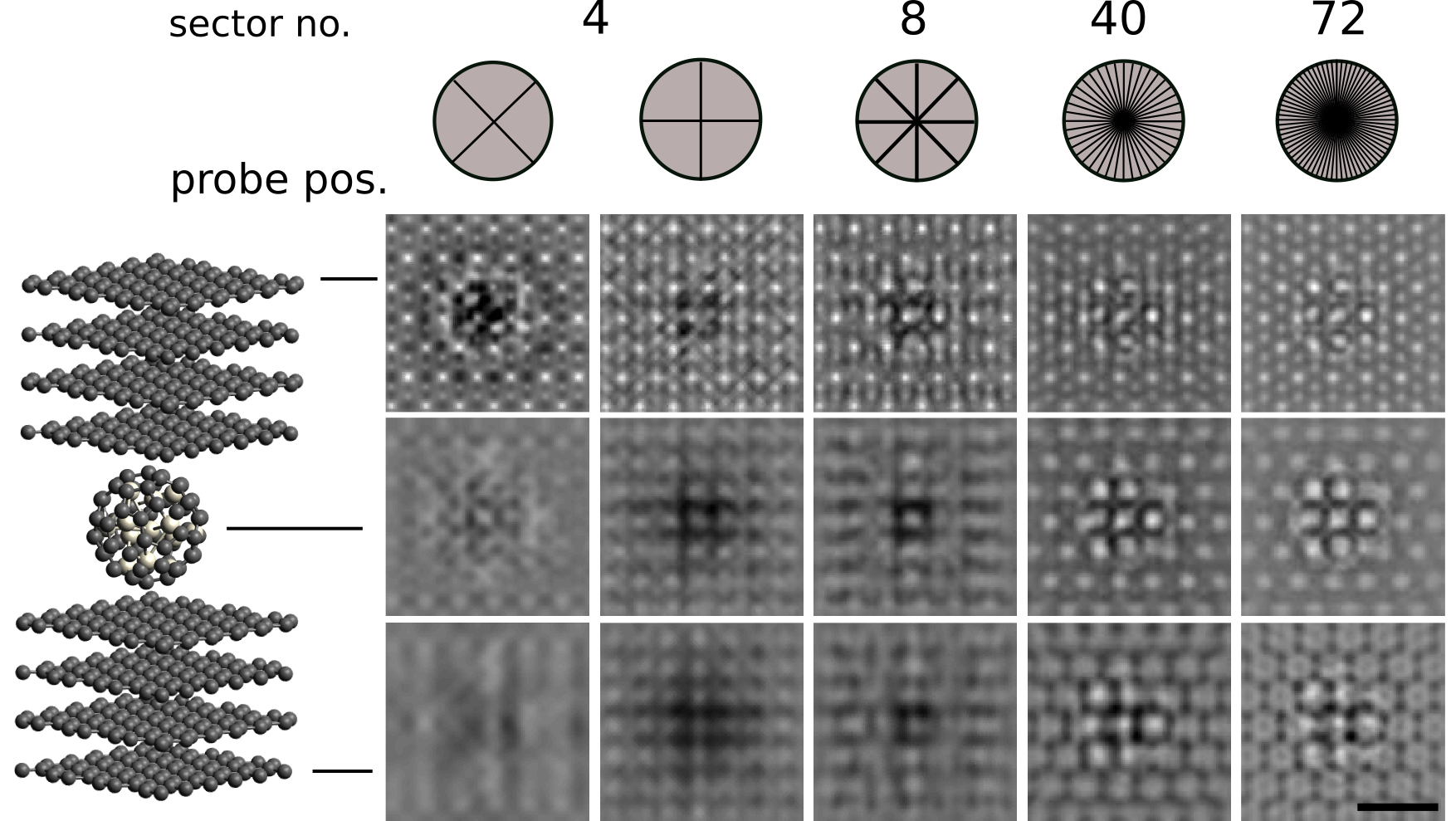
With the help of simulations, we found that by increasing the number of detector segments, it is possible to overcome the limitations of the current IDPC methods, shown in Fig. 1. A hetereostructure consisting of a Pt particle wrapped in a fullerene and then sandwiched between eight-layered graphite is imaged at 80kV in the IDPC mode using different detector geometries. As the probe moves from the upper surface down through the sample, the image contrast and resolution improve when the number of detector segments increases. Experimental realisation for the proposed IDPC-STEM imaging requires an aberration-corrected STEM equipped with a pixelated detector. A set of Ronchigrams corresponding to each scanning position have then to be collected. The posteriorly application of numerical detectors and our novel algorithm will allow optical sectioning for a thick sample.Figure 1 Simulated IDPC images using different numbers of detector segments for an aberration-corrected STEM operating at 80kV. The first two quadrant detector geometries are proposed in [4] and [3], respectively. The sample is a platinum particle wrapped in fullerene, and sandwiched between eight graphene slices. The whole sample thickness is 3.3nm. The images in each row are simulated for a given location of the probe focus. Scale bar: 0.5nm.
- References
[1] H Rose. Optik 39 (1974) 416–436.
[2] N. Dekkers and H De Lang. Optik 41 (1974) 452–456.
[3] H Rose. Ultramicroscopy 2 (1977) 251-267.
[4] I. Lazić, E. G. Bosch, and S. Lazar. Ultramicroscopy 160 (2016) 265–280.
[5] E. Yücelen, I. Lazić, and E. G. Bosch. Scientific reports 8 (2018) 1–10.