Abstracts
PURPOSE: To evaluate the effects of metoclopramide on abdominal wall healing in rats in the presence of sepsis. METHODS: 40 rats divided into two groups of twenty animals, subdivided into two subgroups of 10 animals each: group (E) - treated with metoclopramide, and saline-treated control group. The two groups were divided into subgroups of 10 to be killed on the 3rd day (n = 10) or day 7 (n = 10) after surgery. Sepsis was induced by cecal ligation and puncture. We performed also the section and anastomosis in left colon. The synthesis of the abdominal wall was made with 3-0 silk thread. We measured the breaking strength of the abdominal wall and made the histopathological evaluation. RESULTS: on 3rd day postoperative, the average breaking strength in the E group was 0.83 ± 0.66 and in group C was 0.35 ± 0.46 (p = 0.010). On the seventh day, the breaking strength in group E was11.44 ± 5.07, in group C 11.66 ± 7.38 (p = 1.000). The E7 group showed lower inflammatory infiltration, foreign body reaction, fibrin than control. CONCLUSION: animals treated with metoclopramide had a higher resistance of the abdominal wall on the 3rd postoperative day.
Abdominal Wall; Sepsis; Wound Healing; Metoclopramide; Rats
OBJETIVO: Avaliar os efeitos da metoclopramide na cicatrização da parede abdominal de ratos na vigência de sepse. METHODS: 40 ratos divididos em dois grupos de 20 animais, subdivididos em dois subgrupos de 10 animais cada: grupo (E) - tratado com metoclopramida, e o grupo controle tratado com solução fisiologica. Os dois grupos foram divididos em subgrupos de de 10 para serem mortos no dia 3 (n = 10) ou o dia 7 (n = 10) após a cirurgia. A sepse foi induzida por ligadura e perfuração cecal. Foi realizada também a secção e anastomose em cólon esquerdo. A síntese da parede abdominal foi feita com fio de seda 3-0. Mediu-se a força de ruptura da parede abdominal e foi feita uma avaliação histopatológica. RESULTADOS: No dia 3 pós-operatório, a força média de ruptura no grupo E foi de 0,83 ± 0,66 e no grupo C foi de 0,35 ± 0,46 (p = 0,010). No sétimo dia, a força de ruptura no grupo E foi 11.44 ± 5,07; no grupo C, 11,66 ± 7,38 (p = 1,000). O grupo E7 apresentou menor infiltração inflamatória e reação de corpo estranho do que o controle de fibrina. CONCLUSÃO: Animais tratados com metoclopramida apresentaram uma maior resistência da parede abdominal no 3º dia pós-operatório.
Parede Abdominal; Sepse; Cicatrização; Metoclopramida; Ratos
17 - ORIGINAL ARTICLE WOUND HEALING
Influence of metoclopramide on abdominal wall healing in rats subjected to colonic anastomosis in the presence of peritoneal sepsis induced1 1 Research performed at Laboratory of Experimental Surgery, Faculty of Medicine, University of Brasilia (UnB), Brasilia-DF, Brazil.
Influência da metoclopramida na cicatrização da parede abdominal de ratos submetidos à anastomose colônica na vigência de sepse peritonial induzida
Naiara Galvão da SilvaI; Alexandre Malta BrandãoI; Marcos Vinícius Melo de OliveiraI; Pedro Henrique Alves de MoraisI; Silvana Marques e SilvaII; Fabiana Pirani CarneiroIII; João Batista de SousaIV
IGraduate student, School of Medicine, UnB, Brasilia-DF, Brazil. Involved with technical procedures, acquisition and interpretation of data and statistical analysis and manuscript writing
IIFellow, PhD degree, Postgraduate Program in Medical Sciences, School of Medicine, UnB, Brasilia-DF, Brazil. Involved with technical procedures, acquisition and interpretation of data, statistical analysis, critical revision and manuscript writing
IIIPhD, Associate Professor of Pathology, School of Medicine, UnB, Brasilia-DF, Brazil. Macroscopic and histopathological examinations, interpretation of data and critical revision
IVPhD, Associate Professor, Surgical Clinics, School of Medicine, UnB, Brasilia-DF, Brazil. Responsible for intellectual and scientific content of the study; designed the protocol, involved with technical procedures, interpretation of data, critical revision and manuscript writing
Correspondence 1 Research performed at Laboratory of Experimental Surgery, Faculty of Medicine, University of Brasilia (UnB), Brasilia-DF, Brazil.
ABSTRACT
PURPOSE: To evaluate the effects of metoclopramide on abdominal wall healing in rats in the presence of sepsis.
METHODS: 40 rats divided into two groups of twenty animals, subdivided into two subgroups of 10 animals each: group (E) - treated with metoclopramide, and saline-treated control group. The two groups were divided into subgroups of 10 to be killed on the 3rd day (n = 10) or day 7 (n = 10) after surgery. Sepsis was induced by cecal ligation and puncture. We performed also the section and anastomosis in left colon. The synthesis of the abdominal wall was made with 3-0 silk thread. We measured the breaking strength of the abdominal wall and made the histopathological evaluation.
RESULTS: on 3rd day postoperative, the average breaking strength in the E group was 0.83 ± 0.66 and in group C was 0.35 ± 0.46 (p = 0.010). On the seventh day, the breaking strength in group E was11.44 ± 5.07, in group C 11.66 ± 7.38 (p = 1.000). The E7 group showed lower inflammatory infiltration, foreign body reaction, fibrin than control.
CONCLUSION: animals treated with metoclopramide had a higher resistance of the abdominal wall on the 3rd postoperative day.
Key words: Abdominal Wall. Sepsis. Wound Healing. Metoclopramide. Rats.
RESUMO
OBJETIVO: Avaliar os efeitos da metoclopramide na cicatrização da parede abdominal de ratos na vigência de sepse.
METHODS: 40 ratos divididos em dois grupos de 20 animais, subdivididos em dois subgrupos de 10 animais cada: grupo (E) - tratado com metoclopramida, e o grupo controle tratado com solução fisiologica. Os dois grupos foram divididos em subgrupos de de 10 para serem mortos no dia 3 (n = 10) ou o dia 7 (n = 10) após a cirurgia. A sepse foi induzida por ligadura e perfuração cecal. Foi realizada também a secção e anastomose em cólon esquerdo. A síntese da parede abdominal foi feita com fio de seda 3-0. Mediu-se a força de ruptura da parede abdominal e foi feita uma avaliação histopatológica.
RESULTADOS: No dia 3 pós-operatório, a força média de ruptura no grupo E foi de 0,83 ± 0,66 e no grupo C foi de 0,35 ± 0,46 (p = 0,010). No sétimo dia, a força de ruptura no grupo E foi 11.44 ± 5,07; no grupo C, 11,66 ± 7,38 (p = 1,000). O grupo E7 apresentou menor infiltração inflamatória e reação de corpo estranho do que o controle de fibrina.
CONCLUSÃO: Animais tratados com metoclopramida apresentaram uma maior resistência da parede abdominal no 3º dia pós-operatório.
Descritores: Parede Abdominal. Sepse. Cicatrização. Metoclopramida. Ratos.
Introduction
Nausea and vomiting are the most common complications after surgery, therefore antiemetics are frequently prescribed1. Among these kinds of drugs, the use of peripheral and central anti-dopamine is of great interest. These drugs act on dopamine receptors in the trigger zone avoiding the vomit. They also have prokinetic action by blocking chemoreceptors, stimulating gastrointestinal motility1,2.
Metoclopramide is the most widely used antiemetic in hospital-clinical setting, but their use is very questioned due to potential side effects - drowsiness, extrapyramidal seizures and agitation2,3..There are few studies relating the use of this drug with its peripheral actions in wound healing, especially in the presence of sepsis.
The widespread infection stems from a severe sepsis in which there is an intense oxidative stress by the releasing of reactive oxygen species and proinflammatory cytokines4,5. It interferes in the process of healing5 and is a risk factor for dehiscence.
Healing is a complex tissue and vessel process that begins with hemostasis5,6 and is followed by the inflammatory response, there is important participation of macrophages and polymorphonuclear cells7 and formation of granulation tissue by a proliferation of fibroblasts that secrete collagen - the main protein in the acquisition of resistance8.
The use of drugs, the tension in the suture line, nutritional status and infection are important factors that interfere with tissue repair wound5. The dehiscence is a worrying factor in the hospital and it is associated with high rates of morbidity and mortality. The study of aggravating or risk factors for this complication is therefore of extreme clinical importance.
Thus, the evaluation of the interference of metoclopramide in the healing of surgical wounds in adverse conditions - as sepsis - is of great importance.
Methods
We used 40 rats, Rattus norvegicus, Wistar, male and healthy, kept for two weeks in the preoperative period in cages housing animals in the Laboratory of Experimental Surgery at the University of Brasilia. It were provided a standard diet and water ad libitum, under 12 hours light and 12 hours of darkness.
The animals were divided into two groups of twenty animals each, subdivided in two subgroups of 10 animals each:
*group (E) - post-operative administration of metoclopramide, animals euthanized at day 3 (n = 10) or day 7 (n = 10) after surgery;
*control group (C) - post-operative administration of saline, animals euthanized on day 3 (n = 10) or day 7 (n = 10) after surgery.
General anesthesia was performed with xilasine hydrochloride 10 mg / kg and ketamine 75 mg / kg intramuscularly. The animals were immobilized on wooden plates with fastening members. Trichotomy anterior abdominal wall and antisepsis with iodopovidine. The median laparotomy of 4 cm in length was started 1 cm above the external genitalia of animals.
The peritoneal sepsis was induced by partial ligation of the cecum with to increase the pressure within this segment of the intestine without causing ischemia, allowing free transit of the contents of the small intestine into the large intestine. Then, there were made perforations in the cecum in 10 random points with venipuncture needle 40 x 13. After ligation and perforation of the cecum, we resected a 0.5 cm segment of the left colon and made a terminal anastomosis with running sutures with 6.0 polypropylene thread.
The synthesis of the abdominal wall was made in two planes with continuous 3.0 silk thread.
After the procedure, metoclopramide was administered to Group E at a daily dose of 5 mg / kg, intraperitoneally until the day of euthanasia, and the animals in group C received identical volumes of saline solution 0.9% by same route of application. The postoperative analgesia was performed with buprenorphine 0.01 mg / kg subcutaneously every 12 hours.
The animals received water and standard diet at will in the days after surgery. We therefore analyzed clinical patterns of apathy, bristling hair, diarrhea, abdominal distension and wound complications such as bruising and signs of surgical site infection, graduated the following scale: 0 - absent 1 - present mild, 2 - presence moderate, 3 - strong presence, 4 - intense presence.
Reoperation of each animal occurred in previously appointed day for each subgroup. The animals were then re-submitted to general anesthesia and antisepsis. Resection of a rectangular area of the abdominal wall of 5 by 6 cm, involving the scar of previouslaparotomy, forwarding it to the analysis of tensile strength and histopathology. After exposure of the abdominal cavity was searched for signs of peritonitis and abscess. Euthanasia was performed by an overdose of tionenbutal.
Analysis of the tensile strength was performed by digital test apparatus called Versa Test (Mecmesin Versa Test, United Kingdom) coupled to a digital dynamometer AGF (MecmesinVersa Test, United Kingdom). The value of rupture was expressed in Newtons (N).
An experienced pathologist examined by light microscopy. We assessed the following parameters: polymorphonuclear infiltrate, mononuclear cells, fibroblasts, neovascularization, collagen amount and edema. Each of these parameters was graded from 0 to 2. It was found also the presence (1) or absence (0) of histopathology abscesses, foreign bodies and fibrin.
For statistical analysis we used SPSS® version 17.0, performing parametric and nonparametric tests, according to the nature of the variables studied. It will be considered a significant value of p <0.05.
Results
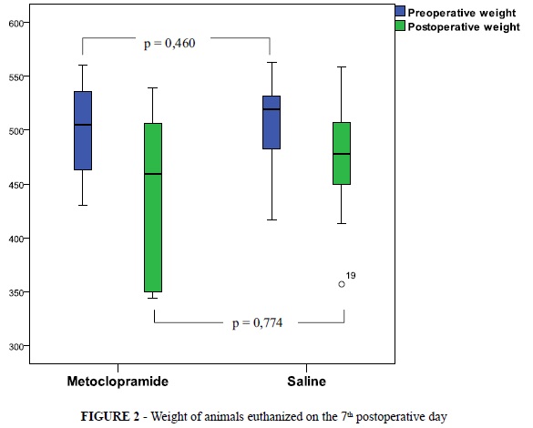
There was no significant difference in weight between groups before and after surgery. All groups of animals in this study had an average weight loss in the postoperative period (Figures 1, 2 and Table 1).
The animals in groups of the third day (E3 and C3) progressed clinically similar. The animals of E7, however showed worst clinical evolution than group C7 comparing by the parameters of apathy and bristling hair in postoperative with p = 0.033 and p = 0.033 in Fisher's exact test, respectively. No rat showed abdominal distension, bruising and signs of surgical site infection. A rat of C7 group died during the study due to sepsis (Table 2).
Those showed signs of peritonitis: four animals in group E3 with a rat containing intra-abdominal abscess; and seven animals in group C3. Six animals in group E3 and three in group C3 had blocked cecum. All animals in groups C7 and E7 had blocked cecum (Table 3).
The maximum and minimum breaking strength of the abdominal wall in the surgical wounds and their averages are shown in Table 4.
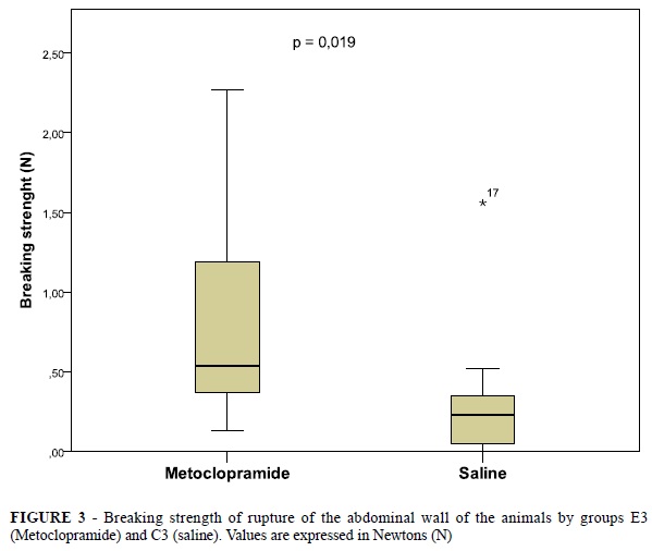
There was significant difference in the tensile strength of the wound between the study group of the third day(E3) and the control group of the third day (C3) with p = 0.019 using the Mann-Whitney test (Figure 3). Effect that were not observed among the groups C7 and E7, whose p = 1.000 by the Mann-Whitney test (Figure 4).
There was no significance between the groups on histopathological analysis as to the parameters of the polymorphonuclear infiltrate, fibroblasts, neovascularization, collagen content, histopathology abscess. The E7 group presented lower infiltration of mononuclear cells when compared to C7 (p = 0.019). The E7 group showed foreign body and fibrin with less frequency than the C7 group (p = 0.001 and p = 0.033). The other groups showed no significant differences regarding the mononuclear infiltrate, foreign body and fibrin (Tables 5 and 6).
Discussion
Metoclopramide is a prokinetic agent routinely used to treat nausea, vomiting and intestinal dysmotility in the post surgical6. In addition, some studies have shown that this drug also effects as an analgesic and antispasmodic6,7,8. Metoclopramide is a benzamide substitute drug, whose main action is to block both dopaminergic receptors 2 (D2): central (CNS) and peripheral (gastrointestinal tract). In the gastrointestinal tract, the D2 receptors negatively modulate the release of acetylcholine at the terminals of cholinergic visceral neurons, thus justifying the prokinetic effect of metoclopramide.
It was reported recently, the presence of D2 receptors in endothelial cells, platelets and lymphocytes, indicating a dopaminergic immunomodulation9,10. However, these same studies suggested a greater involvement of the D1 receptor modulation9,10.
Dopamine, on septic organisms, inhibits the migration and function of immune cells9,10. So that, the blockage by dopamine antagonists, such as metoclopramide, increasesin vitro the expression of pro-inflammatory cells10. In the present study, however, there was less infiltration of mononuclear and no foreign body reaction in the group with metoclopramide when related to the saline group (Tables 5 and 6), indicating that in vivo there is a mechanism by which metoclopramide has an inhibitory effect on the migration of macrophages and lymphocytes.
Overbeck et al.9 demonstrated that metoclopramide increases the release of prolactin. This effect occurs probably by inhibition of central dopaminergic receptors of the pituitary gland9.
Prolactin is a peptide hormone secreted by the pituitary gland that regulates sexual development and reproduction in some animal species. Recently it has been shown that this hormone has also immunomodulatory action, reducing the migration of NK cells, lymphocyte proliferation and maturation and the release of proinflammatory cytokines10. Thus the increased release of prolactin caused by the administration of metoclopramide, may diminish the inflammatory response to sepsis in the surgical wound, as observed in this study (Table 4).
In addition, metoclopramide showed a protective effect in the group E3 compared to C3 when analyzing to breaking strength. The normal healing is a complex process in which there are cell recruitment, extracellular matrix synthesis and cell replication. It is didactically divided into three main stages11: acute response, proliferative phase and remodeling. The first consists mainly of the 4th days following the surgical trauma, characterized by an intense polymorphonuclear infiltrate with few collagen11. Therefore, at this stage, the resistance of the wound depends on the suture. Then metoclopramide could play an analgesic effect, as observed in some studies6,7,8, leading to less contraction of the rat abdominal wall and lower concern for animal to pain. Thus the suture tends to maintain greater traction for a longer time, generating an increase in the tensile resistance of group E3. In addition, there would be a mechanism to contain the inflammation by the metoclopramide's stimulation of prolactin secretion. It could also accelerate the acute phase response anticipating the migration and proliferation of fibroblasts. This anticipation would give greater tensile strength in the abdominal wall compared with the control group E3.
It was observed that the clinical outcome after surgery was more significant in the group of seventh day in which metoclopramide was administered (E7) compared to control (C7). This finding could be explained by the central effect of the drug that blocks dopamine receptors in the central nervous system, leading to a sensitization of the limbic system for the intense inflammatory process caused by sepsis12.
Finally, administration of metoclopramide had no significant effect on the loss or gain of weight in animals in the postoperative period.
Conclusions
The administration of metoclopramide increased the breaking strength of the abdominal wall in the third postoperative day but it did not affect the tensile strength of the the seventh postoperative day. The administration of metoclopramide for 7 days after surgery decreased the migration of mononuclear cells and fibrin deposits in the surgical wound, but it did not affect significant the obtaining of breaking strength by de abdominal wall.
Correspondence:
João Batista de Sousa
Campus Universitário Darcy Ribeiro
Prédio da Reitoria, 2º pavimento, sala B2-16
70910-900 Brasília - DF Brasil
Tel.: (55 61)3307-2201
Conflict of interest: none
Financial source: none
Presented at the XII National Congress on Experimental Surgery of the Brazilian Society for Development of Research in Surgery-SOBRADPEC, 2011 October 26-29 Ribeirao Preto-SP, Brazil.
- 1. Contreras-Domínguez V, Carbonell-Bellolio P. Profilaxia Antiemética em Cirurgia de Abdome Agudo. Estudo comparativo entre droperidol, metoclopramida, tropisetron, granisetron e dexametasona. Rev Bras Anestesiol. 2008;58:1:35-44.
- 2. Bedin A, Pinho MSL, Zanotelli CT, Caldart AS, Turazzi JC, Castro RAC. Dexametasona comparada à metoclopramida na profilaxia de vômitos pós-operatórios em crianças submetidas a procedimentos cirúrgicos ambulatoriais. Rev Bras Anestesiol. 2005;55:4:387-96.
- 3. Gan TJ, Meyer T, Apfel CC, Chung F, Davis PJ, Eubanks S, Kovac A, Philip BK, Sessler DI, Temo J, Trame MR, Watcha M. Consensus guidelines for managing postoperative nausea and vomiting. Anesth Analg. 2003;97:62-71.
- 4. Zhang H, Slutsky AS, Vincent JL. Oxygen free radicals in ARDS, septic shock and organ dysfunction. Intensive Care Med. 2000;26:474-6.
- 5. Sousa JB, Oliveira PG. Cuidados com a ferida operatória: infecção. Clin Cir Bras. 1999;5:215-37.
- 6. Pang WW, Wu HS, Lin CH, Chang DP, Huang MH. Metoclopramida decreases emesis but increases sedation in tramadol atient-controlled analgesia. Can J Anesth. 2002;40:1029-33.
- 7. Kandler D, Lisander B. Analgesic action of metoclopramide in prosthetic hip surgery. Acta Anaesthesiol Scand.1993;37:49-53.
- 8. Hedenbro JL, Olsson AM. Metoclopramida and ureteric colic. Acta Chir Scand, 1988;154:439-40.
- 9. Oberbeck R, Schmitz D, Wilsenack K, Schüler M, Husain B, Schedlowski M, Exton MS. Dopamine affects cellular immune functions during polymicrobial sepsis. Intensive Care Med. 2006;32:731-9.
- 10. Beck GCH, Brinkkoetter P, Hanusch C, Schulte J, Acker K. Clicical review: Immunomodulatory effects of dopamine in general inflammation. Critical Care. 2004;8:485-91.
- 11. Wilgus TA. Immune cells in the healing skin wound: Influential players at each stage of repair. Pharmacological Research. 2008;58:112-6.
- 12. Wichterman KA, Baue AAAE, Chaudry IH. Sepsis and septic shock - a review of laboratory models and a proposal. J Surg Res. 1980;28:189-201.
Publication Dates
-
Publication in this collection
24 Oct 2011 -
Date of issue
2011