-
PDF
- Split View
-
Views
-
Cite
Cite
Radoslaw Zwolinski, Arkadiusz Ammer, Andrzej Walczak, Ryszard Jaszewski, Intrapericardial lipoma: diagnosed unexpectedly and resected during coronary artery bypass surgery, Interactive CardioVascular and Thoracic Surgery, Volume 11, Issue 2, August 2010, Pages 211–212, https://doi.org/10.1510/icvts.2010.233635
Close - Share Icon Share
Abstract
Cardiac lipomas are extremely rare tumors, they usually remain asymptomatic and are detected incidentally, mostly during autopsies. In symptomatic patients, the diagnosis can easily be made by echocardiography, computed tomography, or magnetic resonance imaging. We report a case of pericardial lipoma found unexpectedly during coronary artery bypass grafting (CABG) surgery. The patient underwent a successful resection of the tumor and CABG via a median sternotomy. The patient is currently asymptomatic and has not presented with evidence of recurrence at the 12-month follow-up.
Primary tumors of the heart are rare with an incidence rate between 0.0017% and 0.056% in autopsy reports [1]. Cardiac lipomas represent approximately 10% of all heart tumors [2] and can originate from the endocardium, myocardium and pericardium [3–6]. The majority of them are asymptomatic and are found incidentally at the time of autopsy. Cardiac signs and symptoms depend on the location and size of the tumor. These patients may present with malaise, fatigue, dyspnea, syncope, discomfort in chest, chest pain, palpitation, or sudden death [4–7]. We report an unusual case of a patient who was referred for coronary artery bypass grafting (CABG) surgery due to three vessel diseases and an intrapericardial lipoma, compressing the left atrium, was discovered during the procedure.
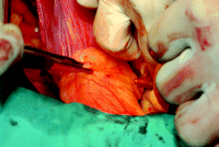
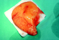
A 72-year-old man was referred to our clinic, presenting with Canadian angina class III angina with mild exertion as well as New York Heart Association (NYHA) class II dyspnea. Cardiac catheterization revealed three vessel diseases, and the ejection fraction was 50% seen on echocardiography (ECHO), and the chest X-ray was within normal limits. We connect 2nd class NYHA with coronary artery disease. Intraoperatively, after a median sternotomy and pericardial incision, a soft, yellow tumor attached to the posterior pericardium beneath the left pulmonary artery was found (Fig. 1 ). We decided to excise the tumor due to its unknown histopathological structure and to avoid future clinical consequences connected to the growth of the tumor, which could have constricted pulmonary artery and left atrium. After the introduction of extracorporeal circulation and delivering of cold crystalloid cardioplegic solution, the tumor was excised and a four coronary artery bypass graft was performed. The tumor measured 6×6×3 cm (Fig. 2 ) and compressed the left atrium. Histopathological examination revealed mature adipocytes confirming our suspicion of a lipoma. The patient was discharged on the ninth postoperative day, and has remained asymptomatic at the 12-month follow-up. ECHO detected no signs of recurrence.
Lipoma attached to the posterior pericardium beneath left pulmonary artery – intraoperative view.
Intraoperatively, it is important to excise all of the tumor with the pedicle to prevent recurrence of the tumor, but the rate of lipoma recurrence, after total and subtotal resection is very low [4–6, 8]. What should be the recommended plan of management for intrapericardial tumor in patients with no need of surgery? We believed that it was impossible to identify the microscopic structure of the tumor to determine the management and prognosis without histopathological examination, and in this case it was impossible to perform a preoperative biopsy, thus the only way to examine the tumor was to excise it and do a histopathological examination. Additionally, the surgical team had to take into consideration the speed of the growth of the tumor which determines the clinical consequences for the patient. The majority of intrapericardial lipomas are mostly asymptomatic due to their slow growth rates and soft nature. Surgical resection is necessary to prevent tumor compression syndromes of the heart and other rare symptoms described in the literature. Therefore, we recommend excision of the tumor in all cases of patients operated on for cardiac artery disease or other cardiac surgeries and very careful and frequent examination and imaging in all cases of patients with no need of surgery to assess the growth of the tumor, clinical consequences for the patient and to estimate the probability of occurrence of the malignant tumor.


