Published online Dec 26, 2018. doi: 10.12998/wjcc.v6.i16.1189
Peer-review started: August 27, 2018
First decision: October 18, 2018
Revised: November 2, 2018
Accepted: November 23, 2018
Article in press: November 24, 2018
Published online: December 26, 2018
Fiberoptic bronchoscopic intubation is the gold standard for endotracheal intubation in difficult or compromised airway situations. However, oxygen insufflation through the working channel of a fiberscope is a controversial method because of the possibility of gastric distention and rupture during an awake fiberoptic bronchoscopic intubation, despite the advantages of preventing fogging of the fiberoptic bronchoscopic lens, blowing oral secretions away, and oxygenation of patients.
Here, we describe a case of cervical instability where we rapidly performed fiberoptic bronchoscopic intubation using oxygen insufflation through working channel of the broncoscopy to administer general anesthesia after two previous failures due to low visibility. A 50-year-old man with a non-specific medical history underwent emergency cervical spine surgery for posterior fusion of the C2 and C3 vertebrae. After two unsuccessful attempts at intubation using the fiberoptic broncoscopy, we performed it successfully using the oxygen insufflation via the working channel, instead of using suction to remove the secretion from the lens.
Oxygen insufflation via the working channel of the broncoscopy is a useful method for assisting with difficult intubation cases.
Core tip: Oxygen insufflation via the working channel is a useful method for assisting with prolonged and difficult fiberoptic intubations. However, we must carefully consider whether conditions are right before making a choice. Required conditions can be summarized as follows: (1) muscle relaxant must be administered to avoid “eating” oxygen; (2) a nasogastric tube must be inserted to reduce pressure in the stomach and to avoid accidental esophageal intubation; (3) cricoid pres sure must be applied to prevent the regurgitation of gastric contents; and (4) the oxygen flow must be used minimally to prevent possible barotrauma to the lung and other oral structures during the induction of anesthesia.
- Citation: Lee D, Baik J, Yun G, Kim E. Oxygen insufflation via working channel in a fiberscope is a useful method: A case report and review of literature. World J Clin Cases 2018; 6(16): 1189-1193
- URL: https://www.wjgnet.com/2307-8960/full/v6/i16/1189.htm
- DOI: https://dx.doi.org/10.12998/wjcc.v6.i16.1189
Difficult airway management is critical during the induction of anesthesia, and airway-related complications are the most frequent causes of morbidity, mortality, and litigation against anesthesiologists[1,2]. Fiberoptic intubation (FOI) is the gold standard for endotracheal intubation in difficult or compromised airway situations[3,4]. Many patients with cervical trauma have difficult or compromised airways because of cervical spine instability and/or limited neck movability. Some of these patients need to undergo surgery under general anesthesia within a few minutes to hours. The success rate of FOI is as high as 67%[5-9], yet, FOI is not a routine practice because of the lack of education and training[10]. Moreover, FOI is often impeded by low fiberscope (FOB) visibility and limited neck extension. Low visibility often results from fogging or oral secretions despite suction via the FOB’s working channel. Here, we describe a case of cervical instability where we rapidly performed FOI using oxygen insufflation through the FOB’s working channel to administer general anesthesia after two previous failures due to low visibility. He has provided informed consent for publication of this case.
A 50-year-old man with severe neck pain presented to the emergency department. He complained of a severe posterior neck pain and a headache in occipital area.
He fell from a height of 5 m while working at the field of construction work. After the accident, he complained of severe neck and occipital pain. He visited the emergency room of our hospital with Thomas-type cervical brace applied by an emergency patient transfer team.
He denied any medical history.
The patient showed an alert mental state and suffered from cervical immobility and instability. A neurological exam in the emergency room showed non-specific findings.
Laboratory testing in the emergency room showed non-specific findings.
Magnetic resonance imaging (MRI) images of the brain showed mild brain concussion. However, MRI images of the cervical spine showed unstable cervical spine fractures with mild spinal cord contusion. The pre-operative abdominal X-ray was shown mild ileus.
After MRI, it was diagnosed as cervical spine unstable fractures with mild spinal cord contusion.
Emergency surgery was commissioned by Neurosuregons for posterior fusion of the C2 and C3 vertebrae. The nil per os time was exactly 8 h and a nasogastric tube was inserted before the patient was brought to the operating room. When the patient arrived at the operating room, a Philadelphia neck collar had already been applied to prevent further cervical injury. For better mask bagging and ventilation, the anterior portion of the neck collar was removed after an intravenous injection of propofol. The posterior portion of the neck collar was retained to constrain further neck movement. To intubate without affecting the neck movement, we chose to use FOB-guided (LF-V, Olympus, Tokyo, Japan) intubation rather than a direct laryngoscope.
In the operating room, pulse oximetry and electrocardiography were performed, and non-invasive blood pressure and bispectral index were monitored before the induction. Following a few minutes of pre-oxygenation, 120 mg propofol was injected intravenously with a continuous infusion of remifentanil. Mask fitting and adequate ventilation were checked after the loss of consciousness and 50 mg of rocuronium was injected. A view of the trachea and esophagus was obtained using the FOB; however, due to sticky and thick secretion particles, a clear view was not obtained. FOB-guided intubation was attempted using intermittent suction via the FOB’s working channel. However, intubation was not successful because the secretion blocked the view of the vocal cords and the peripheral capillary oxygen saturation (SpO2) value was 92%. Mask ventilation was again applied until the SpO2 reached 100%.
Another anesthesiologist made an intubation attempt using an anti-fog FOB lens and intermittent suction using a separated suction tip. Nevertheless, thick secretion particles hindered the view and we noticed that the SpO2 level decreased to 92% again. Mask ventilation was reapplied until the SpO2 returned to 100%.
After two unsuccessful attempts at intubation, we thought of insufflating oxygen via the working channel of the FOB, instead of using suction, to remove the secretion from the lens. FOB-guided intubation using oxygen insufflations through the FOB working channel led to successful intubation, while the SpO2 values were maintained at 100%. O2 was insufflated at 5 L/min. The total time taken for intubation was less than 1 min.
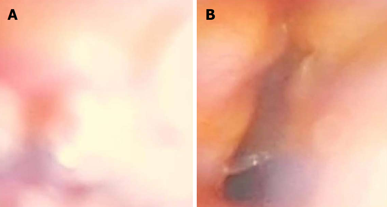
As seen in Figure 1A, a clear view of the larynx was not obtained because of a fogged lens and secretion in spite of intermittent suction. However, in Figure 1B, FOB-guided intubation with 5 L/min oxygen insufflations shows a better view of the larynx than Figure 1A.
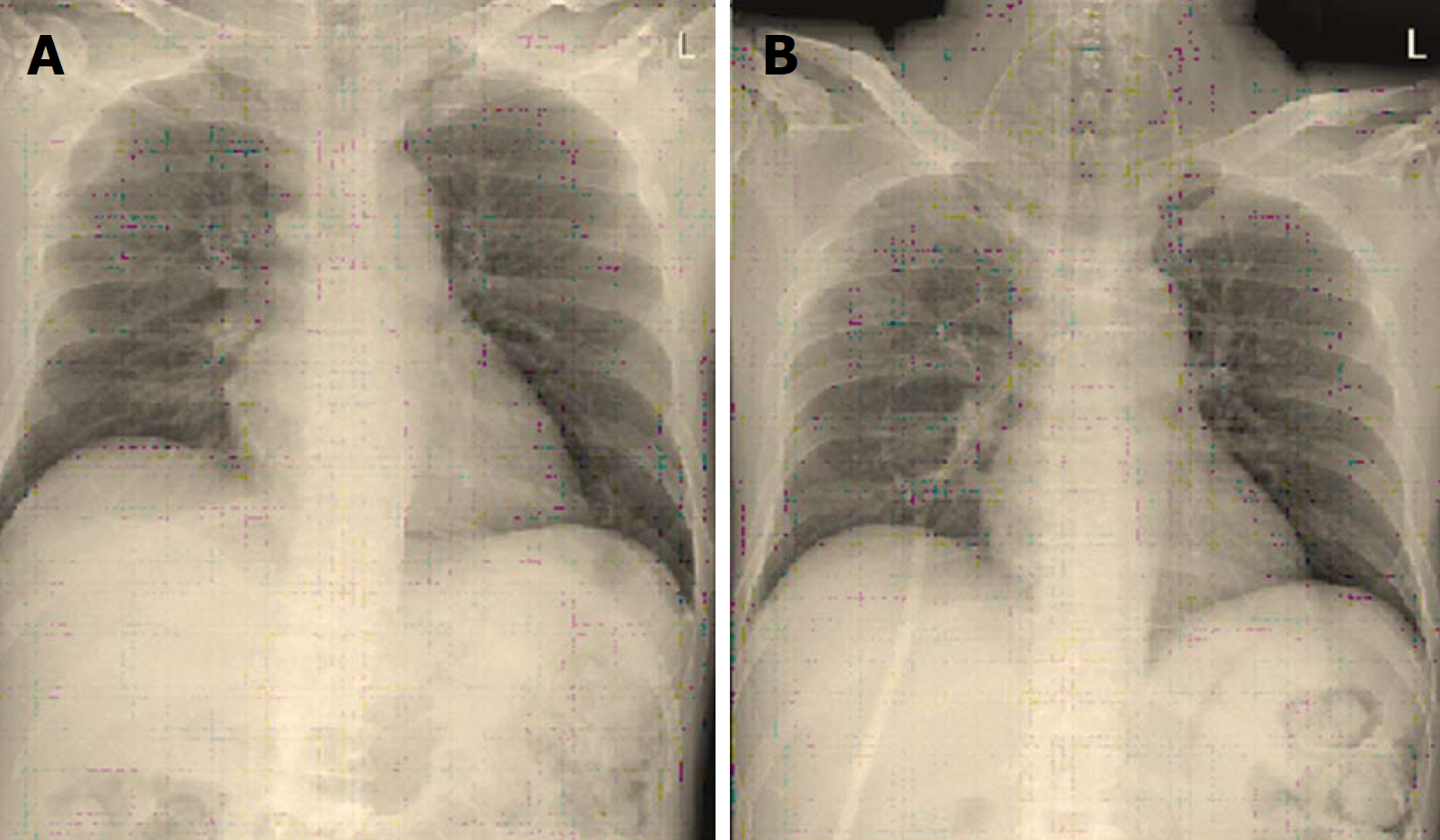
After the intubation, arterial cannulation and an additional intravenous line were provided, and a temperature probe was inserted into the mouth via a bite block. The operation was uneventful. At the end of the surgery, the patient was extubated and he recovered well from anesthesia. He was sent to the recovery room and then to the general ward. A neurological exam in the general ward showed non-specific signs. The post-operative chest X-ray shown in Figure 2B demonstrated no significant change in the gastric gas amount compared to the pre-operative chest X-ray in Figure 2A.
Oxygen insufflation through a FOB working channel is a controversial method. This method had been recommended previously to prevent fogging of the FOB lens, to blow oral secretions away, and to oxygenate the patients[11,12]. In addition, Rudolph et al[13] reported using a Bonfils intubation FOB for a safe and smooth intubation. This provided a better view for a longer time. However, some reports describing gastric distention and rupture during an awake FOI were published[14-16]. This is a rare, but serious complication. However, FOI using suction via a FOB’s working channel can be recommended for a prolonged or difficult FOI if the conditions are appropriate. Unfortunately, all serious complications occur when no muscle relaxant is administered to the patients and no nasogastric tube is inserted. We believe that it is inappropriate to apply oxygen insufflation through a FOB in these conditions. In our case, muscle relaxant was administered and a nasogastric tube was inserted.
When the awake intubation is applied to patients without using muscle relaxants, the patient is not conscious, but the various tracheal reflexes, including gag reflex, remain. Therefore, tracheal reflexes are activated against any kind of flow or stimulation. Hence, patients either spit out or swallow to prevent aspirating oxygen via the trachea. Intake of oxygen through the esophagus might have contributed greatly to stomach distension and rupture in previous case reports of awake intubation.
In our case, we think that the nasogastric tube was helpful in lowering the stomach pressure. It also played a major role in showing the location of the esophageal tract, which helped to avoid intubation or FOB entry into the esophagus.
We performed FOI using Sellick’s maneuver, also known as cricoid pressure, and is used to prevent gas insufflation to the stomach and regurgitation and to assist with the visualization of the glottis[17]. This well-known technique makes it easier to prevent stomach distension and regurgitation of gastric contents. Cricoid compression is also effective in obliterating the esophageal lumen in the presence of a nasogastric tube[18]. Aspiration of gastric contents often occurs in patients with a full stomach or difficulty with gastric emptying. Patients with spinal cord injury or even spinal contusion injury can have difficulty with gastric emptying[19]. In patients with unstable cervical spine, the application of Sellick’s maneuver may cause secondary neurological damage, but its application is relatively small and has been reported to have an effect of supporting the posterior cervical spine[20].
We often perform rapid sequence intubation in patients who have difficulties in gastric emptying to proceed to general anesthesia. The success rate depends on rapid intubation technique using a direct laryngoscope. However, patients with cervical spine trauma usually suffer from cervical instability for which intubation using direct laryngoscope is seldom recommended[21]. Recent studies have shown that the use of FOB is superior not only in aspects of neurological impairment but also hemodynamic stability[22,23]. A light wand or FOB can be a chosen to preform rapid intubation. If an FOB is chosen, a clear view can provide better and faster intubation. FOI with oxygen supplied through the working channel could be a good method in these cases.
Ovassapian et al[24] reported that this method can cause lung barotrauma or rupture and gastric distention and rupture. However, no evidence has been provided yet. We administered 5 L/min oxygen via the suction port for less than a minute by the intermittent thumb occlusion of the FOB channel post. We believe that our method is good for patients with acute spinal cord contusion because of possible difficulties with stomach emptying. In other words, prolonged and repetitive FOI attempts could cause aspiration pneumonia because of regurgitation of gastric contents.
In a previous report, an inadequate intubation was performed using a FOB, which was in the esophagus for about 60 s[15] and could have caused the gastric rupture. According to published reports, FOI complications when administered with oxygen supplied through the working channel include gastric rupture after distension[14-16,24], subcutaneous emphysema[25] and lung barotrauma and rupture[24]. However, we think that this method is still useful under appropriate conditions.
To sum up, this method quicker and easier due for the following reasons: no fogging of the FOB lens, oral secretions are blown away, and the patient is oxygenated[11,12]. Therefore, we think that FOI with an oxygen supply through the working channel can be advantageous for patients with cervical spine trauma.
In our case, the conditions were appropriate. Required conditions can be summarized as follows: (1) muscle relaxant must be administered to avoid “eating”oxygen; (2) a nasogastric tube must be inserted to reduce pressure in the stomach and to avoid accidental esophageal intubation; (3) cricoid pressure must be applied to prevent the regurgitation of gastric contents; and (4) the oxygen flow must be used minimally to prevent possible barotrauma to the lung and other oral structures during the induction of anesthesia.
In conclusion, oxygen insufflation via the working channel is a useful method for assisting with prolonged and difficult FOIs. However, we must carefully consider whether conditions are right before making a choice.
Manuscript source: Unsolicited manuscript
Specialty type: Medicine, research and experimental
Country of origin: South Korea
Peer-review report classification
Grade A (Excellent): 0
Grade B (Very good): 0
Grade C (Good): C, C
Grade D (Fair): 0
Grade E (Poor): 0
P- Reviewer: Ajmal M, Doyle J S- Editor: Ma RY L- Editor: A E- Editor: Wu YXJ
| 1. | Peterson GN, Domino KB, Caplan RA, Posner KL, Lee LA, Cheney FW. Management of the difficult airway: a closed claims analysis. Anesthesiology. 2005;103:33-39. [PubMed] [DOI] [Cited in This Article: ] [Cited by in Crossref: 573] [Cited by in F6Publishing: 477] [Article Influence: 25.1] [Reference Citation Analysis (0)] |
| 2. | Cheney FW, Posner KL, Lee LA, Caplan RA, Domino KB. Trends in anesthesia-related death and brain damage: A closed claims analysis. Anesthesiology. 2006;105:1081-1086. [PubMed] [DOI] [Cited in This Article: ] [Cited by in Crossref: 193] [Cited by in F6Publishing: 202] [Article Influence: 11.9] [Reference Citation Analysis (0)] |
| 3. | American Society of Anesthesiologists Task Force on Management of the Difficult Airway. Practice guidelines for management of the difficult airway: an updated report by the American Society of Anesthesiologists Task Force on Management of the Difficult Airway. Anesthesiology. 2003;98:1269-1277. [PubMed] [DOI] [Cited in This Article: ] [Cited by in Crossref: 1128] [Cited by in F6Publishing: 879] [Article Influence: 41.9] [Reference Citation Analysis (0)] |
| 4. | Koerner IP, Brambrink AM. Fiberoptic techniques. Best Pract Res Clin Anaesthesiol. 2005;19:611-621. [PubMed] [DOI] [Cited in This Article: ] [Cited by in Crossref: 42] [Cited by in F6Publishing: 42] [Article Influence: 2.3] [Reference Citation Analysis (0)] |
| 5. | Randell T, Hakala P, Kyttä J, Kinnunen J. The relevance of clinical and radiological measurements in predicting difficulties in fibreoptic orotracheal intubation in adults. Anaesthesia. 1998;53:1144-1147. [PubMed] [DOI] [Cited in This Article: ] [Cited by in Crossref: 13] [Cited by in F6Publishing: 13] [Article Influence: 0.5] [Reference Citation Analysis (0)] |
| 6. | Hakala P, Randell T, Valli H. Comparison between tracheal tubes for orotracheal fibreoptic intubation. Br J Anaesth. 1999;82:135-136. [PubMed] [DOI] [Cited in This Article: ] [Cited by in Crossref: 33] [Cited by in F6Publishing: 34] [Article Influence: 1.4] [Reference Citation Analysis (0)] |
| 7. | Brull SJ, Wiklund R, Ferris C, Connelly NR, Ehrenwerth J, Silverman DG. Facilitation of fiberoptic orotracheal intubation with a flexible tracheal tube. Anesth Analg. 1994;78:746-748. [PubMed] [DOI] [Cited in This Article: ] [Cited by in Crossref: 63] [Cited by in F6Publishing: 65] [Article Influence: 2.2] [Reference Citation Analysis (0)] |
| 8. | Ayoub CM, Rizk MS, Yaacoub CI, Baraka AS, Lteif AM. Advancing the tracheal tube over a flexible fiberoptic bronchoscope by a sleeve mounted on the insertion cord. Anesth Analg. 2003;96:290-292, table of contents. [PubMed] [Cited in This Article: ] |
| 9. | Jones HE, Pearce AC, Moore P. Fibreoptic intubation. Influence of tracheal tube tip design. Anaesthesia. 1993;48:672-674. [PubMed] [DOI] [Cited in This Article: ] [Cited by in Crossref: 61] [Cited by in F6Publishing: 62] [Article Influence: 2.0] [Reference Citation Analysis (0)] |
| 10. | Agrò FE, Cataldo R. Teaching fiberoptic intubation in Italy: state of the art. Minerva Anestesiol. 2010;76:684-685. [PubMed] [Cited in This Article: ] |
| 11. | Roberts JT. Preparing to use the flexible fiber-optic laryngoscope. J Clin Anesth. 1991;3:64-75. [PubMed] [DOI] [Cited in This Article: ] [Cited by in Crossref: 32] [Cited by in F6Publishing: 32] [Article Influence: 1.0] [Reference Citation Analysis (0)] |
| 12. | Benumof JL. Management of the difficult adult airway. With special emphasis on awake tracheal intubation. Anesthesiology. 1991;75:1087-1110. [PubMed] [DOI] [Cited in This Article: ] [Cited by in Crossref: 363] [Cited by in F6Publishing: 358] [Article Influence: 10.8] [Reference Citation Analysis (0)] |
| 13. | Rudolph C, Schlender M. [Clinical experiences with fiber optic intubation with the Bonfils intubation fiberscope]. Anaesthesiol Reanim. 1996;21:127-130. [PubMed] [Cited in This Article: ] |
| 14. | Hershey MD, Hannenberg AA. Gastric distention and rupture from oxygen insufflation during fiberoptic intubation. Anesthesiology. 1996;85:1479-1480. [PubMed] [DOI] [Cited in This Article: ] [Cited by in Crossref: 30] [Cited by in F6Publishing: 31] [Article Influence: 1.1] [Reference Citation Analysis (0)] |
| 15. | Ho CM, Yin IW, Tsou KF, Chow LH, Tsai SK. Gastric rupture after awake fibreoptic intubation in a patient with laryngeal carcinoma. Br J Anaesth. 2005;94:856-858. [PubMed] [DOI] [Cited in This Article: ] [Cited by in Crossref: 15] [Cited by in F6Publishing: 16] [Article Influence: 0.8] [Reference Citation Analysis (0)] |
| 16. | Chapman N. Gastric rupture and pneumoperitoneum caused by oxygen insufflation via a fiberoptic bronchoscope. Anesth Analg. 2008;106:1592. [PubMed] [DOI] [Cited in This Article: ] [Cited by in Crossref: 12] [Cited by in F6Publishing: 13] [Article Influence: 0.8] [Reference Citation Analysis (0)] |
| 17. | SELLICK BA. Cricoid pressure to control regurgitation of stomach contents during induction of anaesthesia. Lancet. 1961;2:404-406. [PubMed] [DOI] [Cited in This Article: ] [Cited by in Crossref: 705] [Cited by in F6Publishing: 553] [Article Influence: 8.8] [Reference Citation Analysis (0)] |
| 18. | Salem MR, Joseph NJ, Heyman HJ, Belani B, Paulissian R, Ferrara TP. Cricoid compression is effective in obliterating the esophageal lumen in the presence of a nasogastric tube. Anesthesiology. 1985;63:443-446. [PubMed] [DOI] [Cited in This Article: ] [Cited by in Crossref: 66] [Cited by in F6Publishing: 66] [Article Influence: 1.7] [Reference Citation Analysis (0)] |
| 19. | Qualls-Creekmore E, Tong M, Holmes GM. Time-course of recovery of gastric emptying and motility in rats with experimental spinal cord injury. Neurogastroenterol Motil. 2010;22:62-69, e27-e28. [PubMed] [DOI] [Cited in This Article: ] [Cited by in Crossref: 8] [Cited by in F6Publishing: 23] [Article Influence: 1.6] [Reference Citation Analysis (0)] |
| 20. | Prasarn ML, Horodyski M, Schneider P, Wendling A, Hagberg CA, Rechtine GR. The Effect of Cricoid Pressure on the Unstable Cervical Spine. J Emerg Med. 2016;50:427-432. [PubMed] [DOI] [Cited in This Article: ] [Cited by in Crossref: 3] [Cited by in F6Publishing: 3] [Article Influence: 0.3] [Reference Citation Analysis (0)] |
| 21. | Crosby ET, Lui A. The adult cervical spine: implications for airway management. Can J Anaesth. 1990;37:77-93. [PubMed] [DOI] [Cited in This Article: ] [Cited by in Crossref: 120] [Cited by in F6Publishing: 124] [Article Influence: 3.6] [Reference Citation Analysis (0)] |
| 22. | Gill N, Purohit S, Kalra P, Lall T, Khare A. Comparison of hemodynamic responses to intubation: Flexible fiberoptic bronchoscope versus McCoy laryngoscope in presence of rigid cervical collar simulating cervical immobilization for traumatic cervical spine. Anesth Essays Res. 2015;9:337-342. [PubMed] [DOI] [Cited in This Article: ] [Cited by in Crossref: 9] [Cited by in F6Publishing: 11] [Article Influence: 1.2] [Reference Citation Analysis (0)] |
| 23. | Bao FP, Zhang HG, Zhu SM. Anesthetic considerations for patients with acute cervical spinal cord injury. Neural Regen Res. 2017;12:499-504. [PubMed] [DOI] [Cited in This Article: ] [Cited by in Crossref: 16] [Cited by in F6Publishing: 16] [Article Influence: 2.3] [Reference Citation Analysis (0)] |
| 24. | Ovassapian A, Mesnick PS. Oxygen insufflation through the fiberscope to assist intubation is not recommended. Anesthesiology. 1997;87:183-184. [PubMed] [DOI] [Cited in This Article: ] [Cited by in Crossref: 17] [Cited by in F6Publishing: 18] [Article Influence: 0.7] [Reference Citation Analysis (0)] |
| 25. | Richardson MG, Dooley JW. Acute facial, cervical, and thoracic subcutaneous emphysema: a complication of fiberoptic laryngoscopy. Anesth Analg. 1996;82:878-880. [PubMed] [Cited in This Article: ] |


