Article Text

Abstract
BACKGROUND Gradual telomere erosion eventually limits the replicative life span of somatic cells and is regarded as an ultimate tumour suppressor mechanism, eliminating cells that have accumulated genetic alterations. Telomerase, which has been found in over 85% of human cancers, elongates telomeres and may be required for tumorigenesis by the process of immortalisation. Malignant mesothelioma is an incurable malignancy with a poor prognosis. The disease becomes symptomatic decades after exposure to carcinogenic asbestos fibres, suggesting the long term survival of pre-malignant cell clones. This study investigated the presence of telomerase in pleural malignant mesothelioma, which may be the target for future anti-telomerase drugs.
METHODS Telomerase activity was semi-quantitatively measured in extracts from 22 primary pleural mesotheliomas, two benign solitary fibrous tumours of the pleura, four mesothelioma cell lines, and six short term mesothelial cell cultures from normal pleura using a non-isotopic dilution assay of the telomeric repeat amplification protocol.
RESULTS Twenty of the 22 primary mesotheliomas (91%) and all tumour derived mesothelioma cell lines were telomerase positive. Different levels of enzyme activity were observed in the tumours of different histological subtypes. Telomerase activity could not be detected in the six normal mesothelial cell cultures or in the two mesotheliomas. Both benign solitary fibrous tumours showed strong telomerase activity.
CONCLUSIONS Telomerase activity is found in a high proportion of mesotheliomas and anti-telomerase drugs might therefore be useful clinically. The results are consistent with the hypothesis that telomerase activity may be a feature of carcinogenesis in mesotheliomas and possibly in many other cancers.
- telomerase
- mesothelioma
- TRAP assay
Statistics from Altmetric.com
Telomeres are stretches of repetitive non-coding DNA sequences (TTAGGG) associated with specific DNA binding proteins at the ends of eukaryotic chromosomes. Human telomeres progressively shorten during cell division because DNA polymerase cannot replicate the end of a linear template, and critical shortening is believed to limit the cellular life span. A possible explanation for the attrition of human telomeric buffers is the somatic repression of telomerase. This ribonucleoprotein reverse transcriptase can synthesise telomeric repeats at chromosomal ends using its catalytic subunit (hTRT) and a short template element of an integral RNA (hTR). The “telomeric repeat amplification protocol” (TRAP) assay has been developed to detect the activity of the enzyme. Telomerase activity has been found in germ cells and in stem cells, and in a variety of cell lines and malignant tumours, but only weak or no activity was found in mortal somatic cell cultures and in benign tumours.1 It has recently been shown that forced expression of hTRT in telomerase negative cells resulted in reconstitution of telomerase activity, elongation of telomeres, and extension of replicative life span.2 These data indicate a causal relationship between telomere erosion and cellular replicative senescence and suggest that this mechanism acts as an ultimate powerful tumour suppressor system which creates a barrier to any cell that has accumulated genetic alterations and escaped normal growth control by its environment. The data further lead to the speculation that telomerase may be required for the efficient growth of neoplastic cells and that inhibition of telomerase activity could represent a new therapeutic approach for cancer.3 Since it still is unclear whether telomerase activity commonly occurs in most tumours, we investigated telomerase activity in malignant mesothelioma. This cancer is a highly aggressive malignancy, thought to arise from mesothelial cells, which is resistant to treatment. It is characterised by an extremely long latency period (20–40 years) between exposure to asbestos, a major contributory factor in the development of this cancer, and the onset of disease symptoms, which suggests the long term survival of genetically altered cell clones.4
In this study we have investigated whether mesothelioma shares telomerase activity with other cancers and we further report the absence of telomerase activity in somatic mesothelial cell cultures.
Methods
TISSUES AND CELLS
Specimens of 22 mesotheliomas (nine epithelial, four sarcomatous, nine mixed types) and of two benign solitary fibrous tumours of the pleura were snap frozen in liquid nitrogen at the time of pleuroscopy and stored at –80°C. Short term mesothelial cell cultures were established from stripped pleura from necropsied non-cancerous patients after trypsin disaggregation. Mesothelial cells were grown as primary cultures on fibronectin coated culture flasks in RPMI 1640 supplemented with 20% heat inactivated fetal bovine serum (Gibco BRL, Belgium) and 10 ng/ml hydrocortisone (Upjohn, Belgium). The cells were identified as mesothelial cells by several criteria including immunocytochemical staining with antikeratin antibodies (Dako, Glostrup, Denmark), cell morphology (cobblestone), and the presence of long microvilli observed by transmission electron microscopy. Third passage confluent cultures grown at 37°C and 5% CO2 were harvested for TRAP analysis. Malignant mesothelioma cells were obtained from four human mesothelioma cell lines (UIA-MM 1, 2, 5 and 7) which were isolated from pleural fluid as described elsewhere.5 The Jurkat cell line served as a positive control.
NON-ISOTOPIC TRAP ASSAY
Lysate preparation and the TRAP assay were performed as described by Kim et al 6 with minor modifications. Briefly, homogenates of frozen sections (100 mm3) and cell pellets (106 cells) were prepared with disposable pestles in 200 μl ice cold lysis buffer. After 30 minutes of incubation on ice the lysates were centrifuged at 14 000g for one hour at 4°C and the protein concentration of the supernatant measured according to Bradford (Bio-Rad Protein Assay, Bio-Rad Laboratories GmbH, Germany). Aliquots of a dilution series of each lysate (6 μg, 0.6 μg, and 0.06 μg of total protein) were used for the TRAP assays. After initial incubation at 23°C for 30 minutes telomerase products were amplified using TS and ACX primers (35 cycles at 94°C for 30 s, 53°C for 30 s, and 72°C for 30 s). Assay specificity was confirmed by inclusion of an RNase pre-incubation control step. The presence of Taq polymerase inhibitors in cases lacking the 6 bp ladder was assessed using the TSNT internal control oligonucleotide and the NT internal control primer, generating a 36 bp control band. PCR products were separated by electrophoresis on a 12.5% non-denaturing polyacrylamide gel (19:1). The gel was stained with ethidium bromide or SYBR green I nucleic acid gel stain (FMC, BioProducts, Rockland, Maine, USA) and analysed by the CCD camera coupled Gel Doc 1000 Molecular Analyst Software package (Bio-Rad). Telomerase activity was graded as negative (6 bp ladder not detected), weakly positive (ladder detected with 6 μg protein), moderately positive (ladder detected with 6 or 0.6 μg protein but not with 0.06 μg), or strongly positive (ladder detected at all three protein concentrations).
Results
In a preliminary experiment the sensitivity of our ethidium bromide based procedure was compared with an assay in which we stained the amplified telomerase products with SYBR green I that is known to be as sensitive as classical isotopic TRAP assays.7 One hundred telomerase positive Jurkat cells were found to be sufficient for the detection of telomerase activity in the study using SYBR green. Similar sensitivity was observed in the ethidium bromide based assay (data not shown).
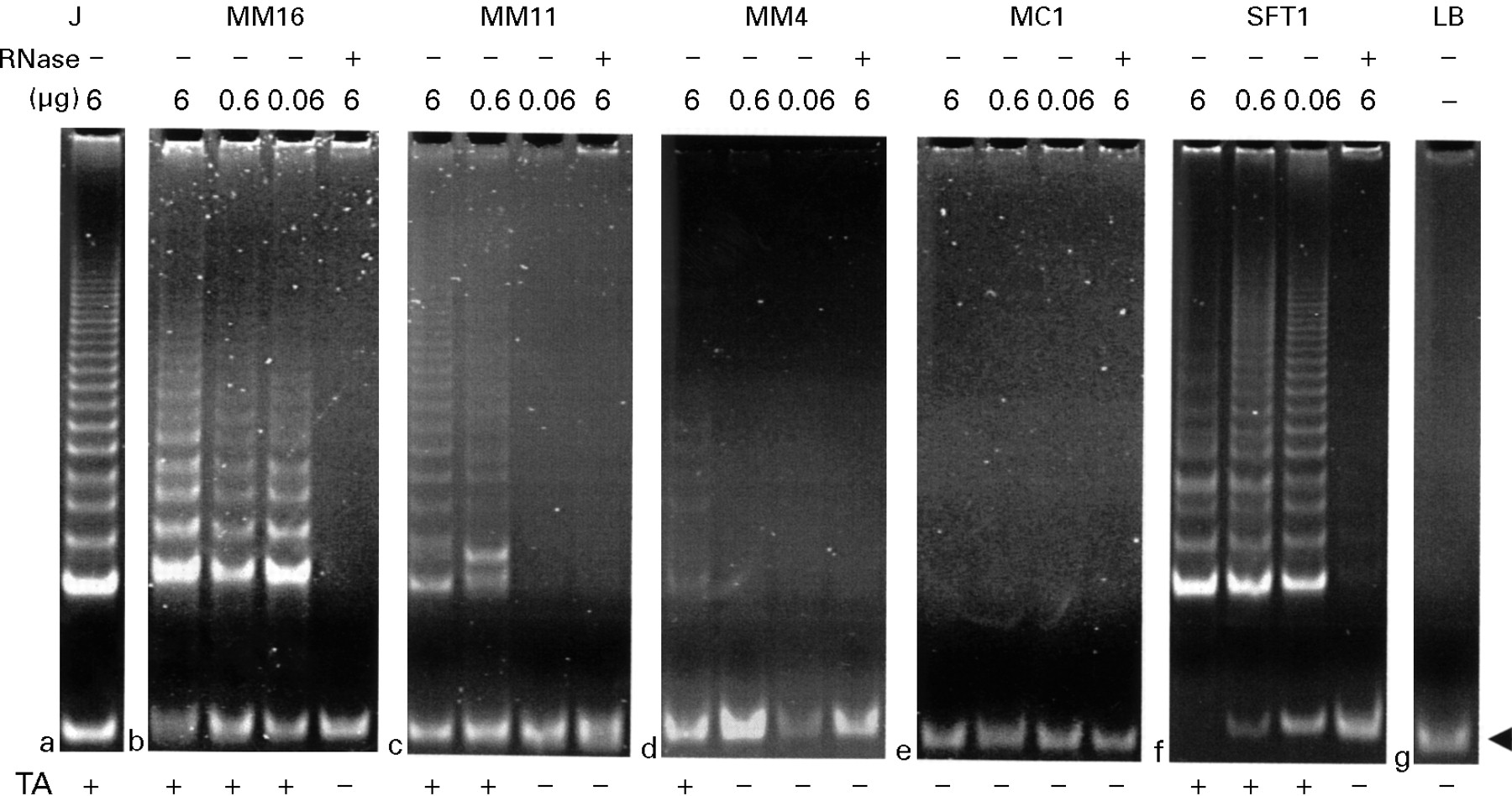
Representative results for serial dilutions of extracts of the analysed materials are shown in fig 1. The TRAP assay was performed with the use of 6 μg protein extracts and varying levels of telomerase activity were observed in 20 (91%) of 22 mesotheliomas, in all four cancer cell lines, and in both solitary fibrous tumours. Signals were abolished after pre-incubation with RNase A. Because the signal intensities varied among samples, we estimated telomerase activity by dilution assay using 10-fold (0.6 μg) and 100-fold (0.06 μg) diluted extracts. Five of 20 telomerase positive mesotheliomas retained signals after the 10-fold dilution (panels b and c) and two of these retained signals after the 100-fold dilution (panel b), indicating that these tumours expressed high levels of telomerase activity. High levels of activity were also observed after 10-fold and 100-fold dilution of the extracts of both solitary fibrous tumours (panel f). The dilution assay could not detect any telomerase activity in the mesothelial cells (panel e) nor in mesothelioma cases MM2 and MM21. False negativity caused by Taq polymerase inhibitors was excluded since 36 bp control products were amplified in all TRAP assays. Table1 summarises the results of the dilution assays of mesotheliomas and solitary fibrous tumours according to signal intensity and histology. No statistical analysis was performed because of the small size of the study.

Representative results of non-isotopic TRAP assays for serial dilutions of extracts from mesothelioma tumour samples (MM4, MM11 and MM16), mesothelial cells (MC1), and one solitary fibrous tumour (SFT1). Jurkat cell extracts (J) served as positive control (a). For every assay a 36 bp internal control band is visible (arrow). Each sample analysis contained an RNase A treated control (+ at top) showing loss of signal. Lysis buffer alone (LB) was applied as negative control (g). PCR products were separated by electrophoresis on a non-denaturing 12.5% polyacrylamide gel, stained with ethidium bromide, and visualised on a UV transilluminator. The assay result is indicated at the bottom of the figure. Telomerase activity (TA) was semi-quantitatively estimated and was graded strong (b), moderate (c), weak (d) or absent (e).
Telomerase activity profiles of individual mesotheliomas and solitary fibrous tumours in relation to histological subtype
Discussion
The finding of telomerase activity in over 90% of mesotheliomas is in agreement with the reported activity in a variety of other cancer types. This result is consistent with the hypothesis that telomerase activity may be a feature of carcinogenesis in mesothelioma and also, perhaps, in many other cancers. Carcinogenesis is recognised as a multistep process resulting from the accumulation of sequential genetic alterations in a cell. The very long latency period characteristic of mesothelioma suggests that multiple cumulative genetic, cytotoxic, and proliferative events occur during the tumorigenic conversion of the progenitor cell. Of the known changes that we and others have investigated in mesothelioma, the most frequent are in the p16 and NF2 genes.8According to the telomere senescence model, such genetically altered pre-malignant cells may finally be eliminated before they can ever develop a full blown malignant phenotype. Conversely, the presence of telomerase activity in mesothelioma may indicate that these cells can overcome the restraint of their finite life span and are no longer hampered in their clonal evolution towards a more malignant phenotype. In view of the finding of telomerase activity in mesothelioma, anti-telomerase drugs might be useful clinically. However, telomerase activity could not be detected in the lysates of two mesotheliomas. It has been reported that some tissue samples contain an inhibitor of Taq polymerase and thus give a false negative result.6 The successful co-amplification in our cases of the TRAP internal control excluded this possibility. At present we therefore cannot exclude the possibility that some mesotheliomas have telomere stabilising mechanisms that are independent of telomerase, or are not immortal at all. This certainly would weaken the efficacy of future anti-telomerase drugs.
We did not detect telomerase activity in mesothelial cells. There is considerable evidence that mesothelioma originates from surface mesothelial cells rather than from a “multipotential” subserosal stem cell, although this has not been established beyond all doubt.4 Assuming the former scenario is correct, the absence of detectable telomerase activity in mesothelial cells, in contrast to the presence of activity in mesotheliomas, could fit with the interpretation that most adult tumours develop from telomerase negative precursors after telomerase re-activation.9 On the other hand, others may interpret these results as evidence for the above mentioned stem cell origin of mesothelioma, arguing that these telomerase positive tumours do, indeed, arise from telomerase positive stem cells without the need for a re-activation step.10
The strong telomerase activity detected in both benign solitary fibrous tumours may support the hypothesis of their origin from telomerase positive, CD34 positive fibroblastic stem cells or suggest unpredictable clinical behaviour. Similarly, Umbricht et al 11 found 19% of benign follicular tumours of the thyroid with detectable telomerase activity and argued that some histologically benign lesions may be precursor lesions of follicular carcinomas. Our observations may also indicate that the use of telomerase activity as a marker for malignancy of serosal lesions needs further validation. It is obvious that larger series need to be studied, which should also include unequivocal benign, mesothelium derived tumours such as adenomatoid tumours.
The “classic model” states that telomerase upregulation is forced by critical telomere erosion beyond the point where cell multiplication normally stops. An in vitro situation is seen during continuous culture of SV40 large T antigen transformed telomerase negative human cells which eventually undergo “crisis” —the condition in which cellular chromosomes are characterised by ultrashort telomeres and that coincides with telomerase activity.12 At present it is not known whether SV40-like viruses, for which DNA sequences have been found in some mesotheliomas, are responsible for an in vivo equivalent of crisis during mesothelioma carcinogenesis.13
The “co-selection hypothesis” states that telomerase is indirectly re-activated as part of a “package” of changes in gene expression that occurs after some other genetic event.14It remains speculative whether asbestos induced DNA damage represents such a genetic event. Similarly, others have suggested that telomerase activation is a mere side effect of de-differentiation.15We observed different levels of enzyme activity in the tumours of different histological subtypes. Because of the limited size of the study we did not perform statistical analysis to find any correlation between the level of telomerase activity in the tissue lysates and the histological subtype. In any case, statistical analysis may be inaccurate because of weaknesses in the TRAP assay. Indeed, both heterogeneity of tumour differentiation within a tissue section and its morphology are lost during an in vitro biochemical assay, indicating the need for alternative methods for detecting telomerase activity at the cellular level. In situ hybridisation techniques with probes directed against hTRT mRNA and hTR, together with immunohistochemistry using antibodies to the various subunits of the holo-enzyme, may help in the further investigation of the expression of telomerase in benign and malignant pleural lesions.
Acknowledgments
The authors would like to thank Petra Boukamp of the German Cancer Research Center, Heidelberg, Germany for constructive comments on an earlier version of the manuscript. This study was supported by a grant from the Belgian ‘Sports against Cancer Society’, 1997.