Article Text

Abstract
Currently, traditional and non-traditional risk factors for cardiovascular disease have been established. The first group includes age, which constitutes one of the most important factors in the development of chronic diseases. The second group includes inflammation, the pathophysiology of which contributes to an accelerated process of vascular remodelling and atherogenesis in autoimmune diseases. Indeed, the term inflammaging has been used to refer to the inflammatory origin of ageing, explicitly due to the chronic inflammatory process associated with age (in healthy individuals). Taking this into account, it can be inferred that people with autoimmune diseases are likely to have an early acceleration of vascular ageing (vascular stiffness) as evidenced in the alteration of non-invasive cardiovascular tests such as pulse wave velocity. Thus, an association is created between autoimmunity and high morbidity and mortality rates caused by cardiovascular disease in this population group. The beneficial impact of the treatments for rheumatoid arthritis at the cardiovascular level has been reported, opening new opportunities for pharmacotherapy.
- autoimmune diseases
- inflammation
- arthritis
- rheumatoid
- cardiovascular diseases
This is an open access article distributed in accordance with the Creative Commons Attribution Non Commercial (CC BY-NC 4.0) license, which permits others to distribute, remix, adapt, build upon this work non-commercially, and license their derivative works on different terms, provided the original work is properly cited, appropriate credit is given, any changes made indicated, and the use is non-commercial. See: http://creativecommons.org/licenses/by-nc/4.0/.
Statistics from Altmetric.com
Key messages
What is already known about this subject?
‘Inflammaging’ is characterised by the presence of persistent low-grade inflammation that develops with age and causes deficient tissue repair and degeneration, and it is reflected as an increase in the plasma levels of inflammatory cytokines and acute-phase reactants.
What does this study add?
The low-grade, persistent pro-inflammatory milieu characteristic for the ageing process triggers morphological and functional changes, most prominently endothelial dysfunction, diffuse intimal–medial thickening and arterial stiffness.
How might this impact on clinical practice?
The pattern of cytokine expression observed in autoimmune diseases, like rheumatoid arthritis, is similar to that seen in ‘inflammaging’ with similar consequences as increase vascular ageing induced by this chronic inflammation.
Background
Age is the strongest independent risk factor for the onset and progression of many of the major non-congenital chronic diseases including cancer, neurodegenerative diseases and cardiovascular diseases (CVDs).1 It has been proposed that these chronic diseases are manifestations of an accelerated ageing process.1 With the recent advance in the understanding of the molecular mechanisms of ageing, an interest in biomarkers capable of differentiating between biological and chronological age has developed, because such biomarkers might improve risk prediction for this group of diseases.1–5 Exploration of the molecular mechanisms has generated the following list of factors that cause ageing: defective repair of DNA damage, imbalance in the production and elimination of cellular debris, defective self-molecules that can be recognised by the innate immune system and induce ‘inflammaging’, mitochondrial dysfunction, and cellular senescence.6
At the vascular level, ageing gradually affects the structure and function of blood vessels, thus causing loss of elasticity of the arterial wall and endothelial dysfunction.2 3 7 As the world population ages, establishing the physiological connection between these features of vascular ageing and the causal molecular mechanisms of ageing is of great value for the prevention of vascular disease.4 In this review, we focus on inflammaging, which is importantly connected with the impact of autoimmune diseases (AIDs) and treatment thereof on the cardiovascular system. We first discuss the theoretical background of this connection, then explore the epidemiological relationship between AID and vascular ageing as a read-out of this connection, and, finally, discuss the options for pharmacotherapy.
Inflammatory origin of ageing ‘inflammaging’ and AIDs
In the year 2000, Franceschi et al introduced the term ‘inflammaging’ (inflammation+ageing) to describe the chronic, sterile, low-grade inflammatory process that develops with age, confers susceptibility to suffering age-related pathologies and facilitates the spread of the effects of ageing at the systemic level.1 8 9 ‘Inflammaging’ is considered to be caused by endogenous signals resulting from an imbalance between the production and disposal of cellular debris and defective self-molecules. Thus, ‘inflammaging’ could be defined as a process of autoimmune/self-reactive origin.1 5 This new theory establishes inflammation as a fundamental component for the physiology of ageing and its associated diseases.1 5
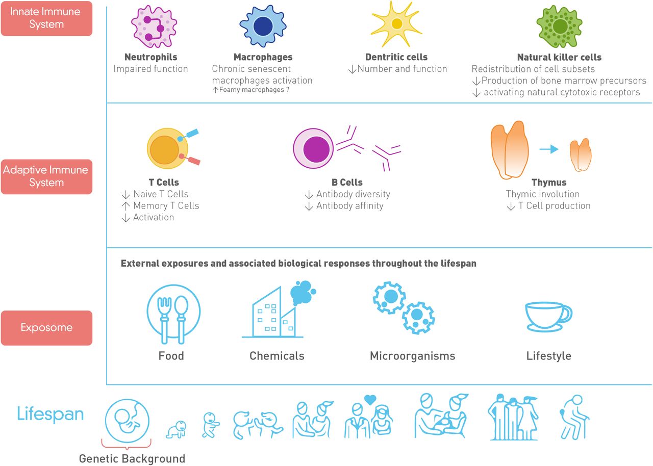
The increase in inflammaging is related to immune senescence, the complex remodelling process the immune system undergoes with ageing.9 10 In the adaptive immune system, this remodelling process is characterised by thymic involution, a reduced number of naïve T cells, a relative increase in memory T cells, and the reduction of antibodies diversity and affinity. Thymic involution is associated with the release of autoreactive T cells that recognise endogenous molecules and contribute to the inflammaging.11 The changes in the innate immune system are less clear, but it has been reported that macrophages and dendritic cells become less responsive to antigens of the innate immune system during ageing8 12 (figure 1). In the circulation, the inflammaging is reflected as altered levels of pro-inflammatory cytokines (interleukin 6 (IL-6), IL-1β, IL-8, tumour necrosis factor alpha (TNF-α), interferon gamma (IFN-γ)), acute-phase reactants (C reactive protein (CRP)), and decreases in IL-10 and IL-1 receptor antagonist (IL-1RA).5

Remodelling of the immune system with ageing. Disturbances in the immunological system are associated with a dysregulation in both the innate and adaptive immune system. Also, the exposome as a cumulative measure of external stimuli and the biological responses throughout the lifespan associated with the genetic background play an important role in the response of the immunological system.
Even in long-lived healthy individuals, ageing occurs secondary to a state of low-grade, chronic subclinical inflammation.13 However, conditions presenting with a high inflammatory burden will favour or accelerate the ageing process. Such is the case of AID, where the dysregulation in innate and adaptive immune systems stimulates autoantibody production and a chronic immune-mediated inflammatory response. This chronic inflammatory process affects different organs and systems including the cardiovascular system.14 15 It has been shown that people with AID present high rates of morbidity and mortality from cardiovascular events.16 17 This is associated with an accelerated degree of arteriosclerosis and remodelling of the blood vessels.14 Arteriosclerosis is a descriptive broad term for the thickening and hardening of the arterial wall. One important cause of arteriosclerosis is atherosclerosis, which is a patchy abnormality in the artery wall frequently associated with ageing, lipid deposits and activation of inflammatory pathways.
Therefore, both the elderly and patients with AID present remodelling of the immune system and chronic inflammation, low-grade in healthy long-lived individuals, that is, in normal ageing, or more severe in patients with AID, that is, in accelerated ageing. Moreover, there are similarities in the pattern of cytokine expression observed in AID and that seen in ‘inflammaging’ with similar effects in the arterial remodelling process as will be discussed later.15
CVD and autoimmunity
AID and cardiovascular risk
CVD is the main cause of morbidity and mortality worldwide, and its incidence has risen by around one-third in the last decade.18 Among the pathophysiological mechanisms of arteriosclerotic CVD, a chronic inflammatory process sustained by a progressive imbalance in the production of pro-inflammatory and anti-inflammatory molecules with age plays a pivotal role. In patients with AID, this chronic inflammatory state starts from early life, explaining the high premature cardiovascular morbidity and mortality.19 20
Indeed, the risk of cardiovascular events and death in patients with rheumatoid arthritis (RA) and other inflammatory arthropathies (ankylosing spondylitis and psoriatic arthritis) is substantially high compared with the general population. This risk is comparable with that in patients with diabetes mellitus, which, like AID, is featured by a chronic pro-inflammatory status. Diabetes mellitus is one of the most important cardiovascular risk factors as shown in studies such as the ‘CARRÉ Investigation’ where it was evidenced that the prevalence of CVD was 5.0% (95% CI 2.3% to 7.7%) in the group without diabetes, 12.4% (95% CI 7.5% to 17.3%) in the group with diabetes mellitus type 2 (DM2) and 12.9% (95% CI 8.8% to 17.0%) in those with RA.21 In RA, the prevalence of CVD ranges from 12% to 67.6% and is considered to be one of the most frequent extra-articular manifestations22; while in controls, this prevalence ranges from 10% to 30%.23 This prevalence varies according to study design, the specific definition of CVD and of composite outcomes (ie, including or not arterial hypertension, stroke, ischaemic heart diseases and so on), the relationship to disease duration (early or established RA) and cardiovascular risk factors (ie, traditional or non-traditional), and inclusion of RA treatment. In addition, there is an increased risk of presenting with acute coronary syndrome, worse short-term outcomes and increased circulatory system diseases mortality (32%) in patients with RA.24 In fact, a meta-analysis25 found that the risk of incident CVD is increased by 48% in patients with RA compared with the general population. In the case of systemic lupus erythematosus (SLE), an AID that affects multiple organs, sometimes from young age on, cardiovascular risk is elevated four to eight times compared with the normal population. This has been shown in some cohorts with a higher absolute CVD risk (23% SLE vs 7% controls; HR 3.8, 95% CI 1.8 to 8) and an estimated 10-year mortality rates of 26% for subjects with SLE vs 19% for comparisons.26 27
Common inflammatory processes in AID and CVD
The relationship between AID and the development of CVD is shaped both by the presence of traditional cardiovascular risk factors (dyslipidaemia, smoking, metabolic syndrome and so on) as well as by non-traditional risk factors such as chronic inflammation, components of the innate and adaptive immune system, subphenotypes of clinical severity, genetic factors associated or not with human leucocyte antigen (ie, TNF super family genes, cytokines and related genes, chemokines, adipokines and nitric oxide synthase genes, among others),20 and the additive protective or adverse effect of some cardiovascular and anti-inflammatory medications used.14 19 In patients with AID, the presence of underlying microvascular and macrovascular lesions, and the production of inflammatory mediators in the perivascular layers have been described. This includes accumulation of lipid particles, autoantibodies, autoantigens, and the production of multiple inflammatory cytokines such as TNF-α and inflammatory cell infiltrates, consisting mainly of lymphocytes in the outer vascular and perivascular layers.14 There is increasing evidence supporting the direct proportional relationship between the inflammatory burden of the underlying disease and the risk of adverse cardiovascular outcomes.28 In the RA case, functional abnormalities of the endothelium and carotid plaque formation have been found associated with duration of disease, degree of disease activity, rheumatoid factor seropositivity and elevated markers of systemic inflammation. On the other hand, in SLE, some factors associated with overt CVD were found more often in SLE cases than controls: lupus anticoagulant, higher cumulative dosage of steroids, high alpha-1 antitrypsin, inflammatory markers (eg, CRP, fibrinogen, IL-6), adhesion molecules and immunological factors (eg, anticardiolipin antibodies, anti-beta II glycoprotein antibodies, antibodies against oxidised low-density lipoproteins (LDLs) and anti-heat shock protein antibodies).28 Furthermore, some studies have shown that the duration of the disease increases this risk, probably due to prolonged exposure to inflammation.29
In this same context, the exaggerated production of molecules such as CRP, fibrinogen and cytokines in patients with AID implies a chronic pro-inflammatory environment that favours endothelial dysfunction and vascular remodelling, and induces other cardiovascular risk factors such as an increased production of lipids (low-density oxidised lipoprotein). These act as a pro-inflammatory stimulus in association with CRP, by forming pro-atherogenic complexes, activating endothelial cells, regulating the production of adhesion molecules and the secretion of chemokines that recruit monocytes or macrophages, and magnifying and perpetuating this process.29 30 Moreover, pro-inflammatory molecules are involved in plaque instability. IFN-γ, a key molecule involved in the rupture of atheromatous plaques, is also upregulated in the inflammation associated with AID.31 32 Upregulation of this molecule in T lymphocytes located at the plaque inhibits the production of extracellular components (mainly collagen) by the muscle cells, contributing to the thinning of the fibrous cap.31 32 Additionally, activated T lymphocytes express CD40L, a molecule that induces foam cells into the production of collagenases and gelatinases that degrade the extracellular matrix. Finally, the thinning of the fibrous cap predisposes to plaque rupture, thrombosis and occlusion of the lumen, a key process in the development of acute CVDs.31
Components of the ‘inflammaging’: common role of pro-inflammatory molecules in AID
Pro-inflammatory cytokines have been identified as a fundamental component of ‘inflammaging’ since they can encourage the expansion of clones of the haematopoietic/immune system depending on the spread of inflammatory signals. In the bone marrow, the immune system of senile individuals is progressively dominated by T and B cell clones, which probably represent an adaptation of the senescent immune system. In fact, these T/B clones represent the result of chronic exposure to antigens that can encompass chronic latent infections, microbiota and autoantigens. The latter can arise de novo during ageing as a self-recognition disorder. Clones of the expanded immune/haematopoietic system are likely to spread this inflammatory signal (cells, nanovesicles and soluble mediators) at the systemic level.33
‘Inflammaging’ is characterised by the presence of persistent low-grade inflammation that leads to deficient tissue repair and degeneration, and that increases plasma levels of inflammatory cytokines and acute-phase reactants. Among the cytokines that have been involved in this process are: IL-6, IL-1β, TNF-α, IFN-γ and IL-1RA.1 5 The analysis of the immunosenescence profile in patients with SLE has shown an increase in IFN-γ levels that has been related to cardiovascular complications. Furthermore, an increase in the IL-6/transforming growth factor beta ratio has also been described in these patients with a consequent overexpression of IL-22 and IL-17. In addition, patients with RA develop features of accelerated ageing, including immunosenescence. These changes include decreased thymic functionality, expansion of late-differentiated effector T cells, increased telomeric attrition and excessive production of cytokines (senescence-associated secretory phenotype).34 Therefore, it should be emphasised that a pattern of cytokine expression is observed in AID similar to that seen in ‘inflammaging’ with similar consequences15 (figure 2).
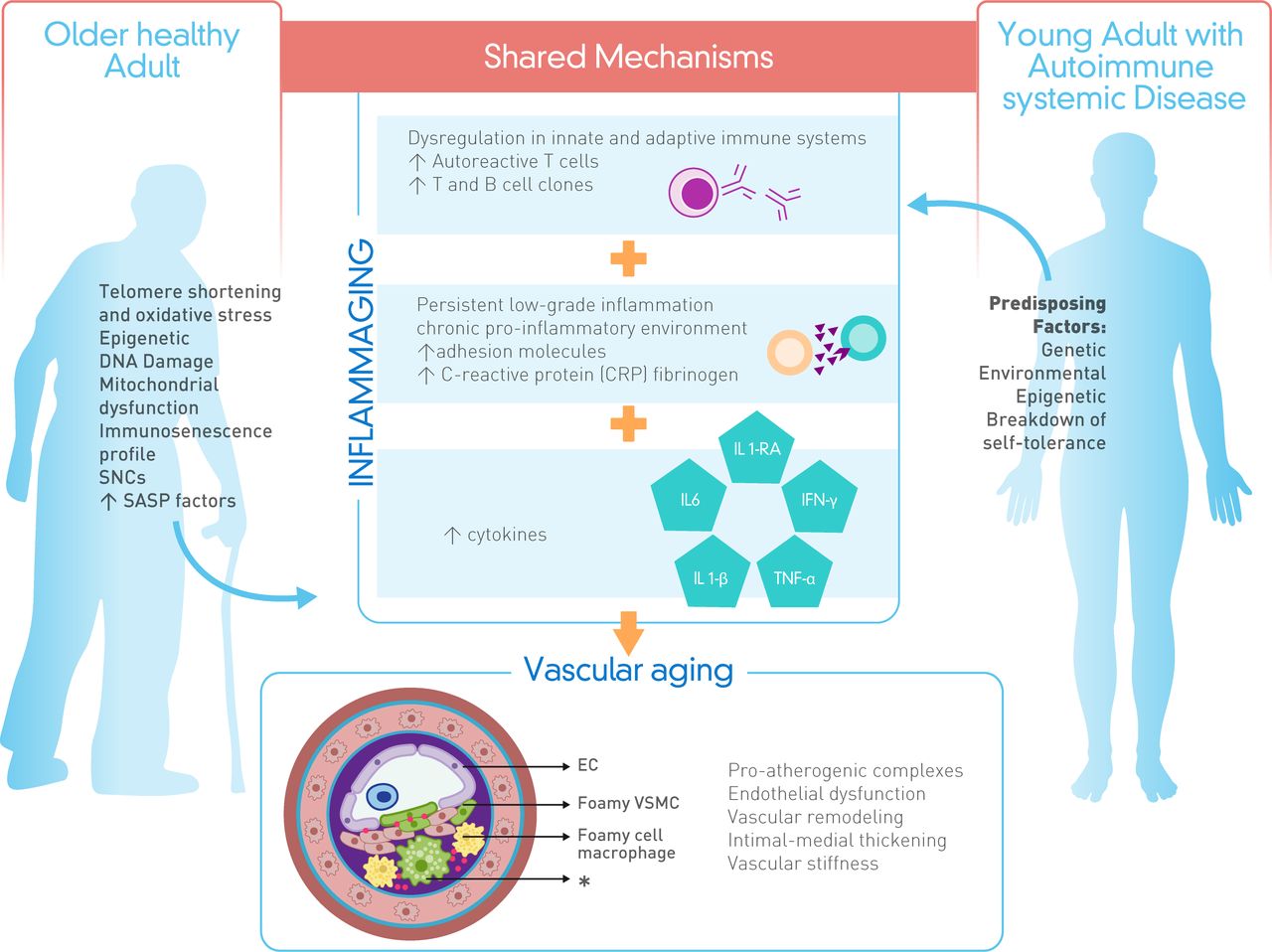
Shared mechanisms in healthy ageing and autoimmune systemic diseases leading to vascular ageing. In the case of autoimmune systemic diseases, inflammatory status (acute-phase reactants, cytokines and adhesion molecules upregulation) and vascular remodelling (high vascular stiffness) are a result of the breakdown self-tolerance due to the mentioned risk factors. While in older healthy adults these immunological and vascular changes are linked with immunosenescence, mitochondrial dysfunction, oxidative stress and DNA damage. *Secondary senescence. EC, endothelial cell; IFN-γ, interferon gamma; IL-1β, interleukin 1 beta; IL-1RA, interleukin 1 receptor antagonist; IL-6, interleukin 6; SASP, senescence-associated secretory phenotype; SNCs: senescent cells; TNF-α, tumour necrosis factor alpha; VSMC, vascular smooth muscle cells.
The relationship between inflammation and vascular stiffness, the main clinical vascular ageing variable
In the clinic, vascular stiffness is the most commonly used measure to determine vascular age. It correlates well with chronological age, it is predictive for various major cardiovascular endpoints, and it is affected by the most important structural and functional vascular changes that occur during ageing.35–38 In the arterial wall, the low-grade pro-inflammatory milieu characteristic for the ageing process triggers morphological, microscopical and functional changes. The entire process of vascular ageing has been recognised as the ‘pro-inflammatory arterial stiffness syndrome’.35 The components of inflammaging have been shown to be associated with the development of this process.7 39 40
These alterations can not only develop with ageing, but also be induced in young animals under experimental pro-inflammatory stimulation.41 When these molecular processes are taken into account, arterial stiffness is understood to be the most important determinant for establishing vascular age, and that is bears relationship with inflammaging.7 42 Accordingly, the relevant features of vascular ageing play a combined role in the outcome of stiffness measurements (ie, pulse wave velocity (PWV))36–38 (figure 3). If AID plays a role in vascular ageing induced by inflammation, vascular stiffness should be correlated with AID.
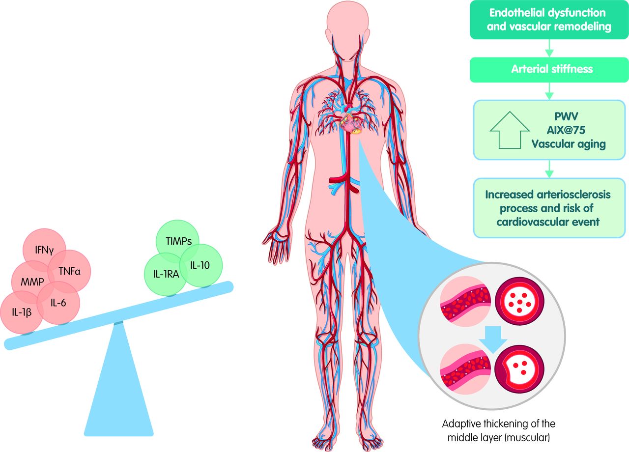
Inflammation and arterial stiffness. A pro-inflammatory milieu contributes to endothelial dysfunction and vascular remodelling. Arterial stiffness, as the main determinant of vascular age, can be measured by PWV or augmentation indexes. AIX@75, augmentation index adjusted to 75 beats per min; IFN-γ, interferon gamma; IL-1β, interleukin 1 beta, IL-1RA, interleukin 1 receptor antagonist; IL-6, interleukin 6; IL-10, interleukin 10; MMP, metalloproteinases; PWV, pulse wave velocity; TIMPs, metalloproteinases inhibitors; TNF-α, tumour necrosis factor alpha.
In the next section, we explore this relationship with an extensive, non-systematic review that is focused on inflammaging, RA and PWV. PWV was chosen because it has been considered gold standard to assess arterial stiffness, there is large epidemiological evidence of its predictive value for cardiovascular events, and it correlates well with vascular age.7 RA has been analysed as a model for research mechanisms linking inflammation with cardiovascular risk and it is considered that studying pathways shared by both RA and atherosclerosis may provide insight into potential mechanisms of risk.29
Non-systematic review of inflammaging, RA and vascular stiffness
Method
An extensive non-systematic review of the literature was done with the Medical Subject Headings (MeSH) terms “vascular stiffness,” and the keywords “arterial stiffness,” “pulse wave velocity,” “PWV” and “inflammaging” that were crossed with the MeSH terms “rheumatoid arthritis” and “Arthritis, rheumatoid” in Pubmed up to 28 July 2020 (see online supplemental table S1). Articles evaluating adult patients with RA and a comparison group of subjects without RA and without any other AID were included (cross-sectional, cohort studies, and cases and controls). The main outcome was the measurement of the carotid–femoral PWV by applanation tonometry, oscillometric method or Doppler ultrasound. Human studies were eligible if they met the following criteria: (1) full-length publications in peer-reviewed journals; (2) case–control, cross-sectional or longitudinal, observational or analytical in design; (3) studies of adult patients with RA fulfilling validated criteria with a control group; (4) provided sufficient data on PWV measurement parameters. No restriction criteria were imposed with regard to the type of the control healthy population studied. Overlapping populations were excluded (ie, same group of authors, same year, only one was considered). In addition, studies were excluded if they did not mention quantitative assessment of the physiological confounders of PWV (ie, blood pressure, heart rate, correction for age or risk factors such as DM2). Narrative or systematic review, case report or series, studies not involving human study or studies contained other autoimmune populations were also excluded. No language or year filters were applied to carry out the search.
Supplemental material
Statistical analysis was done with comprehensive meta-analysis Software. Data from the articles chosen were pooled using the random-effects model (DerSimonian and Laird) due to the studies’ high heterogeneity (sample size, study design, primary objective) to establish an overall effect (mean difference with a CI and the respective p value). Studies evaluating PWV under the same measurement’s techniques were analysed. A meta-regression was done to analyse the influence of age and inflammatory activity on pooled PWV. Evidence level and methodological quality assessment was done by using the Joanna Briggs Institute checklists.
Results
The initial search yielded a total of 712 articles with the following terms and Boolean operators: “Arterial stiffness” and “rheumatoid arthritis”, 206 articles; “Pulse wave velocity” AND “rheumatoid arthritis”, 154 articles; “PWV” AND “rheumatoid arthritis”, 73 articles; “Vascular stiffness” and “rheumatoid arthritis”, 170 articles. For the above searches, the same result was obtained when combined with the MeSH term “arthritis, rheumatoid.” For the search related to ‘inflammaging’ and ‘rheumatoid arthritis’, only 10 articles were found, all corresponding reviews of the literature were not related to RA.
In total, 20 articles met the inclusion criteria, they were evaluated, and the main outcome established. There were no overlapping populations. The global population was 2417 subjects, of which 1104 corresponded to patients with RA and 1099 to subjects without RA. There were no age differences between cases and controls except for one study (see online supplemental table S2). Female sex was the most prevalent in both groups, 84.6% in cases and 77.8% in controls (see online supplemental table S2).
Supplemental material
For the measurement of PWV, the applanation tonometry method was the most frequently used in the study protocols. About 72% of the retrieved publications showed an increase in PWV with a statistically significant difference between patients with RA and the population without RA. Additionally, moderate-to-severe DAS-28 (Composite Disease Activity Score) scores were found in 92.3% of the articles in which a significant increase in PWV values were found compared with controls (see online supplemental table S2). Twelve studies were included in the meta-analysis (those evaluating PWV through applanation tonometry). The pooled analysis under a random-effects model demonstrated a higher PWV in patients with RA (0.712; 95% CI 0.377 to 1.048; Χ2 p<0.000; I=2.82%) compared with controls (see online supplemental figure S1). Meta-regression models showed that age in cases and controls did not have an impact on the PWV difference between groups (coefficient=0.55; p=0.38 and coefficient=0.71; p=0.22, respectively). An additional meta-regression evaluating the DAS-28 impact on PWV difference showed that DAS-28 increase the PWV difference between groups (coefficient=0.69; p=0.038). Evidence level and methodological quality assessment was developed (see online supplemental table S3).
Supplemental material
Supplemental material
Implications of the present and previous reviews
Through this extensive review of the literature, an increase in PWV in patients with RA was made evident compared with populations without RA and without other AID (see online supplemental table S2). In addition, it should be noted that there is a statistically significant relationship between inflammatory activity of the disease (measured through the DAS-28 index) and the increase in carotid–femoral PWV. With PWV, the arterial stiffness increases, and thus the chronological vascular age calculated from it.37 43 Patients and controls had similar age, and this did not influence the PWV difference as shown through the meta-regression models. Although the primary studies did not calculate the vascular age, it could be inferred that the vascular age of the cases would be higher, since they have a higher PWV which, according to the age-adjusted reference curve for PWV, corresponds to a higher vascular age. Therefore, the results of this review of the literature should indicate that individuals with RA have accelerated vascular ageing compared with controls without RA and probably related to the chronic inflammatory state.
In a meta-analysis carried out by Ambrosino et al in 2015, an increase in the greater aortic and brachial PWV as well as in the rate of aortic augmentation adjusted to 75 beats per min was demonstrated when they compared patients with RA with healthy subjects.44 The present study reinforces the findings of Ambrosino et al and updates the search carried out by them, including 10 new studies.
Biomarkers that link AID with vascular stiffness
Södergren et al studied biomarkers associated with early RA (diagnosed in the previous 12 months) and CVD. They found a difference between the levels of the monocyte migration inhibitory factor, a biomarker associated with atherosclerosis, in patients compared with controls.45 However, adjusting this for sex and age of the patients was not correlated with carotid intima–media thickness (cIMT) values. In contrast, Lo Gullo et al demonstrated a direct relationship between higher levels of IL-1β and the expression of CRP, reactive oxygen species, and a higher PWV value. Furthermore, Marder et al have shown a significant positive association between IL-17 levels and PWV values. Bazzichi et al found that patients with AID (RA and scleroderma) had higher levels of PWV compared with healthy individuals, and that this increase was related to osteopontin levels, specifically in individuals with RA. These levels are a predictor of an increase in PWV after an analysis adjusted for confounding variables.46–48
Recently, it has been suggested that the way the process of arterial remodelling, both in its arteriosclerotic component and in endothelial dysfunction, is activated by extracellular matrix metalloproteinases (MMPs) indicates that its inhibition could slow down the vascular pro-inflammation associated with age.35 49 50 Among the molecular patterns known as molecular phenotypes of the ageing arterial wall, the MMP phenotype stands out as one of the most important in this process. This highlights the special participation of MMP 1, 2 and 9 with a fundamental role in vascular ageing process as well as the antagonistic role of tissue MMP inhibitors (TIMPs).35 51 52 In addition, MMP expression and its activity usually correlate with severity in both AID (ie, psoriasis, SLE, RA and so on) and disorders of infectious origins. In fact, the imbalance of pro-inflammatory and anti-inflammatory cytokines seen in pathology of RA may result in part from altered balances in the MMP/TIMP proteolytic axis. Furthermore, in this disease, angiogenesis also occurs at the site of inflammation and it has been shown that MMPs regulate the bioavailability of several angiogenic factors such as vascular endothelial growth factor, fibroblast growth factor receptor and epithelial growth factor.52
Thus, in individuals with hypertension, an increase in PWV has been shown to be in parallel with an increase in MMP 2 and MMP 9 when compared with individuals without hypertension.53 In other studies of individuals with hypertension under an antihypertensive therapeutic regimen, a decrease in arterial stiffness has been shown associated with a decrease in MMP and an increase in TIMP-1.54
Taking all of the above into account, a myriad of pro-inflammatory cytokines and MMPs have been identified as potential effectors of accelerated vascular ageing associated with AID. Therefore, this field of study is likely to be a viable way to result in clinical application.
Therapeutic targets and horizons in the treatment of CVD in patients with RA
CVD risk assessment in RA
An important approach to reduce cardiovascular risk in patients with RA is the tuning between specific risk assessment and AID pharmacotherapy. In clinical practice, some guidelines suggest performing a cardiovascular risk assessment on patients with RA at least every 5 years using tools, for example the Framingham score, that make it possible to predict cardiovascular risk and establish a management plan, as is done in the general population. Note that the Framingham score must be multiplied by 1.5 for patients with RA to obtain the final result. Alternatively, one can use scales specific for this population, such as the Systematic Coronary Risk Evaluation (SCORE), the Expanded Cardiovascular Risk Prediction Scale score for RA and the QRISK2 scale.55 However, although there is biological plausibility for the use of this adjustment or these scales, there is still no clear validation for these generalised recommendations, and cardiovascular risk calculations for patients with RA should rather be done individually and with great caution.20 56 57 In the European consensus for the control of cardiovascular risk in RA and inflammatory arthropathies, emphasis is placed on disease control to decrease cardiovascular risk.55 Therefore, the effect of pharmacotherapy should become an integral part of the risk assessment. This includes effects of both AID as well as cardiovascular drugs.
Current pharmacotherapy in patients with RA and its effect on CVD
The use of statins in patients with RA with primary CVD risk prevention has been shown to reduce arterial stiffness and carotid plaque formation regardless of the reduction in LDLs. Furthermore, in patients with RA who discontinued statin therapy, it was found that at 3 months, the risk of myocardial infarction had increased by 67%, and that it increased an additional 2% for each month after that.57 Its discontinuation is also related to a 60% increase in the risk of cardiovascular mortality and 79% of all-cause mortality in patients with RA.58 Hence, the use of statins in patients with RA is strongly indicated according to national lipid guidelines considering the SCORE CVD risk prediction model if no national guideline is available to stratify the CVD risk in these patients to take the decision.55
The use of statins also modulates non-CVD processes. A study evaluating patients with RA being treated with at least one disease-modifying antirheumatic drug (DMARD) showed that concomitant use of statins reduced all-cause mortality by 21%.57 In patients with RA in which statin is added to the regimen of methotrexate and corticosteroids, the disease activity declines significantly, which could suggest a direct role in disease control.57 58 Indeed, beyond the LDL-lowering action, statins are known to exert anti-inflammatory and immune-modulating effects by inhibiting MMPs, nuclear factor kappa beta (NF-ĸβ), cell adhesion and migration molecules such as selectins and vascular cell adhesion molecule 1. These effects of statins, which have been comprehensively reviewed elsewhere,59 might account for the additive effect on AID drugs.
In contrast, the use of glucocorticoids might increase CVD. Although it is true that glucocorticoids are part of the treatment of patients with AID, and that eventually, in particular cases, non-steroidal anti-inflammatory drugs are used, it is pertinent to closely monitor the therapeutic management since this medication adds greater cardiovascular risk to the underlying disease.60 In particular, sustained glucocorticoid treatment increases risk for CVD through multiple pathways. Although glucocorticoid therapy might promote hypertension, its risk-increasing effect is best explained not by hypertension alone, but rather by the metabolic effects of prolonged glucocorticoid treatment on other pathways, particularly impaired glucose tolerance, including overt diabetes and disturbances in serum lipids.61 In fact, disturbances in plasma cortisol levels predispose to the development of a variety of pathologies with atherosclerotic CVD being a top killer in patients suffering from primary adrenal insufficiency or Cushing’s syndrome/disease.62
Next to glucocorticoids, conventional DMARDs have been used in clinical practice for a long time, and there is information available regarding their cardiovascular profile. With respect to methotrexate, the most widely used synthetic conventional DMARD, there are meta-analyses that show that its use reduces cardiovascular risk by between 21% and 28%. Similarly, the use of hydroxychloroquine and sulfasalazine as well as their combinations has also been associated with a decrease in cardiovascular risk.60 63 Thus, next to statins the use of such DMARDs is indicated in all patients with RA according to the disease activity level in order to control the disease and the consequences of chronic inflammation at the cardiovascular level.55
The use of biological DMARDs (ie, monoclonal antibodies, fusion proteins, TNF inhibitors and non-TNF inhibitors) is significantly associated with the regulation of the chronic inflammatory process and thus, they could be considered potential therapeutic drugs with a direct effect on the ‘inflammaging’ which would reduce the risk of CVD. Table 1 lists studies that evaluated impact of biological DMARDs on cardiovascular risk and function. In particular, the decrease in PWV in patients with RA under treatment with biological DMARDS illustrates the anti-ageing effect in the vasculature. The impact of the introduction of biological medicines in RA has been such that by 2010, in some cohorts, CVD mortality rates in this population decreased progressively and significantly until they reached levels close to that of the general population. This could be due in part to the use of this type of DMARDs.64 65 In fact, a meta-analysis suggested that anti-TNF treatment may have a beneficial effect on aortic stiffness and, therefore, on cardiovascular risk.66 It is appealing to hypothesise that cardiovascular improvement the use of these DMARDs entails is a result of an effect on inflammaging, as can be expected for TNF inhibition.65 Specific in-depth study of this possibility will be needed to confirm this and to provide a rationale for the targeted use of such drugs to decrease CVD risk in patients with AID.
Effects of biological DMARDs on cardiovascular and arterial stiffness in patients with rheumatoid arthritis (RA) and other inflammatory arthropathies
The effects of both biological and conventional DMARDs have been also studied on measures of subclinical atherosclerosis and vascular dysfunction (hence, vascular ageing) besides PWV. The impact of methrotexate, sulfasalazine and hydroxychloroquine on cIMT, and flow-mediated dilation (FMD), surrogate markers of atherosclerosis and endothelial dysfunction, respectively, has been tested.67 After a year of treatment, a significant decrease in cIMT and an improvement in FMD have been found. Additional research has shown similar results.68 69
Regarding biological DMARDs, its beneficial effect has been described on endothelial function and subclinical atherosclerosis in patients with RA. Adalimumab was associated with improvement in FMD without cIMT progression, after 12 months of treatment in patients with refractory RA to conventional treatment.70 In another study, anti-TNF therapy was associated with a reduction in cIMT progression.71
Similar effects have been observed under treatment with other biologics besides anti-TNF agents (table 1). In a study that for the first time evaluated the effect of rituximab on endothelial dysfunction, it has shown a dramatic improvement of FMD at 6 months of treatment.71 However, the research of the potential beneficial effect on CVD (measured through FMD and cIMT) of this and other biological therapies is still needed.
On the other hand, the coronary artery calcium score (CACS), performed by coronary CT angiography, is a strong predictor of cardiovascular events and is potentially useful for improving the cardiovascular risk assessment in asymptomatic people. A higher prevalence of coronary artery calcification has been found in patients with RA,71 but research is still needed to establish the roll of treatment on CACS.
Novel biological drugs make it possible to find additional therapeutic options with adequate efficacy and safety. One possibility is blockade of the complement pathways. Apart from the possible benefits of eculizumab, inhibitor of C5 cleavage, in AID, its use is being considered as a possible therapy for membranous glomerulopathy.72 Another strategy involves metabolic adaptation of immune cells. Given their energy and substrate requirements for differentiation and activation, unravelling the relation of metabolism and immune cell function is now an active field of investigation and will allow the development of therapeutic strategies such as the modulation of pro-inflammatory metabolism.73 Recently, metabolism-modulating therapies are under study in patients with AID. For example, effects of metabolic regulators of mitochondrial function and biogenesis such as metformin (by means of mitochondrial complex I inhibition) are explored in RA and SLE. Agents that inhibit mammalian target of rapamycin (ie, sirolimus, rapamycin and temsirolius), a main metabolic switch that is unusually active in many autoinflammatory and AID, are under investigation as well in RA, SLE, DM2 and multiple sclerosis. Furthermore, effects of some activators of SIRT 1, an important nutrient-sensing protein that suppresses NF-κβ-dependent inflammatory responses, (eg, resveratrol) are under evaluation in patients with RA.74 75 In addition, new therapeutic targets in the inflammatory signalling pathways are under study, and others are considered potential candidates for AID, particularly RA and SLE. Some of these pathways concern extracellular targets such as macrophage granulocyte colony-stimulating factor inhibitors (eg, mavrilimumab) and other intracellular binding sites such as new inhibitors of the JAK pathway (eg, filgotinib), inhibitors of the Bruton’s tyrosine kinase pathway or phosphoinositol 3-kinase, or new targets at the level of dendritic cells or the Fc-gamma receptor.75 76 Again, the facts summarised here highlight the potential role of ‘inflammaging’ pathways with the development of arterial stiffness and endothelial dysfunction in patients with AID.
Conclusions
Pathologies characterised by a continuous state of imbalance between the production of pro-inflammatory and anti-inflammatory molecules, which is what occurs in AID (eg, RA), lead to an accelerated process of arteriosclerosis, vascular remodelling and, therefore, an increased vascular age, resulting in an increase in the morbidity and mortality rate from cardiovascular events.
Inflammaging and its vascular impact in the context of AIDs such as RA open a new field of medical research. This discipline is marked by early, personalised diagnosis and risk stratification, through the combination of routinely used non-invasive vascular function tests as well as AID variables. It offers opportunities for novel approaches in the field of clinical pharmacology and personalised medicine, in which specific combinations of statins with immune-modulating AID drugs play a leading role, and may benefit both CVD as well as AID progression. Thus, inflammaging has become a target for primary and secondary prevention of cardiovascular events in patients with a pathology that is immunological in origin.
Acknowledgments
The language of the paper has benefited from the academic editing services of Cecile Dunn and the support of the 'Call for the improvement of the language in research papers' of the Fundación Universitaria de Ciencias de la Salud (FUCS).
References
Supplementary materials
Supplementary Data
This web only file has been produced by the BMJ Publishing Group from an electronic file supplied by the author(s) and has not been edited for content.
Footnotes
Contributors PS-M—study concepts and design, manuscript and figure preparation, manuscript editing, final approval of the article; GB-A—study concepts and design, manuscript and figure preparation, manuscript editing, final approval of the article. MAM-C—study concepts and design, manuscript and figure preparation, manuscript editing, final approval of the article. AP—study concepts and design, manuscript and figure preparation, manuscript editing, final approval of the article. DE—study concepts and design, manuscript and figure preparation, manuscript editing, final approval of the article. PKB-N—study concepts and design, manuscript and figure preparation, manuscript editing, final approval of the article. AR-V—study concepts and design, manuscript and figure preparation, manuscript editing, final approval of the article. AJMR—manuscript and figure preparation, manuscript editing, final approval of the article.
Funding The authors received financial support from the Ministry of Science (Minciencias) grant number 844-2019 and Fundación Universitaria de Ciencias de la Salud (FUCS) contract 829-2019: 'Pact for the generation of new knowledge through scientific research projects in Medical and Health Sciences'; project code 500784467051. AJMR receives funding from Stichting Lijf en Leven, Rotterdam, Project 60, and TKI-LSH grant # EMCLSH19013.
Competing interests None declared.
Patient consent for publication Not required.
Provenance and peer review Not commissioned; externally peer reviewed.