Article Text

Abstract
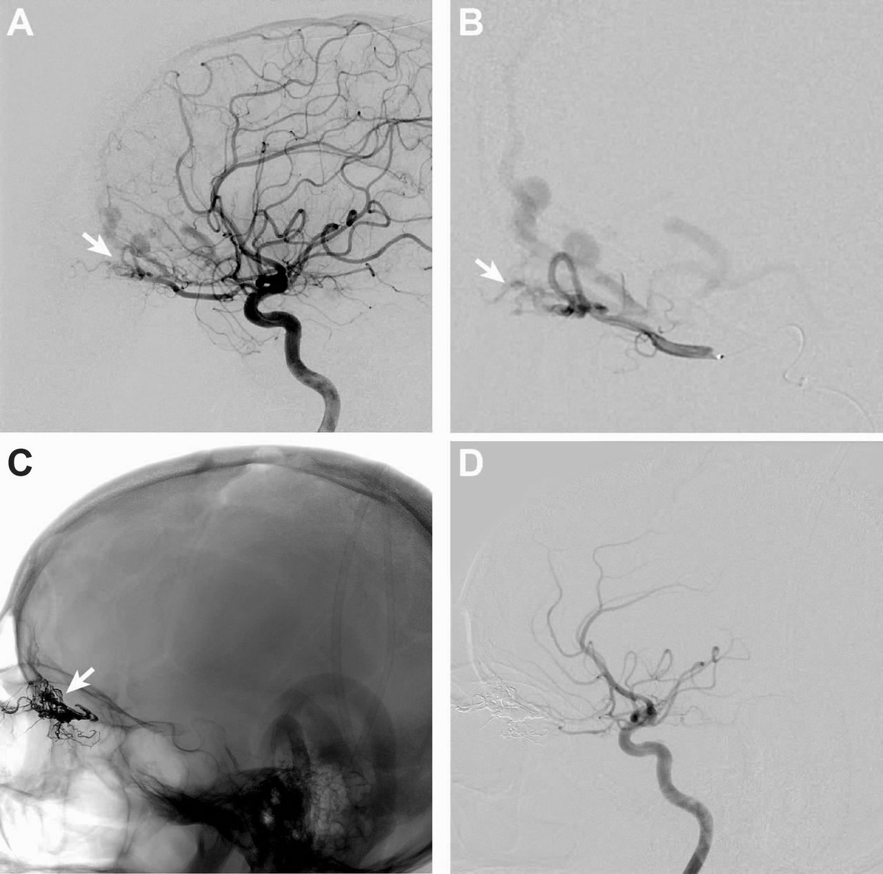
Introduction High flow vascular lesions of the anterior cranial fossa and orbit (HFVL) include arteriovenous malformations and dural arteriovenous fistulae located within the orbit, periorbital region, ethmoid sinuses, the anteroinferior frontal lobes, or within the dura of the anterior cranial fossa. Rupture of HFVL may result in intracranial hemorrhage, so these lesions typically undergo treatment even when discovered for other reasons. HFVL may be treated by trans-arterial endovascular embolization, surgical ligation, radiosurgery, or a combination of these approaches, but the most optimal treatment is not well defined. We determined patient outcomes and HFVL obliteration after treatment.
Methods We retrospectively reviewed all patients referred for diagnostic angiography or endovascular embolization of HFVL at our neurovascular referral center over an 8 year period. Patient demographic, treatment, and outcome data were deterred from the medical record. DSA, CT, and MRI studies were reviewed for HFVL characteristics and obliteration after treatment.
Results HFVL were identified in 11 patients (five females and six males) ranging in age from 8 to 76 years (mean 53 years). Presenting symptoms included headaches (nine patients; 82%), visual symptoms (five patients; 45%), intracranial hemorrhage (two patients; 18%), tinnitus (one patient; 9%), or no symptoms (one patient; 9%). Nine patients (81%) had medical comorbidities, but none had a hypercoagulable disorder or prior trauma. The HFVL were comprised of seven dural arteriovenous fistulae (64%) and four arteriovenous malformations (36%). The ophthalmic artery was the dominant feeding vessel in 10 patients (91%). 10 patients (91%) were treated, including embolization via the ophthalmic artery (four patients; 36%), embolization followed by surgical ligation (3 patients; 27%), embolization followed by radiosurgery (one patient; 9%), surgical ligation alone (one patient; 9%), or radiosurgery alone (one patient; 9%). Endovascular embolization was performed with Onyx (six patients; 75%) or n-BCA (two patients; 25%) HFVL cure was achieved in six patients (55%), including three patients treated by embolization alone, two treated by embolization and surgical ligation, and one by surgery alone. One patient treated by embolization alone developed a post-treatment partial visual deficit, but there were no other complications related to treatment. No patient deaths occurred.
Conclusions HFVL are uncommon lesions that are challenging to treat. Endovascular embolization alone or in combination with surgery results in HFVL obliteration in 50% of patients with an acceptable safety profile. Further studies should determine whether radiosurgery alone or in combination with endovascular embolization results in high rates of HFVL cure.

Disclosures A. Moraff: None. R. Dodd: None. M. Marks: None. H. Do: None. G. Steinberg: None. S. Chang: None. J. Heit: None.
This is an Open Access article distributed in accordance with the Creative Commons Attribution Non Commercial (CC BY-NC 4.0) license, which permits others to distribute, remix, adapt, build upon this work noncommercially, and license their derivative works on different terms, provided the original work is properly cited and the use is non-commercial. See: http://creativecommons.org/licenses/by-nc/4.0/