Article Text

Abstract
Background Myocarditis is a highly morbid complication of immune checkpoint inhibitor (ICI) use that remains inadequately characterized. The QRS duration and the QTc interval are standardized electrocardiographic measures that are prolonged in other cardiac conditions; however, there are no data on their utility in ICI myocarditis.
Methods From an international registry, ECG parameters were compared between 140 myocarditis cases and 179 controls across multiple time points (pre-ICI, on ICI prior to myocarditis, and at the time of myocarditis). The association between ECG values and major adverse cardiac events (MACE) was also tested.
Results Both the QRS duration and QTc interval were similar between cases and controls prior to myocarditis. When compared with controls on an ICI (93±19 ms) or to baseline prior to myocarditis (97±19 ms), the QRS duration prolonged with myocarditis (110±22 ms, p<0.001 and p=0.009, respectively). In contrast, the QTc interval at the time of myocarditis (435±39 ms) was not increased compared with pre-myocarditis baseline (422±27 ms, p=0.42). A prolonged QRS duration conferred an increased risk of subsequent MACE (HR 3.28, 95% CI 1.98 to 5.62, p<0.001). After adjustment, each 10 ms increase in the QRS duration conferred a 1.3-fold increase in the odds of MACE (95% CI 1.07 to 1.61, p=0.011). Conversely, there was no association between the QTc interval and MACE among men (HR 1.33, 95% CI 0.70 to 2.53, p=0.38) or women (HR 1.48, 95% CI 0.61 to 3.58, p=0.39).
Conclusions The QRS duration is increased in ICI myocarditis and is associated with increased MACE risk. Use of this widely available ECG parameter may aid in ICI myocarditis diagnosis and risk-stratification.
- autoimmunity
- immune tolerance
- immunotherapy
- inflammation
- self tolerance
This is an open access article distributed in accordance with the Creative Commons Attribution Non Commercial (CC BY-NC 4.0) license, which permits others to distribute, remix, adapt, build upon this work non-commercially, and license their derivative works on different terms, provided the original work is properly cited, appropriate credit is given, any changes made indicated, and the use is non-commercial. See http://creativecommons.org/licenses/by-nc/4.0/.
Statistics from Altmetric.com
Background
Since the introduction of ipilimumab in 2011, immune checkpoint inhibitors (ICIs) have improved oncological care, offering new therapeutic options for a variety of cancers.1 Through activation of the immune system, ICIs can enhance antitumor activity but may also lead to immune-related adverse events (irAEs).2 3 Myocarditis is an uncommon irAE.4 5 The incidence of ICI-associated myocarditis is unclear, with estimates ranging from 0.09% to 1.1%.3 6–10 There are several potential cardiac adverse effects associated with ICIs but the focus on myocarditis is due to the morbidity and mortality associated with that diagnosis.11 12 Specifically, data from multiple groups report a mortality ranging from 17% to 50%.3 6 7 13 In comparison, the mortality of non-ICI myocarditis is far less than 5%.14 15 Therefore, there is a significant need for improved diagnostic and risk-stratification methods in ICI myocarditis, as well as newly recognized cardiac toxicities related to other forms of immune therapy for cancer.16 17
The QRS duration and the QTc interval are standardized measures routinely available from a 12-lead ECG. The QRS duration corresponds to ventricular depolarization, while the QTc interval predominantly represents ventricular repolarization. Monitoring of the QRS duration is employed in the care of patients who are at risk of developing arrhythmias and heart failure.18 19 Measurement of the QTc interval is also routinely used in both oncology trials and routine oncology care.20 However, there are no data on the utility of measurement of the QRS duration and the QTc interval among patients with ICI myocarditis. Therefore, the goal of this study was to evaluate whether the QRS duration and QTc interval increase with ICI myocarditis and whether these parameters associate with the development of subsequent adverse cardiac events.
Methods
Patients
Data on myocarditis cases (n=140) were obtained from a 23-center international registry specifically designed for collating cases of ICI myocarditis.5 7 21 Cases were included between November 2013 and April 2019. Controls (n=179) were selected patients from a Massachusetts General Hospital registry of patients treated with an ICI during the same time interval for whom ECG data were available. Controls were not selected to match on any parameter with the cases.
ECG data
ECGs were obtained at various timepoints relative to ICI initiation and to the development of myocarditis (among cases). The acquisition of an ECG was not protocol-specified and was performed at the discretion of the medical care team. ECGs were recorded using the preferred system at each registry site and the parameters were derived automatically by those systems. Forty randomly selected ECGs (20 from controls and 20 from myocarditis cases) were also manually interpreted by a reader (DAZ) blinded to the case/control identity and the automated measurements. These manually measured values were then compared with those which had been automatically derived. The primary measures of interest included the PR interval, QRS duration, and the QTc interval. The PR interval represents the time delay between atrial depolarization and ventricular depolarization and was included in the analysis to assess which measures of intracardiac conduction were affected in ICI myocarditis. QT interval correction for heart rate was calculated using the Fridericia formula22 as this is more accurate than the commonly used Bazett formula and considered the most appropriate strategy by the US Food and Drug Administration.23 24 Based on standardized criteria, a QRS duration greater than 110 ms was considered prolonged.25 Similarly, based on standardized criteria, a QTc interval greater than 450 ms in men and 460 ms in women was considered prolonged.26
Clinical data
Clinical data of interest obtained retrospectively from electronic medical records included patient demographics, medications, prior medical conditions, lab values, and cardiovascular risk factors. Data relevant to cancer included the cancer type, prior cancer therapies, and specific ICI treatments.
Definitions and outcomes of interest
Myocarditis was diagnosed through one of the following standardized approaches: (1) histopathological features on endomyocardial biopsy or autopsy or (2) a clinically accepted scoring system designed for suspected myocarditis that uses clinical findings, biomarkers, and imaging features.27 This latter clinical diagnostic algorithm relies on the presence of one or more clinical features (chest pain, heart failure, arrhythmias, or cardiogenic shock) plus one or more diagnostic findings (ECG abnormalities, elevated troponin levels, functional or structural abnormalities on cardiac imaging, or evidence of edema or late gadolinium enhancement on cardiac MRI) occurring in the absence of significant coronary disease or known alternative causes.27 This standardized scoring system was devised because an endomyocardial biopsy is typically performed in less than 15% of myocarditis cases.28 The main outcome of interest was major adverse cardiac events (MACE), which was a composite of cardiovascular death,29 cardiac arrest,30 cardiogenic shock,31 and hemodynamically significant complete heart block.7
Statistical analysis
Continuous variables were summarized as either the mean±SD or as the median and IQR, as appropriate, and categorical variables were presented as percentages. Comparisons between myocarditis cases and controls were made with Fisher’s exact test for categorical variables or with the Wilcoxon rank sum test for continuous variables (age and body mass index (BMI), both of which demonstrated non-Gaussian distribution). Comparisons of ECG parameters by case and ICI exposure status used the Kruskal-Wallis test with Dunn’s correction for multiple comparisons. The relationship between measured ECG parameters and left ventricular ejection fraction (EF) or left ventricular end-diastolic volume (LVEDV) was analyzed with simple linear regression and the associations were tested with the Pearson correlation coefficient. HRs for the association of dichotomized ECG parameters with MACE were determined using Cox proportional hazard models with follow-up time used as the time scale. The assumption of proportionality was verified by the method of Therneau and Grambsch.32 Kaplan-Meier curves and the log-rank test were used to analyze the relationship between QRS duration or QTc interval and MACE-free survival. The χ2 test was used to compare the frequencies of MACE component endpoints among myocarditis cases and subgroups. Multivariable logistic regression was used to address the relationship between multiple covariates, including ECG parameters at the time of myocarditis, and MACE risk. All statistical tests were two-sided and p<0.05 was considered significant. Statistical analysis was performed using R V.3.5.1 (R Foundation for Statistical Computing, Vienna, Austria) and SPSS V.26 (IBM Corporation).
Results
Patient characteristics
One hundred and forty patients with ICI myocarditis and 179 controls were included in the analysis. The median time between ICI initiation and myocarditis diagnosis was 58 days, and 91 cases (65.0%) occurred within the first 90 days of ICI therapy. Of the 140 cases, 70 (50.0%) were diagnosed based on clinical criteria, 58 patients (41.4%) were diagnosed by pathology from an endomyocardial biopsy, and 12 patients (8.6%) were diagnosed based on autopsy pathology. The median duration of follow-up for myocarditis cases was 69 days (IQR 19–153). Myocarditis patients had a higher BMI than controls (28±6.1 vs 26±5.8, p=0.002); otherwise, cases and controls had similar rates of cardiovascular risk factors (table 1). The most common cancer in both groups was melanoma. Renal cell cancer was more common among myocarditis cases (8.6% vs 2.2%, p=0.02). The use of atezolizumab was higher among cases (7.1% vs 1.7%, p=0.02). There was a lower frequency of anti-programmed cell death 1 (PD-1) therapy use in the myocarditis group (87.1% vs 96.1%, p=0.005); conversely, there was a greater frequency of anti-programmed death ligand 1 (PD-L1) therapy use in the myocarditis group (11.4% vs 2.2%, p=0.001). The use of ipilimumab/nivolumab combination therapy was also more common in the myocarditis group (25.7% vs 11.2%, p=0.001). The rates of pneumonitis (24.3% vs 12.3%, p=0.007) and neurological adverse events (8.6% vs 2.2%, p=0.02) were higher in the myocarditis group.
Baseline characteristics
ECG characteristics
We compared computer-derived ECG parameters between controls and myocarditis cases at three different time-points: prior to initiation of ICI (“pre-ICI”), after the initiation of ICI but prior to development of myocarditis (“on-ICI”), and at the time of presentation with myocarditis. For validation of this approach, we also manually measured the parameters from 40 random ECGs (20 control and 20 cases) and found these values to be similar to the computer-derived values (online supplemental file 1). The median time intervals between pre-ICI baseline ECG and initiation of ICI were 103 days (IQR 24–215) and 82 days (IQR 35–172), controls and cases, respectively. The median time intervals between initiation of ICI and on-ICI ECG were 165 days (IQR 41–379) and 44 days (IQR 11–152), controls and cases, respectively. The pre-ICI baseline PR interval, QRS duration, and QTc (corrected for heart rate with the Fridericia formula, subsequently designated QTc-F) interval were similar between controls and cases (figure 1A–C). The on-ICI (prior to development of myocarditis for cases) PR interval, QRS duration, and QTc-F did not change from baseline and were similar between cases and controls. Additionally, among cases prior to the development of myocarditis, there was no increase in these parameters between the pre-ICI values and the on-ICI values. With the development of myocarditis, the PR interval did not increase. In contrast, the QRS duration at the time of myocarditis (110±22 ms) did increase and was greater than the QRS duration of cases pre-ICI (99±20 ms, p=0.001), controls on-ICI (93±19 ms, p<0.001), and cases on-ICI prior to the development of myocarditis (97±19, p=0.009). Cases were stratified by QRS duration using the cut-off of 110 ms, the upper limit of normal in adults.25 A normal QRS duration of ≤110 ms was observed in 74 cases (52.8%), while 66 (47.1%) had a prolonged QRS duration >110 ms. A similar testing strategy was adopted for the QTc-F interval. The QTc-F interval at time of myocarditis (435±39 ms) was increased when compared with controls on ICI (419±28 ms, p=0.02) but not when compared with values from cases prior to the development of myocarditis (422±27 ms, p=0.42). Of 99 male cases, 29 (29.3%) had a normal QTc-F≤450 ms, while 70 (70.7%) had a prolonged QTc-F>450 ms. Among women with myocarditis, 25 (61.0%) had a normal QTc-F≤460 ms, while 16 (39.0%) had a prolonged QTc-F>460 ms. We also analyzed the QTc interval across time points using the Bazett formula to correct for heart rate (subsequently designated QTc-B). With this approach, the QTc-B interval at the time of myocarditis (460±36 ms) was increased when compared with either on-ICI controls (440±28 ms, p<0.001) or on-ICI cases (440±29 ms, p=0.007) (online supplemental file 2).
Supplemental material
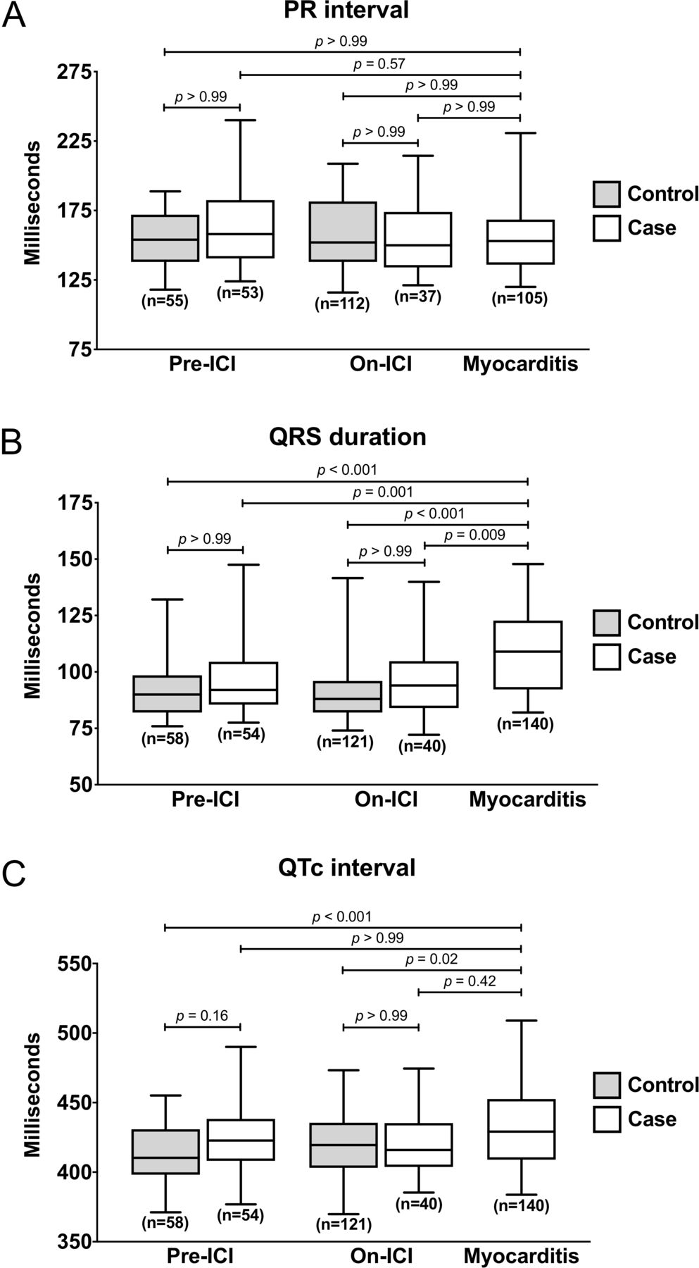
Changes in ECG parameters with ICI myocarditis. (A) PR interval, (B) QRS duration, and (C) QTc-F interval values were derived from ECGs obtained pre-ICI therapy (“baseline or pre-ICI”), after initiating ICI therapy (“on-ICI”), or at the time of myocarditis (for cases only; “myocarditis”). Shown are box-and-whisker plots with the central line indicating the median value, the margins of the box indicating the 25th/75th percentiles, and the whiskers indicating the 5th/95th percentiles. ICI, immune checkpoint inhibitor.
Sensitivity of QRS duration for the diagnosis of ICI myocarditis
Given the significantly prolonged QRS duration observed during myocarditis, the sensitivity and specificity of QRS duration towards myocarditis diagnosis was determined. Applying a cut-off of >110 ms achieved a sensitivity of 48.6% and specificity of 87.0% for the diagnosis of myocarditis; the application of a higher cut-off of >130 ms yielded a sensitivity of 16.4% and a specificity of 92.6% (online supplemental table 1). A receiver-operating characteristic curve applying QRS duration towards myocarditis diagnosis had an area under the curve of 0.73 (online supplemental figure 3). Using the change in QRS duration (ΔQRS; equal to the QRS duration at time of myocarditis minus the QRS duration at baseline prior to myocarditis) yielded a sensitivity of 51.7% when applying a diagnostic threshold of >10 ms; this decreased to 31.0% and 21.6% for >20 ms and >30 ms, respectively.
Association of ECG parameters with left ventricular size and function
We analyzed the relationship between the measured ECG parameters and left ventricular EF and LVEDV among myocarditis cases (figure 2A–C). The QRS duration was not related to either echocardiographic EF (r=−0.10, 95% CI −0.27 to 0.07, p=0.25) or MRI-derived EF (r=−0.095, 95% CI −0.29 to 0.11, p=0.35), but was directly related to the LVEDV (r=0.32, 95% CI 0.13 to 0.49, p=0.001). The QTc-F interval was inversely associated with the echocardiographic EF (r=−0.20, 95% CI −0.36 to −0.02, p=0.026) and the MRI-derived EF (r=−0.20, 95% CI −0.039 to −0.01, p=0.044), but was not related to the LVEDV (r=0.16, 95% CI −0.04 to 0.35, p=0.12).

Relationship between ECG parameters, ejection fraction, and left ventricular volumes. (A) Scatter-plots of QRS duration (left) or QTc-F interval (right) versus echocardiographic left ventricular EF. Shown are the linear regression lines with their 95% CI. (B) Scatter-plots of QRS duration (left) or QTc-F interval (right) versus EF. (C) Scatter-plots of QRS duration (left) or QTc-F interval (right) versus LVEDV. EF, ejection fraction; LVEDV, left ventricular end-diastolic volume.
Association of QRS duration with MACE
Overall, 69 of the 140 cases (49%) experienced a MACE. The median time to a MACE was 34 days (IQR 12–103) and 68% of MACE occurred within the first 90 days after diagnosis of myocarditis. A non-sinus rhythm at presentation with myocarditis was associated with an HR for MACE of 1.94 relative to sinus rhythm at presentation (95% CI 1.27 to 3.43, log-rank p=0.01) (online supplemental figure 4). Using an unadjusted Cox proportional hazard model, a QRS duration >110 ms was associated with a HR for MACE of 3.28 relative to a QRS duration ≤110 ms (95% CI 1.98 to 5.62, p<0.001) (figure 3A). Increased QRS duration at the time of myocarditis was associated with increased MACE risk after adjustment for age, sex, and cardiovascular comorbidities (table 2). Specifically, an increase in the QRS duration of 10 ms conferred a 1.30-fold increase in the odds of MACE (95% CI 1.07 to 1.61, p=0.011). We also assessed the prognostic utility of troponin in ICI myocarditis. Troponin data were available for 138 of the 140 patients. Of these 138, 125 (90.6%) had an elevated troponin. Of the 125 patients with elevated troponins, 68 (54.4%) experienced a MACE, while of the 13 patients with normal troponins, only 1 (7.7%) had a MACE (p=0.002). An elevated troponin was common and was not associated with increased MACE risk in the multivariable model (OR 2.54, 95% CI 0.69 to 10.83, p=0.17).

Association between QRS duration or QTc-F interval and MACE. (A) Kaplan-Meier curves indicate the occurrence of MACE over 120 days from time of diagnosis for myocarditis patients stratified by QRS duration. Similar analyses were performed for male (B) and female (C) myocarditis patients stratified by QTc-F interval as indicated. P value obtained from the log-rank test. MACE, major adverse cardiac events.
Association with major adverse cardiac events (MACE) by multivariable analysis
Rates for each of the four MACE components (cardiovascular death, cardiac arrest, cardiogenic shock, and complete heart block) were higher among patients with a QRS duration >110 ms than among those with a QRS duration ≤110 ms (figure 4). The most common MACE component in each was cardiovascular death (16.2% among those with QRS ≤110 ms, and 39.4% among those with QRS >110 ms).

Frequency of MACE components. Frequencies of the indicated outcome among all myocarditis patients stratified by QRS duration are shown. Numbers at the end of each bar indicate the fraction of patients in each stratum with the indicated event. MACE, major adverse cardiac events.
Association of QTc interval with MACE
In contrast, a prolonged QTc-F interval was not associated with increased risk for MACE among men (HR 1.33, 95% CI 0.70 to 2.53, p=0.38) or women (HR 1.48, 95% CI 0.61 to 3.58, p=0.39) (figure 3B,C). We also tested the association using QTc interval values corrected for heart rate using the Bazett formula. With this approach, among men, a QTc-B of >450 ms was associated with an increased risk of MACE (HR 2.81, 95% CI 1.42 to 5.38, p=0.002) (online supplemental file 5A). Among women, a QTc-B duration of >460 ms was also associated with an increased risk of MACE (HR 3.00, 95% CI 1.08 to 8.32, log-rank p=0.03) (online supplemental file 5B).
Discussion
In this paper, we present novel findings describing increases in the QRS duration that may aid the diagnosis and risk-stratification of ICI myocarditis. This study has several unique and widely generalizable findings. First, we found that ICI myocarditis is associated with an unchanged PR interval but an increased QRS duration when compared with controls or cases at earlier timepoints (both prior to the initiation of ICI therapy and on ICI therapy but prior to the development of myocarditis). The QTc-F interval was prolonged compared with on-ICI controls but not on-ICI cases. Second, the increase in ECG intervals correlated with structural (LVEDV) and functional (EF) changes in the heart. Third, prolongations of QRS duration but not QTc-F interval at the time of myocarditis diagnosis were associated with an increased risk of MACE; specifically, a QRS duration >110 ms was associated with a 3.28-fold increased risk of MACE. This association between prolonged QRS duration and MACE persisted after adjustment for several cardiovascular covariates. Fourth, each of the individual components of MACE occurred more frequently in myocarditis patients with a prolonged QRS duration, with cardiovascular death the most common.
Despite its high morbidity, the diagnosis of ICI myocarditis can be challenging.21 33–36 First, the presentation can be non-specific, both in terms of clinical symptoms and testing abnormalities.7 13 21 34–36 Second, the utility of standard cardiac testing in the diagnosis and risk-stratification of ICI myocarditis is not yet well established.21 35 36 Serum troponin levels, typically a hallmark of myocardial injury/myocarditis, may be only modestly elevated, may be elevated for other reasons, or, in rare cases, even normal.7 13 Collectively, the available non-invasive diagnostic tests (including serum troponin levels, electrocardiography, and echocardiography) all fail to provide high specificity.4 21 34 35 37 Third, the gold standard diagnostic test, endomyocardial biopsy, confers procedural risk that may be prohibitive for some patients and is not commonly performed.35 38 Our study now provides novel data that the QRS duration and QTc interval may be used as part of a diagnostic algorithm. However, changes in these ECG parameters can too have limited specificity, highlighting the fact that no test can be interpreted in isolation. Rather, surveillance of these values may be an effective and rapid means of identifying ICI recipients who should receive additional, more specific, cardiovascular testing. Prospective data will be needed to demonstrate the efficacy of this approach.
Our study also suggests that the QRS duration may be used to risk-stratify ICI myocarditis patients. Prolongations of these values are associated with poor outcomes in a variety of conditions, including heart failure and acute myocardial infarction.39 40 The prognostic significance of a prolonged QRS duration or QTc interval in non-ICI myocarditis is not clear. One study of patients with suspected myocarditis reported that a prolonged QRS duration or QTc interval were each associated with poor outcomes, though only a prolonged QRS duration was found to be an independent predictor after adjustment for covariates.41 However, a more recent study of non-ICI myocarditis patients failed to find an association between QRS duration or QTc interval and adverse outcomes, though the sensitivity to detect an association was limited by a markedly lower adverse event rate.42 As background, ICI myocarditis has a higher adverse event rate compared with other irAEs or non-ICI lymphocytic myocarditis, and therefore the prognostic associations of prolonged ECG parameters may be more easily identified.3 14 15
Cardiac inflammation alters intracardiac conduction patterns and can promote arrhythmogenesis.43 44 This may occur through several non-mutually exclusive mechanisms: (1) interplay between immune cells and cardiac fibroblasts and/or cardiomyocytes, leading to fibrosis45; (2) direct participation of immune cells, namely macrophages, in cardiac conduction46; and (3) the effects of autoantibodies and cytokines on cardiomyocyte ion channels.47 In addition, patients with chronic inflammatory conditions have increased rates of conduction abnormalities, demonstrating that even inflammation outside the heart can affect cardiac conduction.48 49
In our analysis, the QT interval was corrected for heart rate using the Fridericia formula as this is more accurate than the commonly used Bazett formula and considered the most appropriate strategy by the US Food and Drug Administration.22–24 50 The Bazett formula, however, remains frequently used in clinical practice, so we performed the same analysis using the Bazett-corrected QTc interval. With these values, an association between QTc-B interval and MACE was observed for both men and women, suggesting a potential role for the Bazett-corrected QTc interval towards ICI myocarditis risk stratification.
These findings should be interpreted within the overall limitations of the retrospective study design. First, the clinical data collection was not protocolized and was instead derived from medical records at each participating center, which in some cases led to incomplete data. Second, the timing of ECGs relative to ICI therapy initiation was not standardized, thereby leading to variability within each cohort. Third, we identified differences in the use of anti-PD-1 agents, anti-PD-L1 agents, and combination therapy between the control and myocarditis groups which may have confounded our results. Fourth, all controls were derived from a single center.
In summary, we observed prolongation of the QRS duration in the setting of ICI myocarditis. A QRS duration, but not QTc-F interval, greater than normal was associated with increased risk of MACE. These findings illustrate the potential diagnostic and prognostic value of easily obtained ECG parameters in ICI myocarditis.
References
Supplementary materials
Supplementary Data
This web only file has been produced by the BMJ Publishing Group from an electronic file supplied by the author(s) and has not been edited for content.
Footnotes
Twitter @AZafar_MD, @DahliaBanerjiMD, @EduZatarain, @franckthuny, @TomasNeilan
Contributors DZ devised the project idea, designed the study, performed statistical analysis, and wrote the manuscript. MZOH and RMA performed statistical analysis. MA, AZ, LZh, DB, MJ-O’C, SPM, RS, LZu, KLR, and JC provided critical intellectual input to study design and analysis. SSM created the registry of myocarditis cases. SH and HKG provided both administrative support as well as collection of data. CLC, SE, AB, MA, BJF, MCK, ORC-F, MAR, GS, AM, CT, EZ, MM, DG, RS, SG, EHY, LH, FT, ARL, JG, PT, MGF, and AN provided critical intellectual input to study design and analysis and provided data on clinical myocarditis cases. TGN created the registry of myocarditis cases, devised the project idea, designed the study, supervised the statistical analysis and manuscript development, and provided funding for the project.
Funding This work was supported by the National Institutes of Health (P30CA008748 to DG and CLC; R01HL137562, R01HL130539, and K24HL150238 to TGN; and T32HL007208-39 to DAZ).
Disclaimer The study funders had no role in the design of the study; the collection, analysis, and interpretation of the data; the writing of the manuscript; and the decision to submit the manuscript for publication.
Competing interests SSM has received consultancy fees from OMR Globus, Alpha Detail, and Opinion Research Team. RS has been a consultant to Merck and Novartis. LH has received consultancy, advisory board, and speaker fees from MSD, BMS, Roche, Novartis, Amgen, and Curevac. LZu has been a consultant to Merck. JG has received research support from Amgen. AN has received research support from Amgen and has been a consultant for Takeda Oncology and AstraZeneca. TGN has received advisory fees from Parexel, AbbVie, H3-Biomedicine, Aprea Therapeutics, BMS, and Intrinsic Imaging. TGN has received grant funding from AstraZeneca.
Patient consent for publication Not required.
Ethics approval The institutional review board at each center approved the study, and the requirement for written informed consent was waived.
Provenance and peer review Not commissioned; externally peer reviewed.
Data availability statement Data are available upon reasonable request.
Supplemental material This content has been supplied by the author(s). It has not been vetted by BMJ Publishing Group Limited (BMJ) and may not have been peer-reviewed. Any opinions or recommendations discussed are solely those of the author(s) and are not endorsed by BMJ. BMJ disclaims all liability and responsibility arising from any reliance placed on the content. Where the content includes any translated material, BMJ does not warrant the accuracy and reliability of the translations (including but not limited to local regulations, clinical guidelines, terminology, drug names and drug dosages), and is not responsible for any error and/or omissions arising from translation and adaptation or otherwise.