Article Text

Statistics from Altmetric.com
A 74-year-old lady presented with 1 year history of pain in the region of the coccyx, especially worse while seated. She was otherwise fit and well without any other symptoms. Examination revealed severe tenderness at the coccyx but no neurological symptoms in the lower limbs. Differential diagnoses included osteomyelitis, bone tumour (primary or secondary) or soft tissue pathology. MRI pelvis revealed a large sacral cyst causing widening of the central sacral canal (figs 1 and 2). After discussion with the spinal multidisciplinary team, aspiration of the cyst was performed under CT guidance, and this led to significant improvement in her symptomatology, but it lasted only a few weeks. The next step was a simple cystoperitoneal fluid diversion. For this a communication between the cyst and subarachnoid space had to be demonstrated. Hence a lumbar myelogram was carried out, and this revealed no contrast entering into the sacral cyst from the subarachnoid space (fig 3). It was concluded that there was no connection between the sacral cyst and subarachnoid space and hence it was an arachnoid cyst. The patient was offered sacral laminectomy for cyst removal.
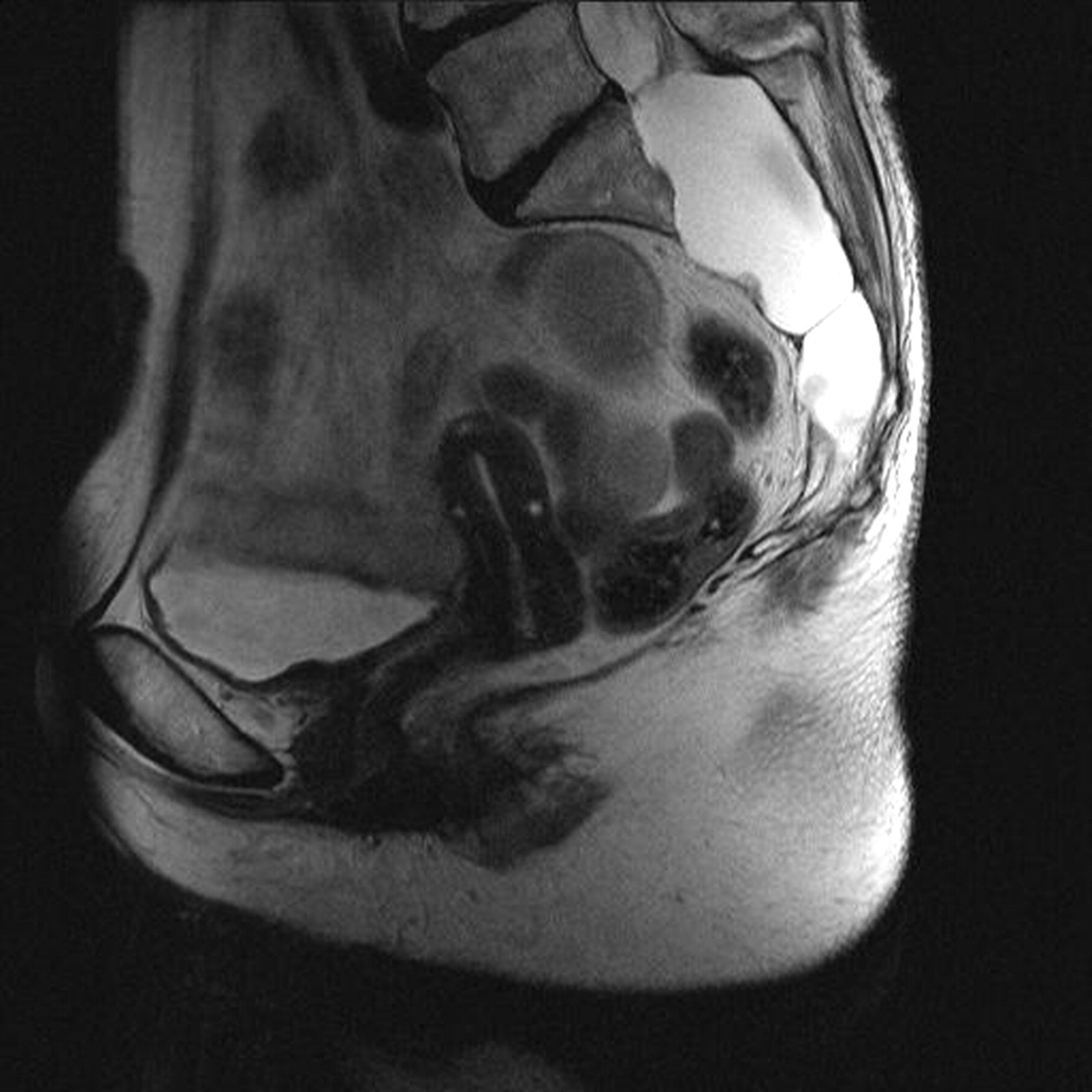
Sagittal T2-weighted MRI pelvis showing a large sacral cyst. The signal in the cyst is exactly the same as cerebrospinal fluid.

Transverse T2-weighted MRI pelvis showing sacral cyst causing widening of sacral central canal.

Lumbar myelogram showing pooling of the contrast within the distal thecal sac. No contrast has entered into the sacral cyst.
Arachnoid cysts are formed from arachnoid membrane expanding through a congenital defect or weakness in the radicular dura towards the epidural space.1 The wall of these cysts does not contain ganglion or nerve fibres. Although the term Tarlov cyst has often been erroneously applied to cystic spinal lesions, the distinctive feature of the Tarlov perineurial cyst is the presence of spinal nerve root fibres within the cyst wall or the cyst cavity itself and communication with the spinal subarachnoid space.2
Intrasacral arachnoid cyst should be considered in the differential diagnosis of low back pain.
Footnotes
Competing interests: none.
Patient consent: Patient/guardian consent was obtained for publication.