Article Text

Statistics from Altmetric.com
Description
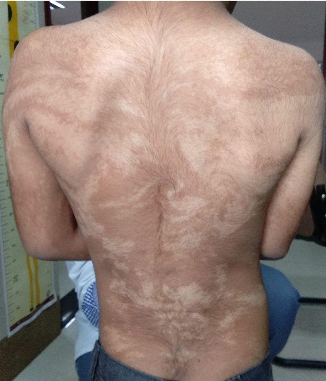
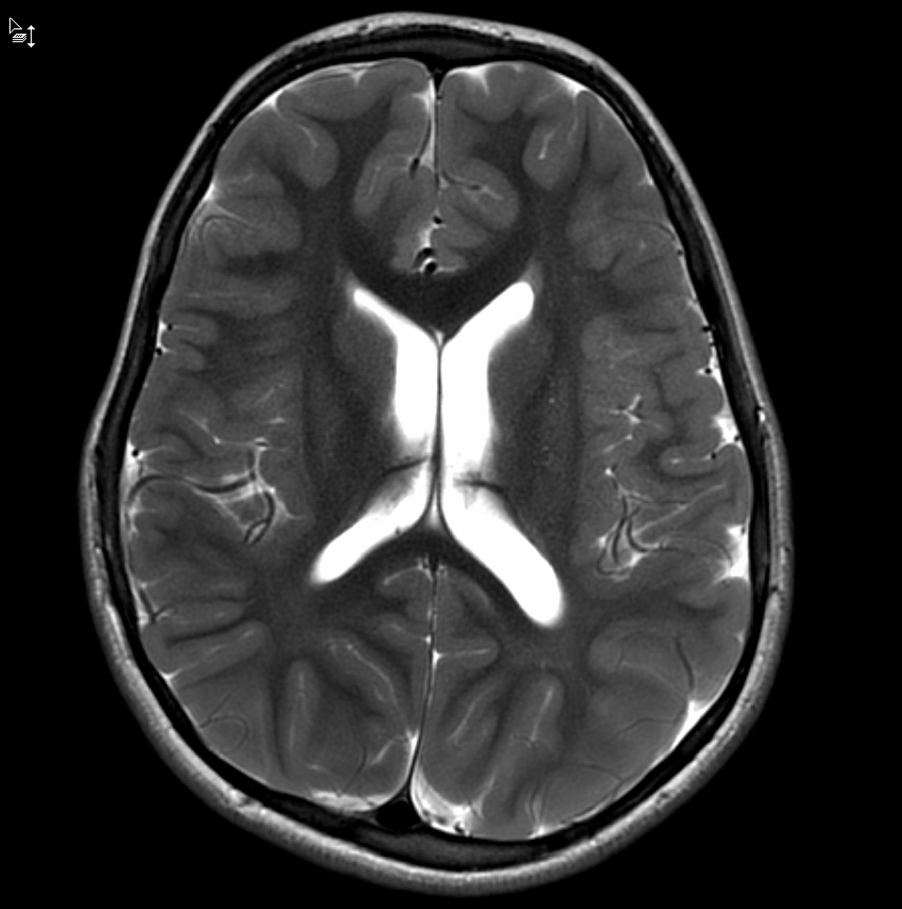
A 6-year-old male child presented to our department with delayed developmental milestones and generalised hypopigmented patches since birth. There was no history of seizures or hearing or visual deficit. Family history was not significant. Examination revealed symmetrical hypopigmented patches arranged linearly, involving predominantly the trunk and upper limbs and were following Blaschko’s lines (figure 1). Anthropometry and the rest of the examination were normal. The child was diagnosed with hypomelanosis of Ito. Hearing assessment was normal. There were no ocular or dental abnormalities. Brain MRI showed mildly enlarged left hemisphere with dilatation of left lateral ventricle (figure 2). The overall IQ of the patient was found to be 46, indicative of moderate level of intellectual disability with specific speech delay. The parents were counselled about the current level of intellectual functioning, asked to give opportunity to the child for learning self-help skills and referred to a speech therapist.

Hypopigmented patches arranged linearly and in whorls along Blaschko’s lines over the back and upper limbs.

Brain MRI showing mild enlargement of left hemisphere with dilatation of left lateral ventricle.
Hypomelanosis of Ito is a rare neuroectodermal disorder often associated with mental retardation and epilepsy. It is characterised by skin abnormalities in the form of unilateral or bilateral cutaneous macular hypopigmented whorls, streaks and patches, corresponding to Blaschko’s lines. Various chromosomal anomalies have been identified in some patients and the current consensus is that the phenotype of hyperpigmentation or hypopigmentation following Blaschko’s lines occurs due to cutaneous mosaicism, either for a monogenic or a chromosomal disorder.1
The nervous system is the most commonly affected system in the form of intellectual disability (70%), seizures (40%), microcephaly (25%) and muscular hypotonia (15%). The musculoskeletal system is the second most frequently involved system, affected by scoliosis and thoracic and limb deformities. Twenty-five per cent of patients have minor ophthalmologic defects (strabismus, nystagmus) and 10% have cardiac defects.2
The brain MRI commonly reveals diffuse white matter abnormalities (>50%) mainly in the parietal, periventricular and subcortical white matter of both hemispheres, either in the form of cystic-like lesions or altered/delayed myelination, generalised cerebral, brainstem, or cerebellar atrophy and/or dilatation of cerebral ventricles and hemispheric asymmetry (both hemimegalencephaly and hemiatrophy).3
Learning points
Cutaneous markers should be looked for in all cases of developmental delay even if there is no history of seizure disorder.
Neurocutaneous disorders like neurofibromatosis, tuberous sclerosis complex, Sturge-Weber syndrome, Von Hippel-Lindau disease, incontinentia pigmenti and hypomelanosis of Ito primarily affect the skin and central nervous system but also affect several other organ systems and can be diagnosed bedside by an observant clinician.
In all neurocutaneous disorders we should screen for musculoskeletal, ophthalmologic, cardiac, hearing, dental and renal abnormalities so that a multidisciplinary approach of management can be planned.
Footnotes
Contributors DK, SS and PG made substantial contributions to the conception and drafting of the work and revising it critically for important intellectual content. All authors gave final approval of the version to be published and agreed to be accountable for all aspects of the work in ensuring that questions related to the accuracy or integrity of any part of the work are appropriately investigated and resolved.
Funding The authors have not declared a specific grant for this research from any funding agency in the public, commercial or not-for-profit sectors. No funding or grant received from any funding agency
Competing interests None declared.
Provenance and peer review Not commissioned; externally peer reviewed.
Patient consent for publication Parental/guardian consent obtained.