
Abstract
Combining multiple materials in a single nanoparticle has gained much attention in recent years. In this work, the optical absorption property of gold-silicon (Au@Si) core–shell nanoparticles (NPs) embedded in a silica matrix were are theoretically demonstrated in the wavelength range from 400 to 800 nm, based on a discrete-dipole approximation method. For a single core–shell nanoparticle, the study revealed the localized surface plasmon resonance (LSPR) showed a regular redshift with an increase in its Si shell thickness. The observed redshifts in the LSPR peaks were in agreement with the experimental results. The optical absorption property was also observed for two Au@Si core–shell NPs separated, on average, by a distance as small as a few nanometers. The results suggest that the shifts in spectral peak position depend on both the interparticle distance and geometric configuration of the nanoparticles. The obtained results also suggest that this nanomaterial, with a strong wavelength-tuneable absorption property, could be an attractive candidate for applications in biomedicine, nanocatalysis, optical devices, and future functional devices.
Export citation and abstract BibTeX RIS

Original content from this work may be used under the terms of the Creative Commons Attribution 4.0 licence. Any further distribution of this work must maintain attribution to the author(s) and the title of the work, journal citation and DOI.
1. Introduction
Combining multiple materials in single nanoparticles has gained much attention in recent years due to the additional functionalities exhibited. Using materials with different properties can result in new materials with characteristics not exhibited in particles of individual materials [1–9]. Spherical nanoparticles (NPs) fabricated from noble metals, such as silver (Ag), gold (Au), and semiconductor materials are a few examples of such composite NPs. They can exhibit extraordinary physical and chemical properties, which find a wide range of applications in nanotechnology [1, 2, 6]. Spherical silicon (Si) NPs, with sizes ranging from 100 nm to 200 nm, show both strong magnetic and electric dipole resonances in the visible and near-IR ranges [10]. Alternatively, noble metals NPs embedded in a dielectric matrix exhibit extraordinary optical resonance, called localized surface plasmon resonance (LSPR). This is due to the collective oscillations of their free electrons with respect to a fixed lattice of positive ions when impinged upon by a light wave. However, these pure metals have some limitations related to high dissipative losses, leading to a large absorption and unwanted heating effects. Therefore, it is interesting to combine Si and Au in one functional nanostructure because Si is photoluminescent while Au is a plasmonic material. The interplay between fluorescence and the plasmonic effect reportedly have significant potential for enhancement of solar cell efficiency [11]. In biomedicine, this system can be used for drug delivery and cancer therapy [12]. However, in this work, the optical absorption property for radiation of wavelengths in the range of 400 to 800 nm of Au@Si core–shell NPs was investigated. First, the effect of Si shell thickness on the absorption property of Au@Si core–shell nanoparticles (NPs) was studied. Furthermore, the effects of interparticle distance between two Au@Si core–shell NPs was also examined. The interparticle distance for the simulation was limited to a few nanometres, as has been previously reported [2]. The discrete-dipole approximation (DDA) method that accounts for optical dipole-dipole coupling between particles was utilized to explain these results. Both single [13, 14] and multiple core–shell nanoparticles [15, 16] have been theoretically investigated by DDA calculations. This study will contribute to a better understanding of the optical absorption property that should be obtained from fabrication of Au@Si core–shell bifunctional nanostructures for use in the emerging fields of nanophotonics, sensing and associated applications.
2. Computational details
The DDA, first introduced by Purcell and Pennypacker [17], is a method for determining the optical properties of materials by subdividing them into an array of point dipoles. In this model, the optical absorption property of the whole structure can be exactly calculated when the coordinates  and polarizabilities of the individual dipoles
  are known. The absorption cross-section can be calculated as [18, 19]:

where  and
and  are electric field and wave vector of the incident light, respectively. While
  is the polarizability of dipole calculated using Kuwata's approximation [20]:

where,  is the volume of the particle,
  is a size parameter,
  for a spherical shape,
  is the wavelength of incident light,
  and
and  are the dielectric functions of pure nanoparticles and the host matrix, respectively. The empirical constants
  and
and  are defined as:
For core–shell nanoparticles,  in equation (2) can be written as [7, 21]:

where,

 and
and  are the dielectric constant of the shell and core, respectively. The geometric parameters,
  and
and  correspond to the core and shell radii of a core@shell particle as denoted in figure 1(a), where core and shell refer to their respective and specific materials. In the DDA method, the target particle was assumed to be a point dipole. Therefore,
  in equation (1) is denoted as the induced polarization of the dipole
  in the presence of a radiating electric field. To obtain this induced polarization at each dipole, the following matrix [18] must be solved:

where the interaction matrix  is given by

and  is the wave vector of the incident light,
  is the dielectric constant of the surrounding medium,
  is the dipole distance between
  and
and 
 is the unit vector along
  and
and  is a 3 × 3 identity matrix. The diagonal element is defined as:


Figure 1. Schematic of (a) a core–shell and the affective sphere and (b) an electric field interacting with two closely spaced Au@Si core–shell NPs; the core has a radius,  and electrical permittivity
  while the shell defined by radius
  and electrical permittivity
 and
and  represent the electrical permittivity of the surrounding medium and particle distance, respectively. The direction of propagation and the polarization of the optical field are also indicated.
Download figure:
Standard image High-resolution imageIn this calculation, the dielectric constants for Au were taken from published reports [22]. The simulations were performed using silica as the dielectric surrounding medium. The modeled samples and parameters are depicted in figure 1.
3. Results and discussion
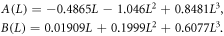
First, the validity of the DDA method for optical absorption spectra was examined as shown in figure 2. The optical absorption produced by a pure single Au spherical nanoparticle with diameters of 10, 30 and 50 nm in silica, shown in figure 2(a), was compared with the exact Mie solution. It was found that the DDA method presents good agreement with the exact Mie absorption efficiency. Resonance absorption peaks were observed at ∼534, ∼536 and ∼546 nm, respectively, for 10, 30 and 50 nm diameter particles. The most significant departure from the exact Mie solution was found with particles larger than 50 nm in diameter. A slight difference in the absorption spectrum was observed using both methods and was observed at wavelengths greater than 600 nm (see figure 2(a)). Moreover, in figure 2, the absorption spectrum of Si nanoparticles that were 10 nm in diameter and embedded in silica are also presented. However, their absorption peak in the visible wavelength range of electromagnetic waves (EMW) was not observed. Enhanced Si visible wavelength range absorption spectra were affected through interactions with Au nanoparticles. First, as shown in figure 2(b), the absorption spectra produced by Au@Si core–shell spherical nanoparticles in the wavelength range from 400 nm to 900 nm of EMW was calculated. The magnitudes of particle core and shell thickness were taken from published experimental data [2]. The Au@Si core–shell nanostructures synthesized by Mohapatra et al [2] show a nanostructure with an average diameter of ∼ 4.6 nm. They consisted of an Au core with an average size (diameter) of ∼ 3.1 ± 5 nm and a nearly spherical Si shell embedded in a silica matrix. In their measured absorption spectrum, they found that the absorption peak exhibits only a single surface plasmon resonance (SPR) peak at around 583 nm. However, the best-fitted curve in the calculation was found in the spectrum produced from Au@Si nanoparticles with a 3.5 nm Au core and a 0.6 nm thick Si shell, shown in figure 2(b). The calculated absorption spectra were clearly in good agreement with the experimental ones in terms of the SPR peak positions. However, the SPR peaks in the measured spectra are broader than those in the calculated spectrum (see figure 2(b)). This is due to the non-spherical and concentrically symmetric core–shell particles. Additionally, there was inhomogeneity in the interparticle distances of the particles of the synthesized materials. Moreover, the irregular shape of the Si shell coating may have impacted the SPR peaks, as shown an earlier transmission electron microscopy study [2]. Moreover, in comparison with pure Au NPs, which exhibited only a single dipole SPR peak at about ∼532 nm, the observed absorption peak shifted to a longer wavelength (from ∼532 nm to ∼584 nm). Furthermore, the correlation between the plasmonic peak position and the dimensions of the Au core, Si shell thickness, and the distance between the two particles are presented. The effect of the Si shell thickness on the plasmonic absorption of Au@Si core–shell NPs (schematic in figure 1(a)) was characterized. The plasmonic absorption spectra of these nanoparticles with a fixed Au core diameter of 3.0 nm with Si shells of varying thickness in figure 3. The absorption cross-section obtained from each sample was normalized by dividing values at various depths by the maximum absorption value along the cross-section ( ). These absorption values are displayed along a wavelength axis. It was found that the spectra all exhibited the same features. The variation of the simulated peak position as a function of the Si shell thickness exhibited an identical trend found in the experimental system (figure 2(a)). Moreover, the SPR peak of the Au@Si core–shell NP presented an obvious redshift to the longer wavelengths of 502, 532, 586, 624 and 654 nm as the thickness of Si shell was increased, respectively, to 0.2 to 0.5, 1.0, 1.5 and 2.0 nm, as is shown in figure 3(a). The wavelength shift can be approximated as the polynomial function (shown in figure 3(b)) given by:

where  is the Si shell thickness. It is well established that the position of the localized SPR is very sensitive to the thickness of the Si shell. Notably, the absorption maximum of pure Au nanoparticles was observed at approximately 532 nm. Coupling theory, developed by Prodan and Nordlander [23, 24], was used to explain the shift in the LSPR peak position. A metallic sphere plasmon from the outer surface of the metallic shell layer was coupled with cavity plasmons produced from the inner metallic shell. This led to a splitting of the LSPR into two new LSPRs, an anti-bonding plasmon (lower wavelength-shifted) and a symmetric plasmon (higher wavelength-shifted).

Figure 2. Calculated absorption spectra of (a) comparison of DDA with exact Mie theory for single Au NPs and (b) pure Si, pure Au, and Au@Si core–shell NPs. The calculated absorption spectrum of pure Si is also presented.
Download figure:
Standard image High-resolution image
Figure 3. Calculated (a) absorbance spectra of a single Au@Si nanoparticle with a fixed 4.6 nm total radius and varying shell thickness of Au from 0.2 nm to 2.0 nm and (b) LSPR peak position as a function of shell thickness.
Download figure:
Standard image High-resolution imageFigures 4 and 5 show the DDA-simulated optical absorption properties of two closely positioned Au@Si core–shell NPs with varying interparticle distances of 5, 6, 7 and 8 nm. The Au@Si core–shell NPs had a fixed Au core of 4.15 nm and Si shell thickness of 0.45 nm. Two different polarization directions of incident EM waves were presented. This plot did not include the data points where the nanospheres were overlapping, which results in the emergence of higher-order resonances. In figure 4, the polarization direction of the incident EM waves was parallel to the interparticle axis (sample in figure 1(b)). In figure 5, it was orthogonal to the interparticle axis (sample in figure 1(b)). It was observed that under parallel polarization, the LSPR slightly redshifted as the interparticle distance was reduced (figure 4 (a)). Conversely, there was a very weak blueshift with decreasing interparticle distance for orthogonal polarization (figure 5(a)). For example, in figure 4(a), it shifted to ∼584, ∼588, ∼592 and ∼600 nm for interparticle distances of 8, 7, 6 and 5 nm, respectively. While in figure 5(a), when the interparticle distance was reduced from 8, 7, 6 and 5 nm, LSPR peak shifted from ∼578 nm to ∼577, ∼576 and ∼574 nm, respectively. Figures 4(b) and 5(b) shown a plot of the simulated LSPR shift as a function of the interparticle distance for the parallel and perpendicular polarizations, respectively. They were found that the absorption peak position shifts nonlinearly with exponential-like decrease function for parallel polarization and exponential-like increase function for the perpendicular polarization. The LSPR shift results from the EM coupling of the single-particle plasmons, the polarization dependence of which can be explained on the basis of a simple dipole-dipole coupling model [25]. To explain the shifting of the resonance peak, the sample sketched in figure 1, was taken into consideration. For the first particle system in figure 1(b), if the polarization direction of the excited electric field was parallel to the particle axis, a weakening of the repulsive forces for the surface charges results. The positive charge of one particle faces the corresponding negative charge of another particle. The induced attractive force between these two charges distributed on different particles weakens the repulsive force within each particle, producing a correspondingly higher resonance wavelength (as shown in figure 4). In contrast, in the particle system of figure 1(b), when the polarization direction of the excited electric field is normal to the particle axis, the charge distributions of both particles act cooperatively to enhance the repulsive action of both particles, thus decreasing the resonance wavelength (shown in figure 5). The second explanation is the plasmon modes coupling theory developed by Prodan et al [23, 24] for an explanation of the physical origin of the tunable plasmon resonance in metal nanoshells and in two NPs systems. In this method, the dimer plasmons can be viewed as bonding and antibonding linear combinations of individual nanosphere plasmons. For the dimer LSPR peaks as a function of interparticle distance, as the interparticle distance is decreased, individual nanosphere plasmons with different angular momentum ( ) hybridize, occurring dimer plasmons with finite dipole moments. This interaction produces extra shifts of the dimer plasmons at small separations, as seen in figures 4(b) and 5(b). For large separations of two composite NPs, the plasmons on the different NPs interact only weakly and the dimer plasmons are essentially bonding and antibonding combinations of the nanoparticle plasmons of the same
  Consequently, it was clearly seen that the decay trend becomes independent of the interparticle distance. Therefore, the calculated results are in good agreement with the dipole-dipole coupling and the plasmon modes coupling theory [23–25].

Figure 4. Calculated (a) absorbance spectra of Au@Si core–shell NPs with varying interparticle distances in the wavelength range of 400–800 nm, and (b) spectral position of the absorption maximum versus interparticle distance of Au@Si core–shell NPs. The polarization direction of the incident light was parallel to the long particle pair axis.
Download figure:
Standard image High-resolution image
Figure 5. Calculated (a) absorbance spectra of Au@Si core–shell NPs with varying the interparticle distances (d) in the wavelength range of 400–800 nm, and (b) spectral position of the absorption maximum versus interparticle distance of Au@Si core–shell NPs. The polarization direction of the incident light was perpendicular to the long particle pair axis.
Download figure:
Standard image High-resolution image
4. Conclusions
The optical properties of plasmonic nanostructures and the optical absorption properties of Au@Si core–shell NPs were studied using the DDA method. First, the calculated absorption spectra showed that the thickness of the Si shell influenced changes in LSPR peak positions. Moreover, the optical absorption property of two Au@Si core–shell NPs, separated a few nanometres, suggests that the shifts in spectral peak position depend on both interparticle distance and geometric configuration. Au@Si core–shell nanocomposite material is expected to be applicable in biomedicine, nanocatalysis, use in optical devices and incorporated into future functional devices.
Acknowledgments
This research project is financially supported by the Integrated Research Group for Energy and Environment (IRGEE), Khon Kaen University, Nong Khai Campus, Nong Khai. The authors also acknowledge the facilities and support of the Faculty of Applied Science and Engineering, Nong Khai Campus, Khon Kaen University and partially funded by the Thailand Center of Excellence in Physics (ThEP) and Faculty of Science, Mahasarakham University, grant number ThEP-61-EQP-MSU2.