


Abstract
SrTiO3 (STO) is one of the key perovskite oxides with fascinating properties in electronics and optoelectronics. The generation and evolution of surface nanostructures induced by femtosecond lasers on STO surfaces are studied, showing distinct features with a clear fluence and pulse number dependence. Unlike regular ripples, which are either parallel or perpendicular to the laser polarization, the ripples coexist in both directions after irradiated with four pulses, and they cover the whole structured area. When increasing the number of laser pulses, the ripples parallel to the laser polarization dominate at a lower fluence regime, while the ripples perpendicular to the laser polarization are the only surface structures formed at a higher fluence regime. The finite-difference time-domain method and the Drude model have been adopted to analyze energy deposition on the STO surface. The results demonstrate that the combination of energy deposition at different excitation levels and the feedback effect is the origin of the formation and evolution behavior of the surface nanostructures on STO.
Export citation and abstract BibTeX RIS
1. Introduction
SrTiO3 (STO, strontium titanate) is one of the key perovskite-structured transition-metal oxides with fascinating properties for fundamental solid-state physics, chemistry, and applications [1, 2]. In the past few years, it has attracted attention after the discovery of 2D electron gas from an LaAlO3/SrTiO3 heterointerface [1, 3], and blue fluorescence light emission at room temperature from an Ar + - irradiated STO [4]. Moreover, STO is a model material to study photocatalytic water oxidation kinetics, such as the first hole transfer process [5]. The interaction between light and STO is crucial to understand these chemical and physical mechanisms.
Femtosecond laser induced periodic surface structures (fs-LIPSS) are a universal phenomenon [6], which have been observed on almost all kinds of materials including metal [7], semiconductors [8–11] and insulators [12]. The formation, evolution and properties of fs-LIPSS as a function of laser parameters serve as a platform to explore the laser-material interaction [13–16]. The interaction between femtosecond/nanosecond lasers and STO has been investigated previously, as well as the formed cracks [17–19]. Femtosecond laser micromachining and controllable self-organized micro-gratings in bulk STO have been realized [20–22]. The dislocation generated in STO by femtosecond laser pulse ablation has been examined [23]. However, the generation, evolution and the mechanism of fs-LIPSS on an STO surface have barely been investigated. Herein, we report the pulse number and fluence dependence of fs-LIPSS on an STO surface. The finite-difference time-domain (FDTD) method and the Drude model have been adopted to discuss the mechanism involved in the surface structures' formation.
2. Experimental setup
A double-sided polished single crystal STO wafer (100, 10 mm × 10 mm × 0.5 mm) was obtained from Shanghai Daheng Optics and Fine Mechanics Co. Ltd. The sample was cleaned by acetone and methanol in turn in an ultrasonic bath for 5 min, followed by distilled water rinsing. Then it was dried with nitrogen blowing prior to laser irradiation. The Ti: sapphire laser output femtosecond laser pulses had about 70 fs duration at a center wavelength of 800 nm and a repetition rate of 20 Hz. The laser beam was focused by a 0.2 m focal length lens irradiated normally to the STO surface. The spot of the beam has a spatially Gaussian intensity distribution with a radius of ωx = 113 µm, ωy = 125 µm (as measured at 1/e2 of the maximum intensity with a beam profiling system, Thorlabs, BC106N-VIS/M) on the sample surface. The laser pulse energy was fixed, which gave a fluence of 20.3 kJ m−2 in the center of the beam. The number of laser pulses was controlled by a mechanical shutter (Thorlabs, SH1). The surface morphology of irradiated STO was analyzed by using a scanning electron microscope (JSM-7500F) and an atomic force microscope (Asylum Research, MFP-3D). The sample was further characterized by a confocal Raman spectrometer (XperRam, Nanobase). A 20 mW 532 nm laser and an objective lens with a magnification of 40 were employed in Raman measurement.
3. Results and discussions
After being irradiated with four femtosecond laser pulses, a structured circular area formed. As the intensity of the laser has a Gaussian distribution, the local fluences changed as a function of position. The surface structures at the periphery and the center of the structured area, which corresponds to a local fluence of 10.6 kJ m−2 and 20.3 kJ m−2, are shown in figures 1(a) and (b), respectively. Figures 1(c) and (d) display the magnification of the selected area in figure 1(a). Horizontal and vertical fs-LIPSS, which are parallel (denotes as ripples//) and perpendicular (denotes as ripples⊥) to the laser polarization with periodicities at 486 ± 50 nm and 426 ± 58 nm, respectively, can be observed. Similar features also present in figures 1(b), (e) and (f), which are the surface structures in the center of the beam. The periodicities are 580 ± 80 nm and 527 ± 53 nm for ripples// and ripples⊥, respectively. The Fourier transforms of figures 1(a) and (b) (insets in figure 1) both show elliptic shaped features. The longer axis is parallel to the laser polarization in both cases, indicating the periodicities of ripples⊥ are smaller. Meanwhile, the periodicity of the fs-LIPSS formed in the center of the structured area (corresponding to a higher excitation level) is greater than those formed at the periphery. We notice that micro-meter-size cracks, which are usually formed by nanosecond laser irradiation because of the poor thermal conductivity of STO [17], can be observed in all of the structured area.

Figure 1. SEM images of an STO surface after being irradiated with four laser shots: (a) periphery and (b) center of the structured area; (c) and (d) magnification of the selected area in (a); (e) and (f) magnification of the selected area in (b). The insets in (a) and (b) are the Fourier transforms of (a) and (b), respectively. (c)–(f) share the same scale bar displayed in (f). The arrow indicates the direction of the laser polarization.
Download figure:
Standard image High-resolution imageAfter being irradiated with ten laser pulses, ripples// with a periodicity at 569 ± 43 nm are the dominate structure on the periphery of the structured area, while ripples⊥ disappears, as shown in figure 2(a). Correspondingly, both ripples// and ripples⊥ with periodicities at 691 ± 143 nm and 498 ± 39 nm, respectively, can be observed in the center of the structured area, as shown in figure 2(b). After being irradiated with 15 laser pulses or more, ripples// with changed periodicities are the only surface structures that exist on the periphery of the structured area. Meanwhile, the surface structures in the center of the structured area change dramatically after being irradiated with 15 laser pulses. Ripples// vanish, while very clear ripples⊥ with a periodicity of about 490 ± 46 nm dominate. The structures in the center of the structured area after being irradiated with 50 femtosecond laser pulses are illustrated in figure 2(d). Straight and smooth ripples⊥ are clearly presented. The corresponding surface structures formed at the periphery and the center of the structured area after being irradiated with 4, 10, 15, 50 femtosecond laser pulses are also analyzed by AFM, shown in figure 3. The same features as the one shown in figures 1 and 2 are clearly presented. Selective height profiles show the periodicities of the formed surface structures.
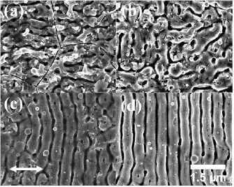
Figure 2. SEM image of the (a) periphery and (b) center of the structures area formed with ten laser pulses; center of the structures area formed with (c) 15, (d) 50 laser pulses. The arrow indicates the direction of the laser polarization.
Download figure:
Standard image High-resolution image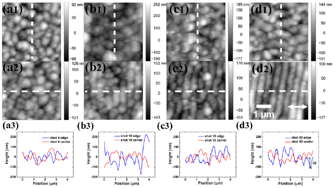
Figure 3. AFM images of the surface structures formed at the periphery after being irradiated with (a1) 4, (b1) 10, (c1) 15, (d1) 50 laser shots; the center after being irradiated with (a2) 4, (b2) 10, (c2) 15, (d2) 50 laser shots; selective profile along the dash line after being irradiated with (a3) 4, (b3) 10, (c3) 15, (d3) 50 laser shots. The arrow indicates the direction of the laser polarization.
Download figure:
Standard image High-resolution imageFigure 4(a) shows the pulse number dependence of the average periodicities of the surface structures both at the periphery and center of the structured area. Unlike the periodicities of ripples observed in the other material, which usually decreases or keeps the same as the number of the pulses increases, the periodicities of ripples// at the periphery shows a unique behavior. It increases when the laser pulses are less than or equal to ten. It declines with more pulses after ten shots. Similarly, the periodicity of the ripples// in the center of the structured area increases as the pulse number increases, when it is less than or equal to ten, until it disappears. The periodicity of ripples⊥ in the center of the structured area decreases as the pulse number increases. The cracks can be observed on all the ripples. Moreover, the structured area increases with more irradiation of the laser pulses. Since ripples// can always be observed at the periphery of the structured area and the fact that the beam has a Gaussian distribution, the increased structured area as the number of pulses increases indicates an incubation effect [24, 25]. The average height of the surface structures (displayed in figure 3(d3)) as a function of the laser pulse numbers are shown in figure 4(b). The surface structures are deeper in the center compared with the one at the periphery of the structured area, formed with four femtosecond pulse irradiations. The height keeps almost constant with more laser pulses in the center of the structured area. It increases in the beginning and decreases when the shot numbers are more than 15 for the structures at the periphery.
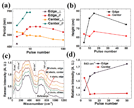
Figure 4. (a) Pulse number dependence of the average periodicity of the surface structures; (b) average height of the surface structures as a function of pulse number; (c) Raman spectra of surface structures formed at different region of the structured area with different laser shot numbers; (d) the intensity at 543 cm−1 of Raman spectra as a function of pulse number.
Download figure:
Standard image High-resolution imageThe Raman spectra of the surface structures formed at the periphery and center of the crater after irradiated with 4 and 50 laser shots are shown in figure 4(a). The Raman spectrum of the un-irradiated STO is shown for comparison. The main features of the Raman spectra of the reference and irradiated STO surface agree well with the predictions that second order scattering should dominate the Raman spectrum of the STO at room temperature, since first order Raman activity is forbidden because all the zone-center optical phonons are of an odd symmetry [26]. Two broad bands in the ranges of 200–400 cm−1 and 600–800 cm−1 can be clearly observed. Additionally, a low frequency band at 79 cm−1 is also presented. Peaks at 251 cm−1, 303 cm−1, 623 cm−1, 688 cm−1, 721 cm−1 originate from additive combinations, and overtones can be identified. After being irradiated with four femtosecond laser shots, a peak at 543 cm−1 and two broad shoulders at 176 cm−1 and 797 cm−1 show up. These three features correspond to TO4, TO2 and LO4 phonons, respectively, which all belong to the first order scattering [27]. The activation of the first order Raman scattering of the irradiated STO surface indicates the breakdown of the inversion symmetry of STO in the structured area because of the defects created in the processes of the surface structure formation. The features become weaker in the center of the structured area, while becoming more intense at the periphery when the laser pulse is increased to 50. The intensity of the peak relative to the reference at 543 cm−1 is extrapolated as an indicator of the density of the defect, shown in figure 4(d). It is more intense in the center after it irradiates with four laser shots and decreases as the pulse number increases to ten. Then it keeps almost constant. This phenomenon illustrates that, for the data shown here, the density of the defect of the surface structures in the center of the structured area irradiated with four laser shots is the highest. The defects become less after being irradiated with successive laser pulses. The intensity of the peak at 543 cm−1 of the Raman spectrum of the surface structures at the periphery increases monotonously as the number of pulses rises, which reveals the increase in the density of the defects.
The classic Sipe-Drude model [28, 29] can well explain the formation of ripples, especially the low spatial frequency fs-LIPSS. The excitation of the surface plasmon polariton has been included in this model [30]. Recently, the combination of the Drude model and the FDTD method has attracted attention because of its flexibility to analyze the formation of the low spatial frequency fs-LIPSS, the high spatial frequency fs-LIPSS and even micrometer-size grooves in the framework of the electromagnetic approach [8, 9, 31, 32]. To further investigate the origin of the fluence and the pulse number dependence of the surface structures on STO, this method has been adopted to discuss the energy deposition at different excitation levels. The FDTD-η maps are utilized to investigate the non-uniform energy deposition when the femtosecond laser light interacts with the material's rough surface to interpret the origin of the ripples. The efficacy factor η in an FDTD-η map describes the efficacy of the inhomogeneous absorption at k (wave vector) at a specific depth.
SrTiO3 has an indirect band gap of 3.25 eV and a direct gap of 3.75 eV. The critical role of the transient change of the dielectric function of the material upon the excitation of a femtosecond laser in the formation of fs-LIPSS has been demonstrated previously [33, 34]. The transient change of the dielectric function caused by the response of free carriers can be calculated by the Drude model [35]
in which  is the dielectric constant in vacuum;
  is 1.17, denoting the optical effective mass of the carrier [36];
  is the mass of the electron; e is the electron charge; ω is the angular frequency; and
  (Drude damping time) is 3.86 fs, calculated by
  , in which µ is the conductivity (5.8 cm2 V−1 s−1) at room temperature [37]. The density of the excited carriers N can be estimated by
 ![$N\approx F(1-R)[\alpha +\frac{\beta F(1-R)}{2\sqrt{2\pi }\tau }]/h\upsilon $](https://content.cld.iop.org/journals/1612-202X/16/5/056007/revision1/lplab139dieqn006.gif) , in which F is the fluence and α and β are the linear and two-photon absorption coefficients, respectively [30]. R is the surface reflectivity and τ is the pulse duration. The refractive index of the unexcited STO, obtained by an angle-dependent reflection measurement and a numerical inversion of the Fresnel formulas procedure, is 3.05 at 800 nm [38]. The linear absorption coefficient is 18.43/m attained by Lambert-beer's law. A single-beam femtosecond laser Z-scan has been conducted [39]. The approach proposed by He has been used to distinguish the order of non-linear absorption [40]. The experimental measured two-photon absorption coefficient is 4.73 × 10⁻¹² m W−1. The change of the dielectric function caused by band-structure renormalization and band filling is neglected.
, in which F is the fluence and α and β are the linear and two-photon absorption coefficients, respectively [30]. R is the surface reflectivity and τ is the pulse duration. The refractive index of the unexcited STO, obtained by an angle-dependent reflection measurement and a numerical inversion of the Fresnel formulas procedure, is 3.05 at 800 nm [38]. The linear absorption coefficient is 18.43/m attained by Lambert-beer's law. A single-beam femtosecond laser Z-scan has been conducted [39]. The approach proposed by He has been used to distinguish the order of non-linear absorption [40]. The experimental measured two-photon absorption coefficient is 4.73 × 10⁻¹² m W−1. The change of the dielectric function caused by band-structure renormalization and band filling is neglected.
Similar fluence and pulse number dependence of surface structures induced by a femtosecond laser have been observed on wider band gap transparent insulators, like silica [41] and glasses [42]. Their theoretical analysis based on the Sipe-Drude model show that energy deposition at different excitation conditions and incubation effects is the origin of the fluence and pulse number dependence. To investigate the energy deposition for the first several pulses on the STO surface, we use the binary function proposed by Skolski et al [32] to generate the random surface roughness. The dimensions of the Yee cells are 40 nm × 40 nm × 20 nm in X, Y and Z directions, respectively. The lateral dimensions of the calculated area are set to 19 µm × 19 µm which contains 23 wavelengths of the femtosecond laser. The filling factor is set to 0.1, consistent with the traditional Sipe-Drude model. We chose a linearly polarized plane wave, propagating along the Z direction as the light source. To reduce the noise in the FDTD-η maps, an average of 10 FDTD-η maps calculated using different binary functions have been performed. Features in the frequency domain are described by the notations proposed by Skolski et al [31, 32], namely type-r, type-s and type-d features. A schematic representation for the FDTD-η map is shown in figure 5(e). The type-s feature corresponds to the formation of ripples⊥ with a periodicity close to the laser wavelength; while the type-r feature represents the energy deposition at the same direction with a much smaller periodicity. The type-d feature can be connected to the periodical structures with a periodicity of λ/Re(n) and the formation of ripples//, in which λ is the wavelength of the laser. Type-g produces features exhibiting periodicity on the order of micrometers.

Figure 5. FDTD-η maps 80 nm below the surface of STO, irradiated with (a) 10.6 kJ m−2; (b) 20.3 kJ m−2; (c) 24 kJ m−2 and (d) 30 kJ m−2 at the rough surface. The black dotted circles indicate the position with a normalized wave vector equal to one and two. The arrow shows the direction of the laser polarization. (e) The schematic representation for the FDTD-η map [32]. (f) The real and imaginary part of the dielectric function of STO upon the excitation at elevated fluences. The dashed line represents −1. (g) The extrapolated periodicity of fs-LIPSS parallel to the laser polarization in FDTD-η maps (corresponding to type-d feature) is a function of fluences.
Download figure:
Standard image High-resolution imageThe FDTD-η map, 80 nm below the STO surface, irradiated with 10.6 kJ m−2 (periphery of the structured area, corresponding to a dielectric function of 7.673 + 0.179i) is shown in figure 5(a). Three different kinds of features can be clearly observed, namely type-s, type-r and type-d features. Type-r and type-d features merge together to form an elliptical structure, similar to the one shown in the inset of figure 1(a). We should notice that the type-s feature also contributes to the formation of ripples⊥. Figure 4(b) shows the FDTD-η map of the STO irradiated with 20.3 kJ m−2 (center of the structured area, corresponding to dielectric functions 3.3249 + 0.6572i). Figure 4(a) shows the similar features and the experimental observation in the inset of figure 1(b). The coexisting of features in both vertical and horizontal directions, shown in figures 5(a) and (b), indicates that the modulation of the energy deposition are in both directions, which well explains the coexisting of ripples// and ripples⊥, as observed in figure 1. We notice that the elliptical structure shrinks at a higher fluence. The peak of the type-d features shift towards smaller wave vectors indicates that the periodicity of the periodical surface structures parallel to the laser polarization increases, which is consistent with the experimental results. The periodicity of ripples⊥ cannot be extrapolated in these two situations because type-s and type-r features have an almost identical intensity and the energy deposition connected with these two features contributes to the formation of ripples⊥.
The Sipe-Drude model, and previously published results based on FDTD simulations, have proved the importance of the inter-pulse feedback mechanism on the evolution of surface structures [13, 31, 43]. The pre-formed surface structures have two main feedback roles in the evolution of surface structures when the consecutive laser pulse irradiates the sample: local fluence enhancement [8, 9] and the modulation of energy deposition [8, 9]. The incubation effect mentioned above, which mainly originates from the defects and color centers induced by femtosecond lasers, can enhance the absorption. This effect is equivalent to a higher effective fluence. The real part and the imaginary part of the dielectric function of STO upon the excitation of different fluences are shown in figure 5(f). The real part of the dielectric function decreases while the imaginary part increases at elevated excitation levels. The real part of the dielectric function is less than −1 when the effective fluence is higher than 26.7 kJ m−2, which indicates the material's behavior is like metal due to the femtosecond laser induced high density of the quasi-free electrons in the conduction band.
The FDTD-η map of STO irradiated with 24 kJ m−2 (corresponding to a dielectric function of 0.95 + 0.92i) is shown in figure 5(c). Type-s and type-r features merge together while the intensity of the type-d features decreases significantly. The type-g feature, which corresponds to the formation of micro-meter-sized grooves, is clearly presented. However, the grooves have not been observed experimentally. When the effective fluence is further increased to 30 kJ m−2 (corresponding to the dielectric function −3.75 + 1.44i), the material becomes metallic. The type-d feature vanishes while the type-s feature dominates, as shown in figure 5(d). These features indicate that ripples⊥ tend to dominate with successive laser pulse irradiations at a higher fluence regime, consistent with the experimental observation that the vertical ripples, which are perpendicular to the laser polarization, are the dominant surface structures formed in the center of the structured area. Since the intensity of the type-d feature is more pronounced in figures 5(a) and (b), the accumulation of the energy deposition of successive laser pulses tends to form ripples//-dominated surface structures, which explains the disappearance of ripples⊥ at the periphery of the structured area.
The discussion above is from the viewpoint of local effective fluence enhancement, while the feedback effect on the modulation of energy deposition because of surface structural change has not been included. To further analyze the feedback effect in the evolution of surface structures, the AFM images, which have the 3D information of the surface structures formed after irradiation of four and ten laser shots, are used for the FDTD simulation. The FDTD-η maps 80 nm below the original surface, which describes the energy deposition of the successive laser pulse on the periphery and center of the structured area formed after the irradiation of four laser shots, are shown in the first two rows in figure 6. The FDTD-η maps are similar at the same effective fluences, indicating that the feedback effect from the surface structures in these two areas is similar. The difference in the evolution of the surface structures, when irradiated with successive laser pulses on the surface structures after four laser shots, are mainly from the difference of the effective fluence. We should keep in mind that the fluence in the flat surface in these two areas are 10.6 kJ m−2 and 20.3 kJ m−2, respectively. In the effective fluence range from 10 kJ m−2 and 20 kJ m−2 shown here, the type-s and type-r features almost vanish, while the type-d feature is most pronounced (compared with those shown in figure 5), suggesting that ripples// dominates, which can well explain the experimental observations that only the ripples parallel to the laser polarization exist after several femtosecond laser pulses irradiate at the periphery. We notice that the type-d feature is more concentrated at a higher effective fluence, which indicates a bigger periodicity, consistent with the simulation shown in figure 5 and the experimental results shown in figure 4(a). The type-s and type-r features merge together when the fluence is 24 kJ m−2, indicating that the energy deposition is modulated perpendicular to the laser polarization. The type-g feature becomes pronounced, but no grooves form experimentally. The FDTD-η maps are featureless in these two cases when the effective fluence increases to 30 kJ m−2. Upon the energy deposition manner discussed above, the surface structures evolve gradually to become the one shown figures 2(a) and (b) after being irradiated with ten laser shots.

Figure 6. FDTD-η maps 80 nm below the original surface of STO irradiated with different effective fluences. The black dotted circles correspond to the position with a normalized wave vector equal to one and two. The arrow indicates the direction of the laser polarization.
Download figure:
Standard image High-resolution image
After the surface structures formed with ten laser shots (as shown in figures 2(a) and (b)), the FDTD-η maps for the subsequent laser pulses are shown in the last two rows in figure 6. At a fluence range from 10–20 kJ m−2, the FDTD-η maps of the periphery of the structured area show similar features to those in figures 6(a1), (a2) and (b1), (b2) in which the type-d feature dominates corresponding to the formation of ripples//. The type-g feature becomes the most pronounced, while the type-s and type-r features disappear compared with figure 6(c3) when the effective fluence increased to 24 kJ m−2. This change is from the different feedback effect from the different surface structures, formed after irradiating with four and ten laser shots. The FDTD-η map is featureless when the effective fluence increased to 30 kJ m−2, as shown in figure 6(c4). For the center of the structured area after irradiation with ten laser shots, we focus on the FDTD-η maps when the effective fluences are 24 kJ m−2 and 30 kJ m−2, as shown in figures 6(d3) and (d4), respectively. Type-g feature dominates while type-s and type-r feature start to occur in figure 6(d3). The type-s feature become the most pronounced when the effective fluence increases to 30 kJ m−2, while the type-g feature becomes weaker, which can well explain the phenomenon that the ripples perpendicular to the laser polarization, with a periodicity around 500 nm, dominate the surface structures formed in the center of the structured area after the irradiation of successive femtosecond laser pulses. We can conclude that, at the edge of the structured area, the effective fluence is the main factor, which determines the generation and evolution of the surface structures. In the center of the structured area, the effective fluence dominates initially and the feedback effect from the pre-formed surface structures gradually becomes more important as the pulse number increases and plays a major role in the formation and evolution of the surface nanostructures. The comparison of the FDTD-η maps from the top to the bottom of figure 6 shows that the feedback effect of the modulation of the energy deposition from the pre-formed surface structures is more sensitive at a higher fluence.
An ablation threshold of about 12 kJ m−2 has been reported for polycrystalline STO [23]. The ablation threshold for our sample is expected to be higher because of the lower two-photon absorption coefficient [44]. The fluence in the center of the structured area is higher, which can reach the ablation threshold prior to the fluence at the edge. Deeper surface structures with higher density of defects are generated in the center of the structured area after being irradiated with four laser shots, as shown in figures 4(b) and (d). The absorption of the subsequent laser pulse increases as the surface structures are formed. The melting time is extended, accompanied by a slower re-solidification velocity, which allows the epitaxy growth of the material to a better crystallinity [45]. Meanwhile, less nanoparticles can be observed on the surface. These two effects both contribute to the reduction of the Raman signal at 543 cm−1 with more irradiation of laser pulses, as shown in the Raman measurement in figures 4(c) and (d). The fluence is lower at the periphery of the structured area. The effective fluence in the valley of the ripples can be enhanced to above the ablation threshold easier than ridges, leading to deeper ripples formed with more defects with follow-up laser pulses (figures 4(b) and (d)) [9].
The periodicity of ripples either decreases or keeps the same in the previously reported results [13, 43]. Inter-pulse feedback from the existed structures has been considered the main mechanism of periodicity shrinkage. However, the pulse number dependence of ripples// shows a different behavior in our work, as shown in figure 4(a). The periodicity increases as the pulse number rises when the pulse number is less or equal to 10 for the whole structured area. As mentioned earlier from the FDTD simulations, the difference of the surface structures is mainly driven by the fluence dependence for the first several pulses. The peak position of the type-d feature in figure 5, which corresponds to the formation of ripples//, has been extrapolated. The corresponding periodicities are shown in figure 5(g), which increases monotonously when the effective fluence increases from 10–24 kJ m−2. The observed experimental increases in the periodicity of ripples// can be understood by considering this result with the fact that the effective fluence increases as the pulse number rises. The inter-pulse feedback mechanism dominates the evolution of the ripples after ten laser shots, in which a decrease of the periodicity can be observed.
4. Conclusion
In summary, we studied the generation and evolution of the surface nanostructures on the STO surface, induced experimentally by a femtosecond laser. The surface nanostructures show distinct features with a clear fluence and pulse number dependence. Unlike the regular ripples, which are either parallel or perpendicular to the laser polarization, the ripples with directions parallel and perpendicular to the laser polarization coexist and cover the whole structured area after irradiated with four laser pulses. When the pulse number is further increased, ripples// gradually dominates at a lower fluence regime, while ripples⊥ are the only surface structures formed at a higher fluence regime. The periodicities of ripples// and ripples⊥ show different behaviors. The simulation results obtained by combining the FDTD method and the Drude model show that the difference of the surface structures for the first several pulses is mainly driven by fluence dependence. In the beginning, energy absorption was modulated in both vertical and horizontal directions, causing the coexistence of ripples// and ripples⊥. The effective fluence increases as the number of the pulse rises by considering the local enhancement and the incubation effect, leading to a different energy deposition manner and surface structures. The inter-pulse feedback effect gradually becomes more important as the pulse number increases, playing a major role in the formation and evolution of the surface nanostructures. This work can benefit in both the fundamental study on surface nanostructure formation and the deep understanding of the mechanism of femtosecond laser-STO interaction.
Acknowledgment
The authors would like to thank Dr. Chao Yang for his contribution on the SEM measurement.
This work was supported by the National Natural Science Foundation of China (Grant Nos. 61705149, 61705148 and 61505129).