

Abstract
Recently, ICRP Task Group 103 developed new mesh-type reference computational phantoms (MRCPs) for the adult male and female by converting the current voxel-type reference computational phantoms (VRCPs) of ICRP Publication 110 into a high-quality/fidelity mesh format. Utilizing the great deformability/flexibility of the MRCPs compared with the VRCPs, in the present study, we established a body-size-dependent phantom library by modifying the MRCPs. The established library includes 108 adult male and 104 adult female phantoms in different standing heights and body weights, covering most body sizes representative of Caucasian and Asian populations. Ten secondary anthropometric parameters with respect to standing height and body weight were derived from various anthropometric databases and applied in the construction of the phantom library. An in-house program for automatic phantom adjustment was developed and applied for practical construction of such a large number of phantoms in the library with minimized human intervention. Organ/tissue doses calculated with three male phantoms in different standing heights (165, 175, and 190 cm) with a fixed body weight of 80 kg for external exposures to broad parallel photon beams from 0.01 to 104 MeV were compared, observing there are significant dose differences particularly for the photon energies <0.1 MeV in which the organ/tissue doses tended to increase with increasing standing height. In addition, the organ/tissue doses of three female phantoms in different body weights (45, 65, and 140 kg) with a fixed standing height of 165 cm were compared, showing a significant decreasing tendency with increasing body weight for the photon energies <10 MeV. For the higher energies, the opposite trend, interestingly, was observed; that is, the organ/tissue doses tended to increase with increasing body weight. The results, despite the limited number of exposure cases, suggest that the use of the body-size-dependent phantom library can improve the accuracy of individual dose estimates for many retrospective dosimetry studies by taking the body size of individuals into account.
Export citation and abstract BibTeX RIS
1. Introduction
The International Commission on Radiological Protection (ICRP), following the most recent recommendations of ICRP Publication 103 (ICRP 2007), released a set of adult male and female reference computational phantoms as described in ICRP Publication 110 (ICRP 2009) to produce dose coefficients of organ/tissue doses and effective doses for external and internal exposures (ICRP 2010, 2015, 2016b, 2017). The ICRP Publication 110 reference phantoms are voxel models constructed from human computed tomography (CT) data and adjusted for consistency with the morphological and physiological parameters of Reference Adult Male and Female provided in ICRP Publication 89 (ICRP 2002). The concept of a 'reference person' used in the ICRP system is beneficial in standardizing prospective dose assessments for radiological protection purposes. In retrospective dose reconstructions, however, the reference phantoms in a fixed anatomy can be used only as an initial-step approximation to estimate radiation doses of exposed individuals, because the estimated doses can be significantly biased when the anatomy of the individuals is largely different from that of the reference phantoms. Meanwhile, individual dose reconstructions are technically challenging, since individual anatomy data such as CT or magnetic resonance (MR) images are mostly unknown. Even if anatomy data are available, constructions of individual phantoms require a labor-intensive and time-consuming process of organ/tissue contouring, which makes it practically difficult to conduct individual dose reconstructions of a large-scale cohort involved in epidemiologic investigation for risk assessment (Geyer et al 2014). It could be considered as a practical solution to pre-generate a number of phantoms in various body heights and weights by modification of existing phantoms, and then to use the one that best fits the individual in dose estimate (Johnson et al 2009, Na et al 2010, Broggio et al 2011, Cassola et al 2011, Segars et al 2013, 2015, Geyer et al 2014, Xie et al 2017, Akhavanallaf et al 2019).
Recently, ICRP Task Group 103 developed new adult male and female mesh-type reference computational phantoms (MRCPs) by converting the voxel-type ICRP-110 reference phantoms to high-quality/fidelity mesh format (Kim et al 2018). The MRCPs are the mesh counterparts of the ICRP-110 reference phantoms, not only preserving the original anatomy but also overcoming the limitations of the voxel phantoms due to their voxel resolutions and the nature of voxel geometry. The MRCPs, for example, represent the micron-scale radiosensitive target and source regions in the skin, lens, urinary bladder, and respiratory and alimentary tract systems (Nguyen et al 2015, Yeom et al 2016a, Kim et al 2017). These micron-scale structures could not be defined in the ICRP-110 voxel phantoms at the given millimeter-scale voxel resolutions. Another notable benefit of the MRCPs is that their mesh geometry is highly deformable/flexible relative to the voxel geometry (Kainz et al 2019); therefore, one can easily modify the reference phantoms to create new phantoms in various postures and/or body sizes, expanding their capabilities for individualized dose reconstructions. The feasibility of this benefit was shown by (Lee et al 2019), who modified the MRCPs in constructing 18 adult male and female percentile-specific phantoms representative of the 10th, 50th, and 90th percentile standing heights and body weights of the Caucasian adult population.
As an extension of the work of (Lee et al 2019), in the present study, we established, by modification of the MRCPs, a comprehensive library of body-size-dependent phantoms comprising a total of 212 adult male and female phantoms (108 males and 104 females) in different standing heights and body weights. The established library covers a wide range of body sizes of most individuals representative of the Caucasian and Asian populations. A subset of the phantom library was implemented into a Monte Carlo radiation transport code to calculate organ/tissue doses for external photon exposures. At this time, unlike previous phantom libraries (Geyer et al 2014, Xie et al 2017, Akhavanallaf et al 2019) presented in the voxel format for implementation in Monte Carlo codes, the phantoms in the new library were directly implemented into Monte Carlo codes, fully maintaining the accuracy of the high-fidelity mesh geometry. The calculated organ/tissue doses among the phantoms in different standing heights and body weights were compared to investigate the impact of body size on dose calculations. In addition, the organ/tissue doses based on the MRCP-based phantom library were compared with those based on another body-size-dependent phantom library (Geyer et al 2014) to investigate dosimetric variation between the two libraries.
2. Materials and methods
2.1. Mesh-type reference computational phantoms (MRCPs)
Figure 1 shows the adult male and female MRCPs that were modified for the construction of the body-size-dependent phantom library. The heights and weights of the MRCPs are consistent with the reference values (i.e. male: 176 cm and 73 kg; female: 163 cm and 60 kg) (ICRP 2002). The MRCPs are in the tetrahedral-mesh format composed of ∼8.2 and ∼8.6 million tetrahedrons for the male and female phantom, respectively. The MRCPs are also given in the polygonal-mesh format for users who are interested in deforming the phantoms; in this case, the male and female phantoms are composed of ∼2.5 and ∼2.6 million triangle facets, respectively. The MRCPs contain 48 organs and tissues with 170 subregions, including all of the target and source regions required for effective dose calculation. The masses of all organs/tissues are matched to the reference values inclusive of blood contents (ICRP 2002, 2016a). Note that the MRCPs in the tetrahedral-mesh format can be directly implemented in general-purpose Monte Carlo codes such as Geant4 (Allison et al 2016), MCNP6 (Goorley et al 2012), and PHITS (Sato et al 2013), which is to say, without the voxelization process conventionally used for other mesh phantoms (Zhang et al 2009, Cassola et al 2010), fully maintaining the advantages of the mesh geometry in dose calculations (Yeom et al 2014, Kim et al 2018). Detailed information on the MRCPs can be found elsewhere (Kim et al 2018).

Figure 1. Mesh-type reference computational phantoms (MRCPs): adult male (left) and female (right).
Download figure:
Standard image High-resolution image2.2. Determination of anthropometric parameters
Target height–weight grids and ten secondary anthropometric parameters were determined based on various anthropometric data. The height–weight grids were determined using the Continuous NHANES (1999–2014) database (https://www.cdc.gov/nchs/nhanes/index.htm) for adult male and female subjects aged from 20 to 50 years, as considered in the ICRP reference data (ICRP 2002). The determination was conducted by using an approach similar to that of (Geyer et al 2014). The subjects were binned by height increments of 5 cm from the 5th to 95th height percentiles in Gaussian distribution. Each height bin was parsed by weight increments of 5 kg, maintaining at least 10 subjects in each bin to assure statistical significance. The height–weight grids based on the NHANES database cannot cover some Asian people with very small body sizes. Therefore, we considered additional height–weight grids that had been determined based on the 7th Korean National Anthropometric Survey data (http://sizekorea.kr/). Figure 2 shows the determined height-weight grids containing 108 and 104 bins for adult males and females, respectively.
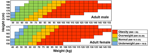
Figure 2. Height–weight grids for phantom library: adult male (upper) and female (lower).
Download figure:
Standard image High-resolution imageTen secondary anthropometric parameters (i.e. sitting height; head height, depth, and breadth; sagittal abdominal diameter; and upper-arm, waist, buttock, thigh, and calf circumferences) were determined by deriving regression equations as a function of height and weight from various anthropometric databases. The equations for the sagittal abdominal diameter and upper-arm, waist, thigh and calf circumferences were derived from the Continuous NHANES (1999–2014) database. The equations for the sitting height and buttock circumference, which are unavailable in the Continuous NHANES (1999–2014) database, were derived from the NHANES III (1988–1994) database. The equations for the head dimensions (i.e. head height, depth, and breadth), which are unavailable in the NHANES databases, were derived from other databases. That is, the data of the PeopleSize 2008 Professional software (https://www.openerg.com/psz/) were used for the head height, and the U.S. Army Anthropometric Survey (ANSUR II) database (Gordon et al 2014) was used for the head depth and breadth. The derived regression equations for all of the parameters are shown in table 1.
Table 1. Regression equations for secondary anthropometric parameters (Y) in centimeters as function of height (Xh) in centimeters and weight (Xw) in kilograms.
| Anthropometric parameters | Male | Female | ||||||
|---|---|---|---|---|---|---|---|---|
 |
 |
 |
 |
 |
 |
 |
 |
|
| Sitting height | 0.3954 | — | 21.98 | 0.5937 | 0.4006 | — | 20.49 | 0.5990 |
| Head height | 0.05322 | — | 13.87 | 0.9977 | 0.04883 | — | 13.70 | 0.9980 |
| Head depth | 0.02380 | 0.01390 | 14.58 | 0.1945 | 0.02811 | 0.01538 | 13.35 | 0.1664 |
| Head breadth | −0.008715 | 0.01524 | 15.65 | 0.1237 | 0.004085 | 0.009938 | 13.43 | 0.05599 |
| Sagittal abdominal diameter | −0.1430 | 0.2012 | 29.97 | 0.8558 | −0.1560 | 0.2090 | 30.79 | 0.8748 |
| Upper-arm circumference | −0.1074 | 0.2102 | 34.89 | 0.8241 | −0.1524 | 0.2599 | 37.02 | 0.8775 |
| Waist circumference | −0.4719 | 0.7801 | 112.5 | 0.9011 | −0.4100 | 0.7758 | 102.1 | 0.8651 |
| Buttock circumference | −0.1275 | 0.5531 | 76.25 | 0.9033 | −0.2088 | 0.7039 | 86.78 | 0.8999 |
| Thigh circumference | −0.1038 | 0.3122 | 46.06 | 0.8188 | −0.1102 | 0.3767 | 43.79 | 0.8168 |
| Calf circumference | −0.0336 | 0.1873 | 29.09 | 0.7896 | −0.0114 | 0.2078 | 24.66 | 0.7728 |
2.3. Construction of 'grid-center' and 'anchor' phantoms
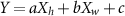
As an initial step in the construction of the phantom library, 'grid-center' and 'anchor' phantoms were constructed as basis/kernel models to be used for deformation to construct the phantom library (see section 2.4). The grid-center phantoms, representing the body size at the grid center (male: 175 cm and 100 kg; female: 165 cm and 95 kg), were constructed by deforming the MRCPs (male: 176 cm and 73 kg; female: 163 cm and 60 kg) following the body-size deformation procedure established by (Lee et al 2019). The grid-center phantoms were then deformed to construct a set of 10 anchor phantoms for each gender in various body-fat percentages (BF%s), covering all BF%s in the phantom library. For the construction of the anchor phantoms, the body shape of the grid-center phantoms was deformed to change the body weight (i.e. the BF%) while maintaining the standing height and all internal organs and tissues except for the breast adipose tissue. The breast adipose tissue, the amount of which was assumed to be in proportion to the BF, was additionally adjusted following the approach of (Lee et al 2019). The body weight according to the target BF% was determined by using the lean body mass (LBM; the body weight minus the body fat) formulas as a function of standing height and body weight, derived by (Deurenberg et al 1991) and (Frankenfield et al 2001). The anchor phantoms represent a typical body shape in accordance with the BF%, which was confirmed by an anatomist. The constructed grid-center and anchor phantoms are shown in figure 3.
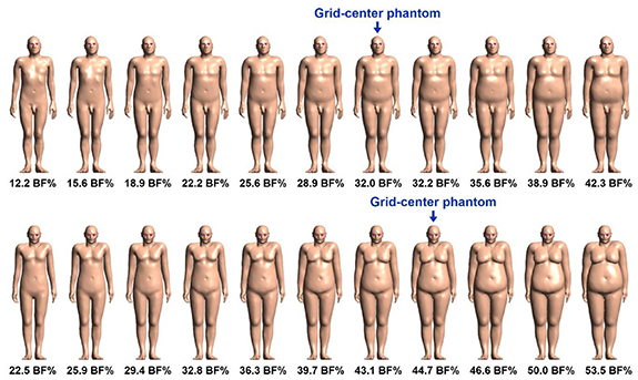
Figure 3. Grid-center and anchor phantoms with their body-fat percentages (BF%s) for adult male (upper) and female (lower).
Download figure:
Standard image High-resolution image2.4. Phantom deformation
The grid-center and anchor phantoms were deformed to construct the phantom library. First, among the grid-center and anchor phantoms, one whose BF% is closest to that of the target phantom in the library was selected. The selected phantom was deformed again by using the deformation procedure of (Lee et al 2019). The deformation procedure can be divided into two main steps: (1) phantom scaling to match the target standing height, sitting height, head dimensions (head height, depth, and breadth), and LBM and (2) phantom adjustment to match the target values of all other anthropometric parameters, to redefine the interior skin structures (e.g. the 50 μm thick radiosensitive target layer), and to match the target mass of the breast adipose tissue. The first step (i.e. phantom scaling) can be easily automated, but this is not the case for the second step (i.e. phantom adjustment). (Lee et al 2019) manually conducted the second step to construct 18 percentile-specific phantoms, thus revealing this manual approach to be labor-intensive and time-consuming, which fact makes it indeed practically challenging to construct ∼200 body-size-dependent phantoms. This issue was fully overcome in the present study by developing and using an in-house C++ program for automatic phantom adjustment.
Figure 4 shows the overall workflow of the adjustment program. For an automatic phantom adjustment, the grid-center and anchor phantoms were segmented into eight regions (i.e. head, foot, upper-arm, waist, hip, thigh, calf, and remaining regions), as shown in figure 5. Accordingly, the phantoms scaled from the grid-center and anchor phantoms were also made of eight regions. First, the exterior body surface segmented into the upper-arm, waist, hip, thigh, and calf regions was adjusted to match the target values of the secondary anthropometric parameters (i.e. upper-arm, waist, buttock, thigh, and calf circumferences and sagittal abdominal diameter). For this adjustment, the facets of the body surface were moved by shifting a vertex in the direction averaged over the normal directions of all of the facets that include the shifting vertex. The movement step was empirically determined as one-fifth of the radius deviation between two circles, the circumferences of which were assumed by the phantom's secondary anthropometric parameters and the target values. For each movement, the program checked to determine if there was any intersection between facets, using the Intersection function in the computational geometry algorithms library (CGAL, https://www.cgal.org/). If an intersection was detected, the involved facets were moved back to the previous locations. The movement was repeated until the phantom's secondary anthropometric parameters were matched to the target values within 5% difference. Likewise, the exterior body surface segmented into the remaining regions was adjusted to match the target body weight within 2% difference; the head and foot regions were not adjusted to maintain the standing heights which had been matched during the first step (i.e. phantom scaling). After the adjustment of the exterior body surface, the adjusted surface was replicated to create three additional surfaces. Then, one of the surfaces was uniformly shrunk to redefine the interior skin surface so that the skin mass was matched to the target value within 1% difference. The other two surfaces also were uniformly shrunk to redefine the skin target layer at a depth of 50–100 μm from the exterior body surface. Finally, the breast adipose tissue was enlarged or shrunk to match the target mass within the 1% difference by repeatedly moving the facets of the breast adipose tissue in the manner done for the adjustment of the exterior body surface. The adjustment program took 16.3 to 45.2 h (24.6 h on average) per phantom with one thread of the Intel® Xeon® CPU E5-2698 v4 (@2.20 GHz with 40 cores (two hyper-threads per core) and 256 GB RAM) in CentOS Linux release 7.3.1611 (Core).
Figure 4. Workflow of developed program for automatic phantom adjustment.
Download figure:
Standard image High-resolution image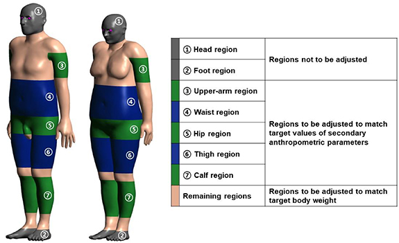
Figure 5. Male (left) and female (right) grid-center phantoms segmented into head, foot, upper-arm, waist, hip, thigh, calf, and remaining regions for automatic phantom adjustment.
Download figure:
Standard image High-resolution image2.5. Monte Carlo dose calculations
Monte Carlo (MC) dose calculations were performed with some selected phantoms from the developed library for external photon exposures in order to determine the influence of body size on dose calculation. To that end, organ/tissue doses calculated using three male phantoms in different body heights (H165W80, H175W80, and H190W80) were compared to see the dosimetric influence of the body height. Organ/tissue doses calculated using three female phantoms in different body weights (H165W45, H165W65, and H165W140) were also compared to see the dosimetric influence of the body weight. The following describes details of MC simulations, according to the RECORDs guideline (Sechopoulos et al 2018). For the MC dose calculations, the selected phantoms in the tetrahedral-mesh format were implemented in the Geant4 MC code (version 10.04, released in December 2017) (Allison et al 2016) by using the G4Tet class, following the implementation procedure of (Yeom et al 2014). The phantoms were assumed to be in vacuum and irradiated by mono-energetic broad-parallel photon beams in three directions (antero-posterior (AP), postero-anterior (PA), and right-lateral (RLAT)). The beam-energy points considered in ICRP Publication 116 (ICRP 2010), ranging from 10 keV to 10 GeV, were used. The number of primary particles varied from 107 to 1010 depending on the energies and body sizes to keep statistical relative errors for all the calculated organ/tissue doses below 5%. Variance reduction techniques were not used in the simulations. The photon beams were simulated, via the G4VUserPrimaryGeneratorAction class, by modeling a 100-cm-radius disk source uniformly emitting photons in the normal direction. Figure 6 shows, as an example, the M_H175W80 phantom irradiated by photon beams in the AP and RLAT geometries. The average absorbed doses for six organs and tissues (i.e. red bone marrow (RBM), colon, lung, stomach, breasts, and gonads) having high tissue-weighting factors (≥ 0.08) (ICRP 2007) were calculated. The absorbed doses for all of the organs and tissues except for the RBM were directly calculated using the G4PSEnergyDeposit class. The RBM absorbed doses were calculated using the dedicated scoring class (Yeom et al 2016b) based on the fluence-to-absorbed-dose response functions (DRFs) derived from micron CT images (Johnson et al 2011). Note that the small and complex structure of the RBM could not be explicitly modeled in the MRCPs (Yeom et al 2016b) used for the construction of the phantom library. The physics library of G4EmLivermorePhysics was used to transport photons and secondary electrons with a secondary range cut value of 1 mm. The simulations were performed on the same server computer with the Intel® Xeon® CPU E5-2698 v4 (@ 2.20 GHz with 40 cores (two hyper-threads per core) and 256 GB RAM) in CentOS Linux release 7.3.1611 (Core). The compiler used was GCC 7.2.0.
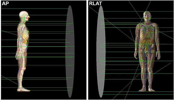
Figure 6. Adult male phantom with standing height of 175 cm and body weight of 80 kg (M_H175W80) irradiated by mono-energetic broad-parallel photon beams in AP (left) and RLAT (right) geometries.
Download figure:
Standard image High-resolution imageAdditional MC simulations were performed to calculate the average absorbed doses for six organs and tissues of the male and female phantoms in three different body weights (male: H175W60, H175W90, and H175W130; female: H165W60, H165W90, and H165W130). In order to determine the dose differences between the two libraries, the calculated organ/tissue doses were compared with those produced by (Chang et al 2018) using the corresponding body-size phantoms from the body-size-dependent phantom library developed in collaboration between the University of Florida and the National Cancer Institute (UF/NCI) (Geyer et al 2014).
3. Results and discussion
3.1. Body-size-dependent phantom library
The body-size-dependent phantom library constructed in the present study is shown in figure 7. The library contains 108 adult male phantoms and 104 adult female phantoms, covering the body sizes of most Caucasian and Asian populations. The male phantoms cover standing heights from 160 to 190 cm and body weights from 50 to 155 kg. The female phantoms cover standing heights from 150 to 175 cm and body weights from 40 to 150 kg. The standing height, sitting height, and head dimensions of the phantoms were matched to the target values within the 0.1% difference. The body weight of the phantoms was matched to the target value within the 2% difference. All of the other anthropometric parameters of the phantoms (i.e. sagittal abdominal diameter and upper-arm, waist, buttock, thigh, and calf circumferences) were matched to the target values within 5% difference. The masses of all of the organs and tissues of the phantoms were automatically determined by the phantom scaling process, except for the skin and breast adipose tissue, the masses of which were matched to the target values within 1% difference.

Figure 7. Constructed phantom library for male (upper) and female (lower).
Download figure:
Standard image High-resolution imageThe variation in organ/tissue masses for 13 randomly selected organs and tissues (i.e. colon, lungs, stomach, breasts, gonads, urinary bladder, oesophagus, liver, thyroid, brain, salivary glands, skin, and residual soft tissue (RST)) is shown in figure 8 by means of organ/tissue mass ratios to adult male and female MRCPs. Most of the organs and tissues, those located in the trunk, show a similar variation, their ratios ranging from 0.73 to 1.57 for males and 0.72 to 1.66 for females; this is because these organs/tissues were scaled strictly in proportion to the LBM. The variation for the brain is the smallest, its ratios ranging from 0.91 to 1.17 for males and 0.92 to 1.16 for females, which is because the organs and tissues located in the head were scaled in proportion to the head dimensions. The variation of the RST is the largest, its ratio ranging from 0.50 to 4.00 for males and 0.55 to 3.98 for females. Note that the RST mainly consists of adipose tissues, showing the greatest variation depending on the body sizes.

Figure 8. Box-and-whisker plots of organ/tissue mass ratios of adult male (upper) and female (lower) phantom library with respect to adult male MRCP (176 cm, 73 kg) and adult female MRCP (163 cm, 60 kg). Filled squares (■) represent mean values. The horizontal line within a box represents the median value. The upper and lower ends of a box represent the 75 and 25 percentile values, respectively. The upper and lower whiskers represent the maximum and minimum values, respectively.
Download figure:
Standard image High-resolution imageFigure 9 shows the variation in organ/tissue depths for 11 randomly selected organs and tissues (i.e. colon, lungs, stomach, breasts, gonads, urinary bladder, oesophagus, liver, thyroid, brain, and salivary glands) from the front, back, and right by means of organ/tissue depth differences with respect to adult male and female MRCPs. It can be seen that the organs/tissues of library phantoms are generally located deeper than those of the MRCPs for both the male and female, which is because the MRCPs have smaller body sizes than most phantoms in the library. Several organs/tissues (e.g. colon and urinary bladder) of all or almost all the female library phantoms tend to be especially deeper from the front than those of the female MRCP; this is because significant modifications were made in several body regions of female library phantoms to match the target values of anthropometric parameters. Note that the MRCPs were developed directly from the CT images of individuals, possibly not satisfying target values of anthropometric parameters corresponding to their standing heights and body weights.

Figure 9. Box-and-whisker plots of organ/tissue depth differences (mm) of adult male (left) and female (right) phantom library with respect to adult male MRCP (176 cm, 73 kg) and adult female MRCP (163 cm, 60 kg) from front (top), back (middle), and right (bottom). Filled squares (■) represent mean values. The horizontal line within a box represents the median value. The upper and lower ends of a box represent the 75 and 25 percentile values, respectively. The upper and lower whiskers represent the maximum and minimum values, respectively.
Download figure:
Standard image High-resolution image3.2. Comparison of organ/tissue doses
Figure 10 shows the ratios of the organ/tissue doses of M_H165W80 and M_H190W80 to those of M_H175W80 for the AP, PA, and RLAT geometries. For the photon energies >0.1 MeV, the ratios tend to be close to unity (mostly between 0.9 and 1.1), which means that the organ/tissue doses among the three phantoms are generally similar within the 10% differences. Relatively large differences up to 30% can be seen at the gonad doses between M_H165W80 and M_H175W80 for the PA geometry and energies >20 MeV. For the photon energies ⩽0.1 MeV, the ratios for most cases are significantly different from unity, and the differences tend to increase as the photon energy decreases. The ratios between M_H165W80 and M_H175W80 are smaller than unity, which means that the organ/tissue doses of M_H165W80 are lower than those of M_H175W80. In contrast, the ratios between M_H190W80 and M_H175W80 are greater than unity, which means that the organ/tissue doses of M_H190W80 are higher than those of M_H175W80. These results demonstrate that the organ/tissue doses tend to increase as the standing height increases, which can be explained by the fact that the thicknesses of the overlying tissues from the body surface to the organs/tissues tend to decrease with increasing standing height, because the standing height with a fixed body weight is in inverse proportion to the BF%.

Figure 10. Ratios of organ/tissue doses of M_H165W80 and M_H190W80 phantoms to those of M_H175W80 phantom for six organs and tissues (RBM, colon, lung, stomach, breasts, and gonads) resulting from external photon exposures in AP, PA, and RLAT irradiation geometries.
Download figure:
Standard image High-resolution imageFigure 11 plots the ratios of the organ/tissue doses of F_H165W45 and F_H165W140 to those of F_H165W65 for the AP, PA, and RLAT geometries. For the photon energies ⩽0.1 MeV, it can also be seen that the ratios are significantly different from unity and that the differences tend to increase by up to several orders of magnitude as the energy decreases. The ratios between F_H165W45 and the F_H165W65 are greater than unity, which means that the organ/tissue doses of F_H165W45 are greater than those of F_H165W65. Contrastingly, the ratios between F_H165W140 and F_H165W65 are smaller than unity, which means that the organ/tissue doses of F_H165W140 are lower than those of F_H165W65. These results clearly demonstrate that the organ/tissue doses tend to decrease as the body weight (mainly body fat) increases, due to the increase in the thicknesses of the overlying tissues from the body surface to the organs/tissues. For the energies >0.1 MeV, the ratios are generally in the range from 0.7 to 2, showing that the dose differences are much less significant than those for the energies ⩽0.1 MeV. Interestingly, for the energies >10 MeV, the opposite dose-difference trend due to body weight was observed; that is, the organ/tissue doses tend to increase even though the body weight increased. This can be explained by the fact that such high energy photons tend to create dose build-up regions by deeper depths and, thus, to deliver greater absorbed doses to the deeper-lying organs/tissues of a heavier person.
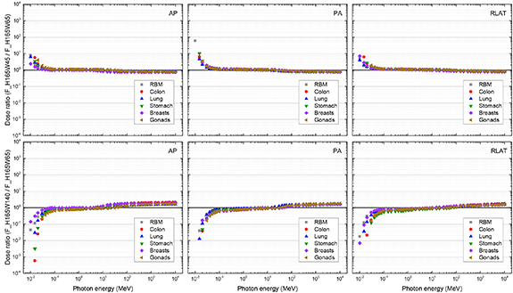
Figure 11. Ratios of organ/tissue doses of F_H165W45 and F_H165W140 phantoms to those of F_H165W65 phantom for six organs and tissues (RBM, colon, lung, stomach, breasts, and gonads) resulting from external photon exposures in AP, PA, and RLAT irradiation geometries.
Download figure:
Standard image High-resolution image3.3. Dose comparison with another body-size-dependent phantom library
Figure 12 plots the ratios of the organ/tissue doses of the phantoms of the same body size (male: H175W60, H175W90, and H175W130; female: H165W60, H165W90, and H165W130) between the MRCP-based phantom library developed in the present study and the UF/NCI phantom library (Chang et al 2018). For the energies >0.1 MeV, the ratios are generally close to unity (mostly between 0.9 and 1.1), meaning that the two libraries provide similar organ/tissue doses for the same body size. Relatively large differences can be seen only for the female breasts, in which the breast doses of the MRCP-based phantoms are greater than those of the UF/NCI phantoms by a factor of about 2 in most cases. For the energies ⩽0.1 MeV, the ratios significantly deviate from unity for most cases, which deviations dramatically increase as the energy decreases by up to several orders of magnitude. That is, the two phantoms, despite having the same body size, provide significantly different organ/tissue doses, which differences are associated with different organ/tissue locations and depths across the different phantom libraries, as stemming from the different original phantoms (MRCPs vs. UF/NCI phantoms). This result raises an important issue: when using the MRCP-based phantom library as well as other existing phantom libraries (Geyer et al 2014, Akhavanallaf et al 2019) for individual dose reconstructions, one should keep in mind that dosimetric uncertainties due to anatomical differences between the phantoms and individuals always exist and would be large, especially for exposure scenarios involving kilovoltage photon beams.

Figure 12. Ratios of organ/tissue doses of phantoms in same body sizes (male: H175W60, H175W90, and H175W130; female: H165W60, H165W90, and H165W130) between library developed in present study and UF/NCI phantom library (Chang et al 2018) for six organs and tissues (RBM, colon, lung, stomach, breasts, and gonads) resulting from external photon exposures in AP and RLAT irradiation geometries.
Download figure:
Standard image High-resolution image
4. Conclusion
In the present study, we established a comprehensive body-size-dependent phantom library based on the new ICRP mesh-type reference computational phantoms (MRCPs) including 108 adult male and 104 adult female phantoms in a wide range of standing heights and body weights representative of most individuals in the Caucasian and Asian populations. The dosimetric influence due to body-size variation was investigated by comparing the organ/tissue doses calculated with the selected phantoms in different standing heights and body weights for external exposures to broad parallel photon beams. It was observed that the calculated organ/tissue doses were indeed strongly associated with the different body sizes, especially for the photon energies ≤0.1 MeV. This finding, despite the limited number of exposure cases, suggests that the use of the body-size-dependent phantom library, relative to MRCPs limited to the reference body size, can significantly improve the accuracy of individual dose estimates for many retrospective dosimetry studies by taking the body size of individuals into account.
Acknowledgments
The authors would like to thank Beom Sun Chung from the Department of Anatomy in Ajou University School of Medicine for his anatomical consultation. This work was supported by the Nuclear Safety Research and Development (NSR&D) Program through the Korea Foundation of Nuclear Safety (KoFONS) funded by the Nuclear Safety and Security Commission (NSSC), by the Basic Science Research Program through the National Research Foundation of Korea (NRF) funded by the Ministry of Science, ICT & Future Planning, and by the Korea Institute of Energy Technology Evaluation and Planning (KETEP) grant funded by the Korea government (MOTIE) (Project No.: 1705006, NRF-2019M2D2A1A02059814, 20191510301040). Two of the authors (Chansoo Choi and Haegin Han) were supported by the Global Ph.D. Fellowship program (Project No.: NRF-2017H1A2A1046391, NRF-2018H1A2A1059767). One of the authors (Yeon Soo Yeom) was supported by a grant of the Korean Health Technology R&D Project through the Korean Health Industry Development Institute (KHIDI), funded by the Ministry of Health & Welfare, Republic of Korea (Project No.: H18C2257).